Clinical presentation
A 19-year-old male was involved in a motor vehicle accident, and was admitted to the hospital secondary to cervical and pelvic fractures and a pulmonary contusion. He had no cardiac history, and his primary-care provider had previously documented normal cardiac physical examinations. Upon admission, the patient was haemodynamically stable with no evidence of haemopericardium. Initial physical examination revealed a heart rate of 50 beats/minute and 3/6 systolic murmur. An electrocardiogram was ordered and it revealed complete – third-degree – heart block. A transthoracic echocardiogram revealed a defect in the membranous ventricular septum with left ventricular-to-right atrial shunt, consistent with a Gerbode ventricular septal defect. Owing to the size of the left ventricular-to-right atrial shunt, surgical repair of his ventricular septal defect before discharge was thought to be indicated. Once he had recovered from his non-cardiac injuries, he underwent surgical ventricular septal defect closure and epicardial pacemaker placement. Intraoperative transoesophageal echocardiogram and surgical findings were consistent with a traumatic Gerbode ventricular septal defect. The patient was discharged following surgical recovery and is doing well.
Imaging findings
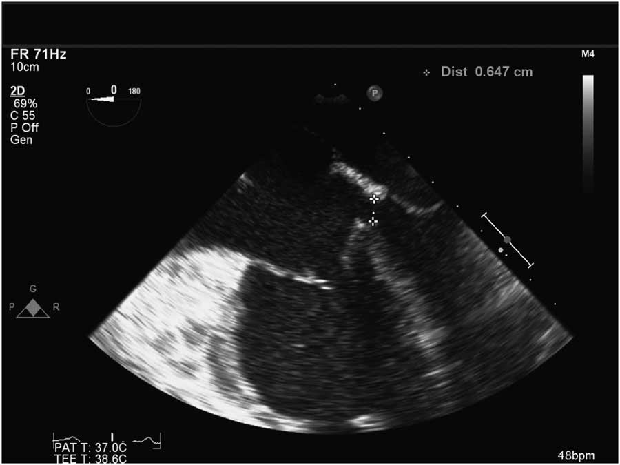
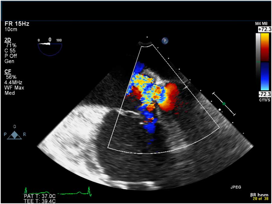
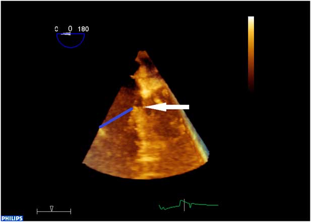
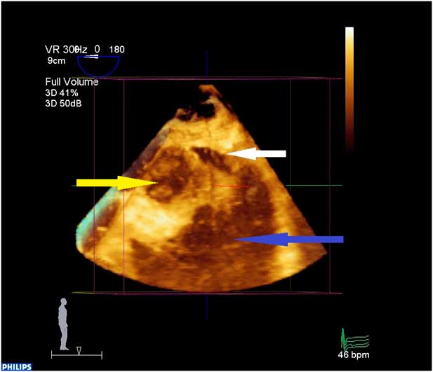
A transthoracic echocardiogram revealed a membranous ventricular septal defect with left ventricle-to-right atrium shunt as well as right atrial and ventricular dilation. A preoperative transoesophageal echocardiogram visualised a moderate ventricular septal defect in the membranous septum between the annular planes of the tricuspid and mitral valves (Figs 1 and 2, Supplementary movies 1 and 2). The peak instantaneous left ventricle-to-right atrium velocity measured by continuous wave Doppler was 4.5 m/second. Three-dimensional imaging confirmed the diagnosis of a traumatic Gerbode ventricular septal defect and allowed for excellent visualisation of the defect and its margins (Figs 3 and 4, Supplementary movies 3 and 4)
Four-chambered, two-dimensional transoesophageal echocardiogram image revealing a Gerbode ventricular septal defect in the membranous ventricular septum between the planes of the tricuspid and mitral valves. The defect measures 6.47 mm in the superior and inferior plane. Margins indicated by callipers.

Four-chambered, color Doppler transoesophageal echocardiogram image revealing a left ventricle-to-right atrium shunt through a Gerbode ventricular septal defect.

Transoesophageal echocardiogram: three-dimensional, four-chambered anterior view. The Gerbode ventricular septal defect is indicated by the white arrow and is located in the membranous septum above the plane of the tricuspid valve (blue line). The defect is fenestrated in nature.

Transoesophageal echocardiogram: three-dimensional view of the ventricular septum as seen from the left ventricle (blue arrow). The aortic valve is indicated by the yellow arrow. The ventricular septal defect is indicated by the white arrow.

Discussion
Gerbode ventricular septal defects are defined as a defect in the membranous ventricular septum between the planes of the tricuspid and mitral valve annuli, resulting in a left ventricle-to-right atrium shunt.Reference Gerbode, Hultgren, Melrose and Osborn 1 Gerbode ventricular septal defects are rare and can be congenital or acquired.Reference Wasserman, Fann, Atwood, Burdon and Fadel 2 , Reference Sasaki and Shimoyama 3 Acquired defects can be caused by trauma, endocarditis, myocardial infarction, or postoperative complications of mitral and aortic valve replacements.Reference Sinisalo, Sreeram, Jokinen and Qureshi 4 This ventricular septal defect was felt to be traumatic, given the patient’s previous history of normal physical examinations and the irregular appearance of the defect’s superior margin. Traumatic heart block is also rare and can occur in isolation or in combination with a traumatic ventricular septal defect.Reference Eckart, Falta and Stewart 5 This case illustrates the presentation of two rare findings occurring in combination. In this case, echocardiography provided prompt and accurate diagnostic information. Three dimensional transoesophageal echocardiography allowed for excellent intraoperative visualisation and characterisation of the defect.
Acknowledgements
None.
Financial Support
This research received no specific grant from any funding agency or from commercial or not-for-profit sectors.
Conflicts of Interest
None.
Ethical Standards
The authors assert that all procedures contributing to this work comply with the ethical standards of the relevant national guidelines on human experimentation and with the Helsinki Declaration of 1975, as revised in 2008.
Supplementary materials
To view supplementary material for this article, please visit http://dx.doi.org/10.1017/S1047951116001359