Respiratory distress is one of the common manifestations of primary pulmonary hypertension of the newborn (PPHN). Reference Stenhorn, Abman, Gleason and Juul1 Conditions that produce high-output cardiac failure viz., vein of Galen, hepatic arteriovenous malformation, and congenital umbilical arteriovenous malformation (UAVM) may mimic PPHN. Reference Murray, Meyerowitz and Hutter2 These high cardiac output conditions can present as early as within 24 hours of life. UAVM is a rare condition of high-output failure and management includes medical stabilisation and surgical repair. Reference Graham, Seashore and Markowitz3–Reference Meyer and Barsness6 Transcatheter closure is an alternative therapy in selected cases. We report the diagnostic and therapeutic challenges of congenital UAVM in a neonate.
Case report
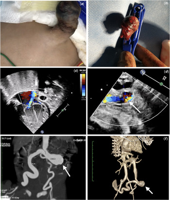
An 8-day-old neonate was presented with respiratory distress and diagnosed as PPHN on functional echocardiogram. Examination showed high-volume pulse, tachycardia, SpO2 92%, hepatomegaly, and 3/6 systolic murmur at the lower sternal border. Careful assessment showed enlarged (Fig 1a) and pulsatile umbilical stump with a bruit over the periumbilical region. Chest X-ray showed cardiomegaly with mildly increased vascularity. Patient deteriorated rapidly and developed renal failure. Hence, the patient was intubated and mechanically ventilated. Transthoracic echocardiogram showed dilated cardiac chambers (Fig 1c), moderate tricuspid regurgitation with a gradient of 64 mmHg. There was hyper pulsatile aorta and dilated external iliac artery. Colour Doppler showed a large umbilical arteriovenous fistula leading to an enlarged umbilical vein (Fig 1d). The ductus venosus was patent and dilated. CT angiogram was done after an improvement from renal failure, and it showed a large communication between the right umbilical artery and vein and dilated right external iliac artery (Fig 1e and f). In view of refractory heart failure, the patient was planned for transcatheter closure of UAVM after informed consent.
(a) Enlarged umbilical cord on day 8 after birth. (b) Dried spontaneously fallen umbilical stump. (c) Transthoracic apical four-chambered view showing dilated right atrium and ventricles with tricuspid regurgitation. (d) Dilated intraabdominal part of the umbilical vein with fistulous communication (arrow). (e, f) CT angiogram showing dilated umbilical vein and right umbilical artery (RUA) and fistulous communication (Nidus arrow). LUA=left umbilical artery.

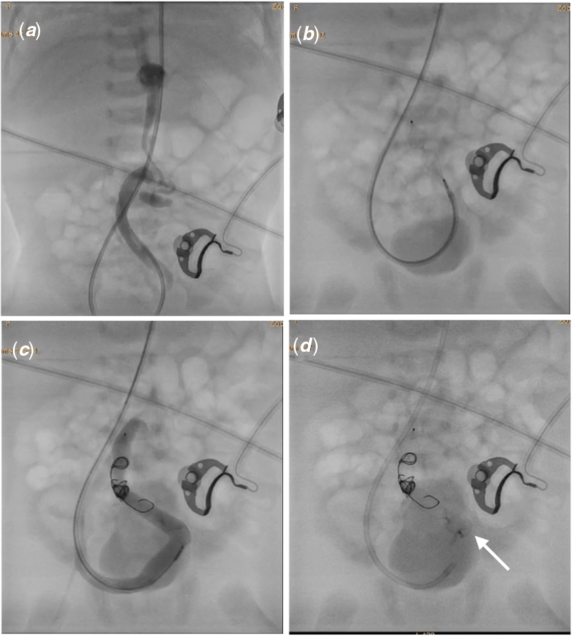
The right axillary and femoral artery were accessed using 5F, 4F sheaths for the interventional procedure. Unexpectedly, the umbilical stump had fallen during the procedure and caused haemorrhage from the umbilicus (Fig 1b). The patient became unstable for a few minutes. But soon purse-string sutures were taken on the umbilical stump and was stabilised with blood transfusion. A 4F Judkin’s right coronary catheter was introduced through the axillary artery using 0.032” hydrophilic angled guidewire. Contrast injection in the right iliac artery showed a large fistula (6.2 mm) communicating between the umbilical artery and vein (Fig 2a). A 5F Judkin’s right coronary guide catheter with a haemostatic check valve was exchanged over the guidewire. 8 mm Amplatzer Vascular Plug 4 (AVP4) (Abbott Vascular, California, United States of America) (Fig 2b) and multiple stainless-steel coils (Cook Medical LLC, Bloomington, Indiana, United States of America) were deployed to close the exit point. But there was significant residual shunt even after 30 minutes. Therefore, a 7/5 KONAR-MFTM (Lifetech Scientific, Shenzhen, China) occluder was placed through the same guiding catheter within the right umbilical artery. Finally, the shunt was abolished (Fig 2c and d). During post-procedure, TTE showed mild tricuspid regurgitation with a gradient of 24 mmHg. Patient showed dramatic improvement and asymptomatic at 6 months of follow-up.
(a) Angiogram in anteroposterior projection showing umbilical arteriovenous malformation. (b) Deployment of Amplatzer Vascular Plug 4 in the distal segment of arterial side of communication. (c) Plain cine picture showing multiple coils and KONAR-MFTM device (broken arrow). (d) Final angiogram still pictures showing insignificant residual leak.

Discussion
Umbilical cord develops due to the fusion of omphalomesenteric yolk stalk and allantoic ducts in the early embryonic period. Graham MS et al. speculated that this anomaly is due to abnormal anastomoses between unabsorbed portion of the ventral segmental arteries and right umbilical vein. Reference Graham, Seashore and Markowitz3 Congenital UAVM is an extremely rare condition. It presents with high-output cardiac failure, PPHN, or haemorrhage from the umbilical stump. Reference Gozar, Gozar and Badiu4,Reference Han, Kim and Jung5 In our case, the neonatal pulmonary vascular resistance didn’t fall due to the high cardiac output state. The direct communication of right iliac artery (right umbilical artery) with the umbilical vein caused high volume through the ductus venosus (DV) into right atrium. The bruit over the periumbilical area clinched the diagnosis of UAVM. There is scanty literature on the management of UAVM. Medical treatment has no significant role. Surgical management is one of the options and carries morbidity. Reference Han, Kim and Jung5–Reference Marcelo Boglione, Alessandro and Reusmann7 In our case, the anatomy appeared suitable for the transcatheter closure. In this case, AVP4 and coils could partially block the shunt, and therefore another device was needed to achieve complete occlusion. The KONAR-MFTM, a double-disc device has the feasibility to deploy through 5F Judkin right coronary guide catheter, hence it was chosen. Heart failure improved after the abolition of UAVM.
Endovascular treatment appears promising as seen in our case. The major advantage of the transcatheter procedure is speedy recovery with less morbidity.
Conclusion
Congenital umbilical arteriovenous malformation is a rare anomaly and needs a high degree of clinical suspicion. Transcatheter endovascular therapy appears to be a good alternative treatment in selected cases. Periprocedural management is crucial to avoid complications like haemorrhage.
Acknowledgements
We thank Dr. Kiran KR Vemireddy, Dr Bhargavi Dhulipudi, Dr. Sujata Patil for helping in managing the case.
Financial support
This research received no specific grant from any funding agency, commercial, or not-for-profit sectors.
Conflict of interest
Dr Nagesewara Koneti receives a royalty from Lifetech Scientific, Shenzhen, China as an inventor of KONAR-MF.
Ethical standards
Informed consent taken from family.