Introduction
In mammals, the ovary is a heterogeneous organ that contains follicles at various stages of development. In order to produce mature oocytes, follicles are activated from the pool of dormant primordial follicles and develop through primary and secondary stages before acquiring an antral cavity. At the antral stage, most follicles undergo atretic degeneration, whereas a few follicles grow further and may reach the pre-ovulatory stage under cyclic gonadotropin stimulation, which occurs after puberty (McGee & Hsueh Reference McGee and Hsueh2000). The development of in vitro conditions to promote primordial follicle development and the study of early folliculogenesis under in vitro circumstances may be important to reduce follicular demise that usually occurs in vivo. In the last three decades, many researchers have dedicated themselves to this topic using different mammalian species (murine: Gao et al., Reference Gao, Wang and WU2007; human: Telfer et al., Reference Telfer, McLaughlin, Ding and Thong2008; bovine: Shimizu et al., Reference Shimizu, Murayama, Sudo, Kawashima, Tetsuka and Miyamoto2008; ovine: Andrade et al., Reference Andrade, Seneda, Alfieri, de Oliveira, Bracarense, Figueiredo and Toniolli2005; goat: Matos et al., 2010; for reviews see Fortune, Reference Fortune2003; Van den Hurk & Zhao, Reference van den Hurk and Zhao2005; Picton et al., Reference Picton, Harris, Muruvi and Chambers2008; Van den Hurk & Santos, Reference van den Hurk and Santos2009; Smitz et al., Reference Smitz, Dolmans, Donnez, Fortune, Hovatta, Jewgenow, Picton, Plancha, Shea, Stouffer, Telfer, Woodruff and Zelinski2010).
Follicles are cultured in defined media, like minimal essential medium (MEM; Martins et al., Reference Martins, Celestino, Saraiva, Chaves, Rossetto, Silva, Lima-Verde, Lopes, Campello and Figueiredo2010), Waymouth medium (Bolamba et al., Reference Bolamba, Floyd, McGlone and Lee2002) or McCoy medium (Duleba et al., Reference Duleba, Pehlivan, Carbone and Spaczynski2001). The type of medium used in in vitro studies has a significant influence on the survival and growth of mammalian follicles. MEM has been used to culture tissues from various species, including bovine (Braw-Tal & Yossefi, Reference Braw-Tal and Yossefi1997) and caprine (Silva et al., Reference Silva, van den Hurk, Costa, Andrade, Nunes, Ferreira, Lôbo and Figueiredo2004) ovarian tissue as well as murine preantral follicles (Zhao et al., Reference Zhao, Tavene, van der Weijden, Bevers and van den Hurk2001). To prevent follicles from atresia, these media mostly include a protein source (like albumin, fetal calf serum or homogeneous serum), antibiotics, antimycotics, transferring (to bind metal ions) and selenium (to bind free radicals), glutamine, pyruvate (energy sources) and occasionally one or more growth factors (like insulin-like growth factor (IGF)-1, epidermal growth factor (EGF), activin-A, growth differentiation factor-9 (GDF-9) and fibroblast growth factor (FGF)-2), hormones (like insulin, follicle stimulating hormone (FSH), growth hormone, thyroid stimulating hormone (TSH) and thyroxine) and/or steroids (like oestradiol, testosterone or androstenedione), sometimes added in a timely manner (for reviews, see Telfer, Reference Telfer1998; Fortune et al., Reference Fortune, Kito and Byrd1999; Van den Hurk et al., Reference van den Hurk, Abir, Telfer and Bevers2000; Picton et al., Reference Picton, Danfour, Harris, Chambers and Huntriss2003; Smitz et al., Reference Smitz, Dolmans, Donnez, Fortune, Hovatta, Jewgenow, Picton, Plancha, Shea, Stouffer, Telfer, Woodruff and Zelinski2010). Ascorbic acid is a protective substance that can be added to the culture medium of early-stage follicles. At physiological concentrations, ascorbic acid, or vitamin C, is a potent free radical scavenger, protecting cells against the damage caused by reactive oxygen species (ROS; Carr & Frei, Reference Carr and Frei1999). The antioxidant properties of ascorbic acid are attributed to its capacity to significantly reduce the damage from ROS, forming ascorbate as a stable free radical (Lutsenko et al., Reference Lutsenko, Carcamo and Golde2002). Ascorbic acid accumulates in granulosa cells, the inner theca, luteal cells, and oocytes (Thomas et al., Reference Thomas, Leask, Srsen, Riley, Spears and Telfer2001). In addition, during follicular growth, ovulation, and corpus luteum formation, the basement membrane and extracellular matrix undergo constant remodeling, thus requiring a great deal of collagen, by which ascorbic acid plays a role in the process of collagen synthesis (Li & Schellhorn, Reference Li and Schellhorn2007). It was furthermore demonstrated that this antioxidant is able to reduce apoptosis in follicles from cows (Thomas et al., Reference Thomas, Leask, Srsen, Riley, Spears and Telfer2001) and rats (Murray et al., Reference Murray, Molinek, Baker, Kojima, Smith, Hillier and Spears2001), and to maintain follicular survival and to promote the in vitro activation and growth of goat primordial follicles (Rossetto et al., Reference Rossetto, Lima-Verde, Matos, Saraiva, Martins, Faustino, Araújo, Silva, Báo, Campello, Figueiredo and Blume2009). To our knowledge, there are no reports describing the dose-dependant effects of ascorbic acid on bovine early-stage follicles cultured in vitro.
The objective of the present study of in vitro cultured bovine ovarian tissue fragments was to evaluate the effects of different concentrations of ascorbic acid on the growth and the viability of the ovarian tissue enclosed early-stage follicles.
Materials and methods
Chemicals
Unless stated otherwise, the culture media, ascorbic acid, and other chemicals used in this study were purchased from Sigma Chemical Co. (St. Louis, MO, USA).
Source of ovaries
Ovarian cortical tissues (n = 10 ovaries) were collected at a local abattoir from five adult, non-pregnant Nelore cows (Bos indicus). The animals were cyclic and in good body condition. Immediately postmortem, the ovaries were washed in 70% alcohol for 10 s followed by two washes with minimum essential medium (MEM) supplemented with 100 μg/ml penicillin and 100 μg/ml streptomycin. The pairs of ovaries were transported in MEM at 20°C, within 1 h to the laboratory.
Experimental protocol
In the laboratory, the ovaries from each animal were stripped of surrounding fat tissue and ligaments and then cut in half. The medulla, large antral follicles and corpora lutea were removed. Subsequently, the ovarian cortex of each pair of ovaries was divided into 25 fragments approximately 3 × 3 × 1 mm. The tissue pieces were then either fixed directly for histological, immunohistochemical and ultrastructural analysis (fresh control) or placed in culture for 2, 4, 6 or 8 days.
Bovine tissues were transferred to 24-well culture dishes containing 1 ml of culture medium. The culture was performed at 39°C in 5% CO2 in a humidified incubator, and all the media were incubated for 1 h prior to use. The basic culture medium (control medium) consisted of MEM (pH 7.2–7.4) supplemented with insulin 6.25 ng/ml, transferrin 6.25 ng/ml and selenium 6.25 ng/ml (ITS), 0.23 mM pyruvate, 2 mM glutamine, 2 mM hypoxanthine, 1.25 mg/ml bovine serum albumin (BSA), and antibiotics (200 UI/ml penicillin and 200 mg/ml streptomycin), and this medium was called MEM+. For the experimental conditions, the medium was supplemented with ascorbic acid at five different concentrations (5, 25, 50, 100 or 200 μg/ml). Ovarian tissue was processed for classical histology, transmission electron microscopy (TEM) and immunohistochemical demonstration of proliferating cell nuclear antigen (PCNA). Each treatment was repeated five times, and the culture medium was replenished every other day.
Morphological analysis and assessment of granulosa cell proliferation by PCNA
Either immediately after recovery or after culture for 2, 4, 6 or 8 days, pieces of ovarian tissue were fixed by immersion in Bouin's fluid for 12 h, dehydrated in a graded series of ethanol solutions, clarified with xylene and embedded in paraffin wax. Thereafter, 5-μm sections of each piece were cut and mounted on glass microscope slides. The majority of these slides was stained with periodic acid Schiff (PAS) and hematoxylin, although every fifth slide was instead stained with antibodies against PCNA (DAKO, Carpinteria, CA, USA), as described previously (Oktay et al., Reference Oktay, Schenken and Nelson1995). For PAS staining, the tissue sections were first deparaffinized using xylene and then rehydrated using 70% ethanol before being stained with periodic acid (Sigma, St. Louis, MO, USA) for 5 min and Schiff reagent for 10 min. For immunohistochemistry, the sections were similarly deparaffinized and rehydrated but then incubated for 8 min at 93–98°C in citric buffer (pH 6). Next, these sections were incubated in 3% H2O2 for 30 min at room temperature (30°C) to block endogenous peroxidase activity and, after washing, bathed in normal rabbit serum for 20 min at room temperature to inhibit non-specific binding. Immunostaining per se was performed by incubation in a 1:100 dilution of the primary antibody (anti-PCNA, Zymed) for 18 h at 8°C followed by washing and incubation in a rabbit anti-mouse second IgG (DAKO, Carpinteria, CA, USA) at a 1:200 dilution for 30 min. For control sections, the primary antibody was replaced by Tris-buffered saline. Finally, streptavidin–biotin complex was added for 30 min at room temperature, followed by diaminobenzidine (DAB) for a further 7 min. After completion of the reactions, the sections were counterstained with hematoxylin. Approximately 150 follicles were randomly evaluated in each medium and culture period.
The developmental stages of the follicles have been defined previously (Silva et al., Reference Silva, van den Hurk, Costa, Andrade, Nunes, Ferreira, Lôbo and Figueiredo2004) as primordial (one layer of flattened granulosa cells around the oocyte) or growing (intermediate, one layer of flattened to cuboidal granulosa cells; primary, one layer of cuboidal granulosa cells; and secondary, two or more layers of cuboidal granulosa cells around the oocyte). Each follicle was evaluated according to the following morphological parameters: (1) integrity of oocyte and granulosa cells; (2) presence or absence of pyknotic bodies; (3) ooplasmic retraction; (4) organisation of granulosa cells; and (5) presence of a non-undulated basement membrane, firmly attached to the granulosa cells. Based on this evaluation, preantral follicles were classified as normal, when a morphologically normal oocyte with a non-pyknotic nucleus was surrounded by granulosa cells organized in discrete layers. Degenerated follicles were defined as those with a retracted oocyte, with a pyknotic nucleus and/or surrounded by disorganized granulosa cells, which were detached from the basement membrane. An average of 150 follicles were evaluated for each treatment (ca. 30 follicles per treatment in one repetition × five repetitions = ca. 150 follicles).
To evaluate follicular activation and growth, only intact follicles with a visible oocyte nucleus were recorded and the proportion of primordial and growing follicles were calculated at day 0 (control) and after 2, 4, 6 or 8 days of culture in the various media tested. Care was taken to count each follicle only once; each follicle was examined in every section in which it appeared and matched with the same follicle on adjacent sections to avoid double counting, thus ensuring that each follicle was counted only once, regardless of its size.
Ultrastructural analysis
Small pieces (1 mm3–from the same fragments used in other analysis) of bovine ovarian tissue were fixed in 2% paraformaldehyde and 2.5% glutaraldehyde in 0. 1 M sodium cacodylate buffer (pH 7.2) for 4 h at room temperature. After fixation, fragments were post-fixed in 1% osmium tetroxide, 0.8% potassium ferricyanide, and 5 mM calcium chloride in 0.1 M sodium cacodylate buffer for 1 h. Subsequently, the samples were dehydrated through a gradient of acetone solutions and the tissues were embedded in Spurr. Semi-thin sections (3 μm) were cut on an ultramicrotome (Reichert Supernova, Heidelberg, Germany) and stained with toluidine blue for light microscopy studies. The ultrathin sections (60–70 nm) were contrasted with uranyl acetate and lead citrate, and examined under a Jeol 1011 (Jeol, Tokyo, Japan) transmission electron microscope. Parameters such as the density and integrity of ooplasmic and granulosa cell organelles, vacuolization, and basement membrane integrity were evaluated.
Statistical analysis
Pair-wise comparisons were done using Tukey's procedure. Chi-squared test was used to compare percentages of follicles with PCNA-positive granulosa cells. All analyses were performed with the Statistical Analysis System (SAS Institute, Cary, NC, USA) and values were considered statistically significant when p < 0.05.
Results
Bovine early-stage follicle survival and activation during in-vitro culture
In total, 3750 early-stage follicles were analyzed: 2013 primordial, 1275 intermediate, 398 primary and 64 secondary follicles. Histological analysis showed that normal and degenerated follicles were found in fragments of ovarian cortex before and after in vitro culture. In degenerated follicles, the main characteristics observed were a retracted oocyte, a pyknotic nucleus and disorganized granulosa cells, with low cellular density.
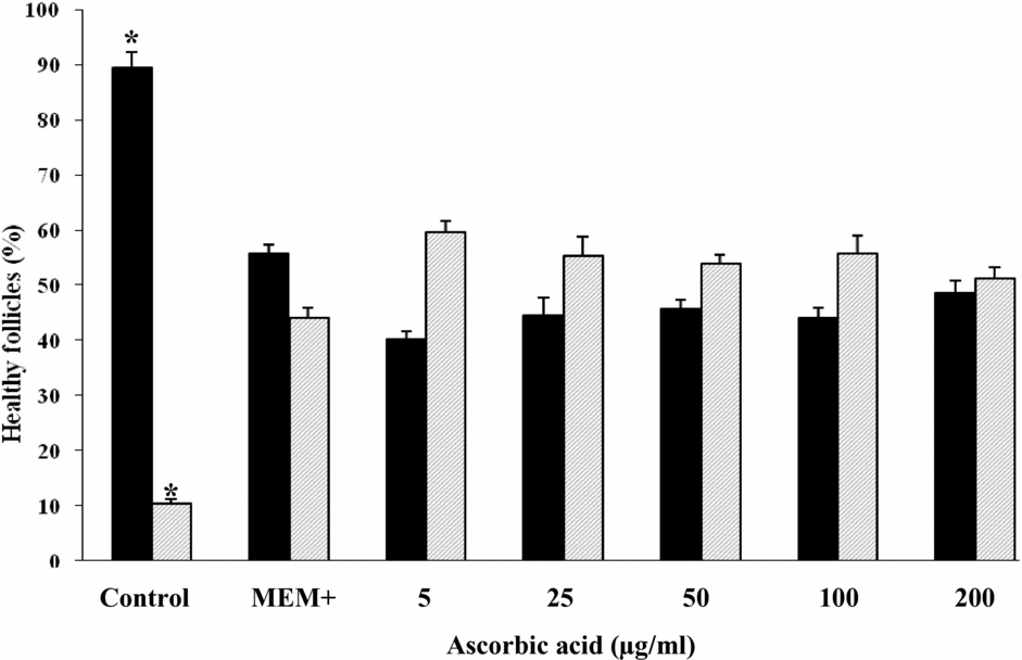
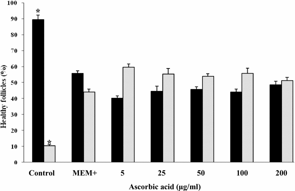
Non-cultured ovarian tissue fragments (day 0) largely contained primordial follicles (89.6%) near to a small percentage of developing follicles (10.4%) (Fig. 1). These percentages had significantly (p < 0.05) decreased and increased, respectively, when ovarian tissues were cultured for 8 days in MEM+ or MEM+ supplemented with five different concentrations of ascorbic acid. Among the ovarian tissues that have been cultured in MEM+ and MEM+ supplemented with five different concentrations of ascorbic acid no significant differences in percentages of primordial follicles nor in those of developing follicles were observed.
Mean ± standard error of the mean (SEM) percentages of healthy early-stage bovine follicles that were at primordial (█) and developing ( ) stages in non-cultured ovarian tissue (control) and in ovarian tissue after in vitro culture for 8 days in minimum essential medium (MEM+) or in MEM+ supplemented with different concentrations (μg/ml) of ascorbic acid. For both primordial-stage and developing-stage follicles, there were differences (p < 0.05 for each) in percentages of healthy follicles between the control group and all cultured groups, but no significant differences in this regard among the cultured groups. *Different (p < 0.05) from other treatments.
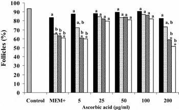
Data for follicular viability are shown in Fig. 2. The percentage of viable follicles was significantly lower (p < 0.05) than that (93.7%) of the control group in ovarian tissues that have been cultured in MEM+ for 4, 6 and 8 days and MEM+ supplemented with 5 or 200 μg/ml of ascorbic acid for 6 or 8 days. The other ascorbic acid treatments did not result in viability percentages that were significantly different from that of follicles from the control group. Regarding the effect of the duration of performed cultures, the percentage of healthy follicles had decreased significantly (p < 0.05) from culture day 4 onwards in MEM+, and from culture day 6 onwards in MEM+ supplemented with 5 or 200 μg/ml of ascorbic acid.
Mean ± standard error of the mean (SEM) percentages of early-stage bovine follicles that were healthy (viable) in non-cultured ovarian tissue (control) and in tissue after culture for 2 days (█) 4 days ( ), 6 days (
 ) or 8 days (□) in MEM+ or MEM+ supplemented with different concentrations of ascorbic acid (μg/ml). *Significant different (p < 0.05) from the Control group. a,bWithin treatments, adjacent bars without a common superscript differ (p < 0.05).

Ultrastructural analysis of bovine early-stage follicles
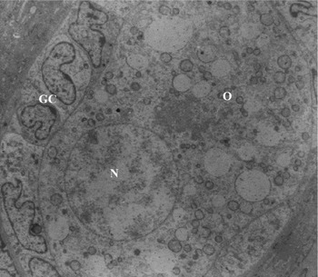
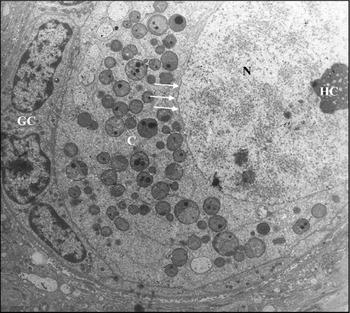
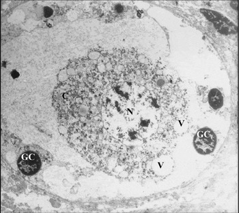
For a better evaluation of follicular integrity, ultrastructural analysis was performed with tissues from fresh control as well as with tissues that had been cultured for 8 days in MEM+ containing 25, 50 or 100 μg/ml of ascorbic acid, all of which showing satisfactory results at histological evaluation with regard to follicular health, growth, and activation. Ultrastructural features of follicles evaluated in fresh control (Fig. 3) or in those cultured for 8 days with 50 μg/ml of ascorbic acid were similar and showed intact oocyte and nuclear membranes and large oocyte nuclei (Fig. 4). Organelles were uniformly distributed in the ooplasm, especially in mitochondria and the endoplasmic reticulum. The homogeneous cytoplasm furthermore contained numerous rounded mitochondria with peripheral cristae and continuous mitochondrial membranes, although there were occasional elongated forms with parallel cristae. The oocyte nucleus had uncondensed chromatin and the nucleolus could generally be observed. In addition, granulosa cells were ultrastructurally normal, showing an elongated and large nucleus with an irregular membrane and a high proportion of nucleus to cytoplasm. The nuclei were irregularly shaped, with loose chromatin in the inner part and small peripheral aggregates of condensed chromatin. In contrast, follicles cultured with 25 or 100 μg/ml of ascorbic acid (Fig. 5) showed signs of degeneration. Such follicles showed an oocyte extremely vacuolated, with vacuoles often being fused thus producing a large vacuolated area. Organelles were more randomly distributed throughout the cytoplasm and signs of endoplasmic reticulum proliferation and damage to mitochondrial membranes and cristae were observed. The oocyte nucleus appeared misshapen and retracted and had a wavy membrane. Granulosa cells look swollen, while the density of cytoplasmic organelles was low. Frequently, the connection between the oocyte and their surrounding granulosa cells had disappeared, while organelles were no longer identifiable.
Electron micrography of a normal bovine preantral follicle (control) showing an oocyte (O) with intact nucleus (N), surrounded by an organized layer of granulosa cells (GC).

Electron micrography of a normal bovine preantral follicle cultured for 8 days with 50 μg/ml of ascorbic acid. Note the presence of an intact nuclear envelope (arrows) between nucleus (N) and cytoplasm (C) and heterochromatin (HC) in the nucleus. GC: granulosa cells.

Electron micrography of a degenerated bovine preantral follicle cultured for 8 days with 100 μg/ml of ascorbic acid. Note the presence of large vacuoles (V) in the oocyte cytoplasm (C) and the swollen granulosa cells (GC). N: nucleus.

Evaluation of bovine granulosa cell proliferation using PCNA
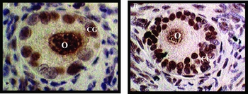
Only intact follicles were used for PCNA evaluation (Fig. 6). In general, marked primordial follicles had only one PCNA-positive granulosa cell, while intermediate and primary follicles showed one to four positive granulosa cells. Secondary follicles presented a higher number of PCNA-labelled granulosa cells (up to 15 cells). In non-cultured bovine ovarian cortex only 5.7% of the primordial follicles had a PCNA-positive granulosa cell, while 38.4% of the developing follicles (i.e. intermediate, primary and secondary follicles) showed at least one PCNA-positive granulosa cell (Table 1). Only the percentages of primordial and developing follicles with stained granulosa cells after in vitro culture with 50 μg/ml of ascorbic acid were significantly higher than those in non-cultured ovarian tissue and in tissue cultured in presence of 25 or 100 μg/ml ascorbic acid (p <0.05); the tissues from the last two treatments showed comparable percentages of PCNA-positive granulosa cells as found in those of the control group (p < 0.05).
Sections of bovine ovarian tissue after in vitro culture, showing (A) a primary and (B) a secondary follicle with granulosa cell nuclei that stain brown, i.e. positive for PCNA. O: oocyte nucleus; GC: granulosa cells.

Percentages of primordial and developing follicles with proliferating cell nuclear antigen (PCNA)-positive granulosa cells in non-cultured ovarian tissue and tissue cultured for 8 days in minimal essential medium (MEM+) (control) and MEM+ supplemented with 25, 50 or 100 μg/ml of ascorbic acid.

a In vitro culture for 8 days.
b Significant different (p < 0.05).
Discussion
This study has demonstrated a positive effect of ascorbic acid on the survival, activation and growth of bovine early-stage follicles during 8 days of in vitro culture.
In vivo, the transition of primordial to primary follicles and subsequent development to the secondary stage is characterized by a slow transformation of granulosa cells from a flattened to a cuboidal shape and a subsequent slow proliferation of these cells over approximately 25 (sheep: McNatty et al., Reference McNatty, Smith, Hudson, Heath, Tisdall and Braw-Tal1995), 30 (cow: Braw-Tal & Yossefi, Reference Braw-Tal and Yossefi1997) or 120 (woman: Gougeon, Reference Gougeon1996) days. The non-growing population of primordial follicles is located in a thin, relatively avascular layer outermost in the ovarian cortex, just beneath the tunica albuginea, whereas the growing follicles are always found at the richly vascularized cortico-medullary border. This situation suggests that activation and growth of primordial follicles depends on blood-borne nutrients, hormones, and/or growth factors (McNatty et al., Reference McNatty, Fidler, Juengel, Quirke, Smith, Heath, Lundy, O'Connell and Tisdall2000). Culture in vitro appears to induce activation of primordial follicles, probably because the media are richer in nutrients, oxygen, insulin, etc than the outermost ovarian cortex. Such environmental changes may stimulate follicle development either directly, or indirectly via the release of stimulatory factors or cessation of production of inhibitory factors by stromal, granulosa or pre-thecal cells within the cultured ovarian cortical tissue.
In the present study, tested media did not seem to be equally effective in maintaining the survival and development of bovine early-stage follicles. Based on histological results, after 8 days of in vitro culture, MEM+ supplemented with 25, 50 or 100 μg/ml of ascorbic acid showed better results than the medium supplemented with MEM+ only or MEM+ supplemented with 5 or 200 μg/ml of ascorbic acid. In correspondence with our results, Thomas et al. (Reference Thomas, Leask, Srsen, Riley, Spears and Telfer2001) previously demonstrated the maintenance of follicular integrity and survival after in vitro culture of isolated bovine early-stage follicles for 12 days in medium containing 50 μg/ml of ascorbic acid. The in vitro culture results in higher oxygen concentrations than in vivo environments, leading to an increased level of reactive oxygen species (ROS; Luvoni et al., Reference Luvoni, Keskintepe and Brackett1996). Polyunsaturated lipids of cellular membranes are very sensitive to peroxidation by ROS. At physiological concentrations, ascorbic acid is an antioxidant that protects cells against the damage caused by ROS (Carr and Frei, Reference Carr and Frei1999). Among the actions of ascorbic acid are the prevention of DNA mutations induced by oxidation (Lutsenko et al., Reference Lutsenko, Carcamo and Golde2002; Sweetman et al., Reference Sweetman, Strain and McKelvey-Martin1997) and protection against lipidic peroxidation (Barja et al., Reference Barja, Lopez-Torres, Perez-Campo, Rojas, Cadenas, Prat and Pamplona1994; Kimura et al., Reference Kimura, Yamada, Morita, Ikeda and Matsuo1992). Ascorbic acid accumulates in the granulosa cells, inner theca, luteal cells, and oocytes (Thomas et al., Reference Thomas, Leask, Srsen, Riley, Spears and Telfer2001). Murray et al. (Reference Murray, Molinek, Baker, Kojima, Smith, Hillier and Spears2001) observed that, at a concentration of 50 μg/ml, ascorbic acid promoted an increase in basement membrane remodelling and granulosa cell survival in isolated mouse late secondary follicles cultured in vitro for 6 days. During follicular growth, ovulation and corpus luteum formation, the basement membrane and extracellular matrix undergo constant remodelling and therefore need a large amount of collagen. In this way, ascorbic acid acts intensively in follicular growth through its action in the biosynthesis of collagen, which influences the process of cellular membrane remodeling (Li & Schellhorn, Reference Li and Schellhorn2007).
After 8 days of culture, the use of MEM+ only, 5 or 200 μg/ml of ascorbic acid reduced the percentage of normal early-stage follicles in comparison to that present in the ovarian cortical tissue of the control group. When using concentrations of 25, 50 or 100 μg/ml, the percentage of normal follicles remained similar to that of the control group. The knowledge that some antioxidant substances act as pro-oxidants when used in high concentrations (Andrade et al., Reference Andrade, Melo-Sterza, Seneda and Alfieri2010), could explain that ascorbic acid at concentrations higher than 100 μg/ml is toxic to cattle early-stage follicles cultured in vitro. Furthermore, high concentrations of ascorbic acid can inhibit important physiological processes in the ovary, resulting in follicular degeneration (Murray et al., Reference Murray, Molinek, Baker, Kojima, Smith, Hillier and Spears2001). Moreover, high concentrations of ascorbic acid may promote oxidative damage to cellular DNA, especially in the presence of Cu2+ and Fe2+ ions, which react with hydrogen peroxide and lead to the formation of the highly reactive hydroxyl radicals that cause damage (Li & Schellhorn, Reference Li and Schellhorn2007). Olson and Seidel (Reference Olson and Seidel2000) reported that co-supplementation of ascorbic acid at a concentration of 100 mM each had a deleterious effect on the embryo culture medium. The balance between antioxidants and pro-oxidants must be preserved in the cells. An excess of antioxidant compounds can increase the redox state, which may have deleterious effects on embryos (Guérin et al., Reference Guérin, El Mouatassim and Ménézo2001). In the present study, the ascorbic acid free medium (MEM+) and the medium supplemented with the lowest (5 μg/ml) or highest (200 μg/ml) dose of ascorbic acid were not effective in maintaining follicular viability during in vitro culture.
Several authors have emphasized the importance of TEM after in vitro culture of early-stage follicles, as it gives close insight into the ultrastructural characteristics of follicles, allowing a better evaluation of their quality (Van den Hurk et al., Reference van den Hurk, Spek, Hage, Fair, Ralph and Schotanus1998; Zhao et al., Reference Zhao, Dorland, Taverne, van der Weijden, Bevers and van den Hurk2000; Salehnia et al., Reference Salehnia, Moghadam and Velojerdi2002). In the present study, bovine early-stage follicles that have been cultured for 8 days in MEM+ supplemented with 50 μg/ml of ascorbic acid appeared ultrastructurally normal, which confirmed the results obtained in the histological studies from this test group. In contrast, most follicles from ovarian tissues cultured in presence of 25 or 100 μg/ml ascorbic acid, i.e. groups that also largely consist of histologically normal follicles, showed various ultrastructural signs of initial degeneration after 8 days of culture and besides exhibited more obvious degenerative features, like ooplasm vacuolization. Cytoplasmic vacuoles are characteristic signs of degeneration in oocytes (Silva et al., Reference Silva, Lucci, Carvalho, Báo, Costa, Santos and Figueiredo2000), granulosa (Hay et al., Reference Hay, Cran and Moor1976) and cumulus cells (Assey et al., Reference Assey, Hyttel, Roche and Boland1994) and may represent endoplasmic reticulum swelling (Tassel & Kennedy, Reference Tassel and Kennedy1980) or altered mitochondrial structure (Fuku et al., Reference Fuku, Xia and Downey1995). Therefore, we conclude from our histological and ultrastructural findings that ascorbic acid exerts its optimal effect on bovine follicular integrity and survival, when added to culture media in a concentration of 50 μg/ml. This concentration is exactly the dose used in the studies of Thomas and co-workers (2001) with isolated bovine follicles, who based their choice for the ascorbic acid concentration on results from experiments with mouse late secondary follicles (Murray et al., (Reference Murray, Molinek, Baker, Kojima, Smith, Hillier and Spears2001).
The PCNA staining of granulosa cells was indicative of their proliferation. PCNA staining in oocytes may be due to DNA repair (Downey et al., Reference Downey, Tan and So1990) during the intense RNA transcription that occurs in growing oocytes (Lintern-Moore & Moore, Reference Lintern-Moore and Moore1979). In agreement with our findings, the onset of bovine primordial follicle growth was also accompanied by PCNA expression in granulosa cells and oocytes (Wandji et al., Reference Wandji, Eppig and Fortune1996), which shows that mitotic activity of granulosa cells is initiated as soon as the primordial follicles leave the resting pool and start to grow. In the current study, the proportion of follicles stained with PCNA had significantly increased after 8 days of culture, but only when ascorbic acid was added to the culture medium in a concentration of 50 μg/ml. Thus, only at this particular concentration ascorbic acid appears able to stimulate the proliferation of granulosa cells. Such an increased proliferation of granulosa cells, in in vitro cultured developing bovine follicles corresponds with that observed in previous in vivo studies with other mammals (humans: Oktay et al., Reference Oktay, Karlikaya, Akman, Ojakian and Oktay2000; rats: Oktay et al., Reference Oktay, Schenken and Nelson1995; goats: Silva et al., Reference Silva, van den Hurk, Costa, Andrade, Nunes, Ferreira, Lôbo and Figueiredo2004) and indicates that the in vitro cultured follicles grow through proliferation of granulosa cells. In contrast to growing follicles, primordial follicles show a low percentage of PCNA expression in granulosa cells. Similar results were described in rats (Meredith et al., Reference Meredith, Dudenhoeffer and Jackson2000) and monkeys (Gougeon & Busso, Reference Gougeon and Busso2000), while mitotic activity had increased when granulosa cells become cuboidal. Cell–cell interactions among oocyte, granulosa and stromal cells have been shown to be essential for the activation of primordial follicles (reviewed by McGee & Hsueh (2000); Picton (Reference Picton2001); and Eppig (Reference Eppig2001)).
In summary, dose dependently, ascorbic acid has an important role in bovine early folliculogenesis in vitro. At a concentration of 50 μg/ml, ascorbic acid realizes optimal early-stage follicle survival, based on histological and ultrastructural features, and promotes the activation of primordial follicles and further follicle growth.
Financial support
E.R. Andrade and A.A. Alfieri are recipients of a PRODOC/CAPES fellowship.