




No CrossRef data available.
Article contents
Purification and characterization of a saccharide-binding protein from penetration glands of Diplostomum pseudospathaceum – a bifunctional molecule with cysteine protease activity
Published online by Cambridge University Press: 09 October 2003
Abstract
A β-1,3-glucan-binding lectin from the penetration glands of Diplostomum pseudospathaceum cercariae was isolated by affinity chromatography using yeast glucan and curdlan as affinity matrices. Further purification to homogeneity was performed by cation-exchange chromatography. The protein migrated as a double band around 24 kDa in gels after SDS–PAGE. The protein is of strongly basic nature – its pI shown by native IEF was around 10. The mass of the protein determined by MALDI-TOF mass spectrometry was 23·9 kDa. N-terminal sequence as well as some internal sequences showed significant alignments with several cysteine protease sequences found in databases. The protein bound a biotinylated synthetic analogue of the irreversible inhibitor of cysteine proteases, E-64 and, moreover, its proteolytic activity was demonstrated in substrate gels. The enzymatic activity could be inhibited by the cysteine protease inhibitor E-64; therefore, the investigated protein was considered to be a bifunctional molecule possessing both lectin and enzyme activities. Glycanohydrolytic activity was not proved. The detected characters of this molecule lead to a hypothesis on its role in penetration of Diplostomum cercariae into fish hosts – that of binding to the carbohydrates of fish mucus and concurrent cleaving of protein components of the mucus and skin.
Information
- Type
- Research Article
- Information
- Copyright
- 2003 Cambridge University Press
INTRODUCTION
Protein–carbohydrate interactions in biological systems represent an important part of recognition on a molecular basis. Carbohydrate-binding proteins, namely lectins, act in cell–cell recognition, cell adhesion to extracellular matrix, cell signalling, discrimination between self and non-self by the immune system etc (for reviews see Gabius & Gabius, 1997; Sharon & Lis, 1989). Among them, glucan-binding proteins (GBPs) occupy an important position (e.g. Söderhäll & Thörnqvist, 1997). They recognize glucan components of bacterial and fungal cell walls, opsonize pathogens and mediate their phagocytosis by effector immune cells. GBPs occur as soluble and cell membrane-bound molecules. Both forms were found in many invertebrates, e.g. annelids (Beschin et al. 1998), crustaceans (Duvic & Söderhäll, 1990), insects (Ochiai & Ashida, 1988), and also in vertebrates (Czop & Austen, 1985). Here they can serve as activators of the innate immune response – the prophenoloxidase/phenoloxidase cascade leading to melanization in invertebrates (see review by Söderhäll, 1992) and the complement system in vertebrates (e.g. Honda et al. 1986).
For the isolation of β-1,3-glucan-binding proteins from invertebrates, few approaches employing affinity techniques have been used. Successful attempts were made using affinity precipitation with a soluble β-1,3-glucan laminarin (Duvic & Söderhäll, 1990), insoluble glucans of different origin (Sritunyalucksana, Lee & Söderhäll, 2002) and soluble glucan immobilized on Sepharose beads (Söderhäll et al. 1988).
In our previous work it has been shown that a GBP occurs in penetration glands of cercariae of the bird schistosome Trichobilharzia szidati (Horák et al. 1997) and the fish eye fluke Diplostomum pseudospathaceum (Mikĕs & Horák, 2001). Although the function of the protein has not yet been fully elucidated, it is unlikely to have similar functions as GBPs in immune systems of invertebrates. According to the localization in penetration glands and its binding specificity for saccharide chains containing β-1,3(4)-linked D-glucose and D-galactose and their derivatives such as sugar acids and aminoacetyls, it is believed to play a role in cercarial penetration into the host, probably as a tissue recognition or antibody neutralizing factor (Mikĕs & Horák, 2001). The fact that β-1,3-glucan (laminarin) was a high-affinity ligand for the Diplostomum lectin can be explained to a certain extent by its structural similarity with glycosaminoglycan chains which seem to be natural ligands of the lectin.
This work has focussed on the purification of the partially described Diplostomum lectin by means of affinity chromatography and ion-exchange chromatography as well as further characterization of its physico-chemical properties and some functional features. As some carbohydrate-binding proteins work as multifunctional molecules, i.e. they possess an active enzyme site besides the carbohydrate-binding domain (e.g. Topferpetersen et al. 1990), attention was paid to conceivable enzymatic – namely histolytic – potential of the studied protein, particularly proteolytic and glycanohydrolytic activities.
MATERIALS AND METHODS
Parasite and reagents
Diplostomum pseudospathaceum Niewiadomska, 1984 cercariae were obtained from Lymnaea stagnalis snails collected in ponds near the towns Třeboň and Blatná, Czech Republic. The snails were separated in 100 ml beakers and placed under the light to enhance cercarial emergence. Cercariae were collected and treated as described previously (Mikĕs & Horák, 2001). Supernatants of homogenates were prepared either with a cocktail of protease inhibitors, 200 μMN-α-p-tosyl-L-lysine chloromethyl ketone (TLCK), 20 μM trans-epoxysuccinyl-L-leucilamido-(4-guanidino) butane (E-64) and 200 μM leupeptin, or as inhibitor-free (for enzyme studies). The biotinylated analogue of the cysteine protease inhibitor E-64 (DCG-04) was a kind gift of Dr Caffrey and Dr Greenbaum, Tropical Disease Research Unit, University of California, San Francisco. Bio-Lyte ampholytes were purchased from Bio-Rad. Other chemicals were from Sigma, unless otherwise noted.
Affinity chromatography
Insoluble yeast β-1,3-glucan (from Saccharomyces cerevisiae) and curdlan (insoluble β-1,3-glucan from Alcaligenes faecalis) were washed 3 times in 20 mM MOPS buffer, pH 7·0, containing 150 mM NaCl (MBS) and 0·05% Triton X-100 (T-MBS). Typically, 300 mg (dry weight) of each polysaccharide was used as a batch. Washed and decanted glucans were resuspended in Diplostomum samples (2 mg/ml, 12 ml in a 15 ml vol. Falcon tube) and gently shaken for 90 min at 8 °C. The glucans with bound proteins were centrifuged (500 g, 2 min) and washed 8 times with T-MBS. Two final washes with 3 ml of MBS were performed using 0·22 μm Ultrafree-CL filtration devices (Millipore) centrifuged at 400 g. Proteins were eluted with 20 mM MOPS buffer, pH 7·0, containing 1 M NaCl. The eluates were desalted and transferred to 20 mM MOPS, pH 7·0, by centrifugation on Centricon-10 filters (Amicon).
Ion-exchange FPLC
Cation-exchange chromatography was performed on BioLogic FPLC apparatus (Bio-Rad) using the UNO S1R column (Bio-Rad) equilibrated with 20 mM MOPS, pH 7·0. Proteins were eluted using a linear gradient of ionic strenghth (0–1 M NaCl in the same buffer). Fractions of 0·5 ml were collected, pooled if needed, concentrated and transferred into 5 mM MOPS buffer, pH 7·0, by centrifugation on Centricon-10 filters.
Electrophoresis and zymography
Individual protein fractions were characterized by SDS–PAGE under reducing (2-mercaptoethanol) and non-reducing conditions (Laemmli, 1970). Samples in sample buffer were boiled for 3 min. Separation was performed in 13% slab gels, 0·75 mm thick, at a constant voltage 150 V using MiniProtean-3 apparatus (Bio-Rad). Typically, proteins were loaded as follows: 15 μg of homogenate supernatants, 5 μg of affinity eluates and 2 μg of FPLC-purified proteins per well. For Edman degradation and mass spectrometry, the reducing agent in the sample buffer was 4 mM dithiothreitol (DTT) and 3 μg of purified protein were loaded. For detection of potential enzymatic activities by zymographic analysis, whole homogenates were used and the same system was employed as described above except that the samples were not boiled but incubated with the sample buffers (either non-reducing or reducing with 4 mM DTT) for 30 min at RT. The gels were copolymerized with substrates – 0·002% hyaluronic acid or 0·1% gelatin. Trypsin (0·1 μg/well) and sheep testicular hyaluronidase (0·2 μg/well) served as positive controls. Following electrophoresis, the gels were washed in 0·1 M MES buffer, pH 6·0, containing 1% Triton X-100 (30 min, 4 changes) and incubated overnight in a moist chamber at 37 °C. Then the gels were briefly washed with water. Gelatin gels were stained with Coomassie Brilliant Blue R-250 (CBB). Areas of proteolysis appeared as colourless bands on a blue background after destaining. Gels with hyaluronic acid were soaked in 50% formamide (30 min) and stained overnight in the dark with Stains-all (Sigma) solution (0·1 mg/ml in 50% formamide). Areas of lysis appeared as pink bands on a blue background after destaining in water.
Isoelectric focussing
Native IEF using the MiniProtean-3 apparatus cooled on ice was performed in 0·75 mm thick polyacrylamide gels (5%) containing 10% (v/v) glycerol and either 6% (v/v) Ampholine 3·5–10 or 10% (v/v) Bio-Lyte 8–10+1% (v/v) Ampholine 3·5–10. Samples (2 μg of protein) were mixed with an equal volume of sample buffer (60% glycerol and either 12% Ampholine or 20% Bio-Lyte+2% Ampholine) and loaded. Electrode solutions were 25 mM sodium hydroxide (catholyte) and 20 mM acetic acid (anolyte). The gels were run at constant voltages 200 V for 90 min and 400 V for another 90 min. Gradients of pH in gels were measured with Glass Micro-pH Combination Electrode (Orion Research Inc., MA). Finally, the gels were fixed in 10% trichloroacetic acid (TCA) for 10 min followed by 1% TCA overnight, washed in a solution of 0·25% SDS in ethanol[ratio ]acetic acid[ratio ]water (33[ratio ]10[ratio ]57) and stained with CBB.
Electrotransfer
For Edman degradation, proteins were electroblotted to PVDF membrane using a SEMI-PHOR (Hoeffer Scientific Instruments) semi-dry transblotter at constant current density 1·2 mA/cm2 for 60 min. The membrane was stained with CBB, destained in 40% methanol+10% acetic acid for 1 h, washed with distilled water and air-dried.
In order to determine potential protease active site in the molecule of the Diplostomum lectin, a ligand blotting technique was employed; DCG-04 – a biotinylated synthetic analogue of the irreversible cysteine protease inhibitor E-64 [trans-epoxysuccinyl-L-leucylamido-(4-guanidino) butane] – was used as a ligand (Greenbaum et al. 2000). Cercarial homogenate (4 μg) was treated with 5 mM DTT (final conc.) for 10 min. Then, E-64 was added (only to the control – 100 μM, 20 min) followed by DCG-04 (5 μM, 30 min, all samples). Finally, the samples were incubated with reducing (2 mM DTT) sample buffer at 60 °C for 5 min. SDS–PAGE was performed as described above. Proteins were electroblotted onto NC-2 nitrocellulose membrane (Serva) at constant current density 1 mA/cm2 for 90 min. After transfer, the membrane was washed 3×5 min with 20 mM Tris–HCl, pH 7·8, +150 mM NaCl (TBS) containing 0·1% Tween-20 and 1×5 min with TBS+0·05% Tween-20 (T-TBS). Blocking was performed using 5% BSA in T-TBS for 1 h. The membrane was then incubated with avidin-peroxidase conjugate (2·5 μg/ml, 30 min), washed 3×5 min in T-TBS and developed in substrate solution (0·1 M Tris–HCl, pH 7·6, 0·6 mM 3,3′-diaminobenzidine, 0·01% H2O2).
Dot tests for enzyme activities
Selected fractions of purified proteins were tested for glycanohydrolytic and proteolytic activities using a dot-method on gels with copolymerized substrates (0·002% hyaluronic acid and 0·1% gelatin, respectively). Polyacrylamide gels (10%) were prepared with either 20 mM acetate buffer, pH 5·0, or 20 mM HEPES buffer, pH 7·5. Polymerized gels were placed into a moist chamber and the samples were applied onto their surface (0·1 μg in 1 μl). Sheep testicular hyaluronidase (0·06 μg in 1 μl) and trypsin (0·03 μg in 1 μl) served as positive controls. For protease studies, one set of samples was also pre-incubated with 2 mM DTT; samples mixed with irreversible inhibitors of serine and cysteine proteases (100 μM TLCK and 10 μM E-64) served as controls. After overnight incubation at 37 °C, the gels were washed 2×5 min in distilled water. Gelatin gels were stained with CBB. Hyaluronan gels were soaked in 50% formamide, stained with 100 μg/ml Stains-all dye in 50% formamide and destained in water.
Automated N-terminal sequencing
The more intense lower band (designated L2) was excised from PVDF membrane and inserted into the cartridge of the Protein Sequencer LF 3600 (Beckman). The sample was subjected to 20 cycles of automated Edman degradation.
Sample preparation for mass spectrometry
CBB-stained protein bands (the less intense upper band designated L1 and the lower L2) were cut from the gel and washed several times with 10 mM DTT, 0·1 M 4-ethylmorpholine acetate (pH 8·1) in 50% acetonitrile (MeCN). After complete destaining, the gel was washed with water, shrunk by dehydration with MeCN and reswollen again in water. The gel was partly dried using a SpeedVac concentrator and then reconstituted with Cleavage Buffer containing 0·01% 2-mercaptoethanol, 0·1 M 4-ethylmorpholine acetate, 10% MeCN, 1 mM CaCl2 and 50 ng/μl sequencing grade trypsin (Promega). Digestion was carried out overnight at 37 °C, the resulting peptides were extracted with 40% MeCN/1% trifluoroacetic acid (TFA). One half of the volume was subjected to mass spectrometric analysis by matrix-assisted laser desorption/ionization time-of-flight mass spectrometry (MALDI-TOF MS) and by high performance liquid chromatography – electrospray ionization tandem mass spectrometry (μ-HPLC/nano-ESI-MS/MS). The second half of the sample was modified by ethylesterification with ethanolic solution of acetyl chloride, dried and subjected to mass spectrometric analysis. The sample obtained after affinity chromatography and ion-exchange chromatography (designated Lsol, corresponding to peak A) was further purified using size-exclusion HPLC and the protein from the corresponding sharp peak was desalted, transferred into the Cleavage Buffer and digested in solution. Peptides were analysed as mentioned above. The purity of the protein in solution was previously confirmed by MALDI-TOF experiments with non-digested sample.
Mass spectrometry analysis
Mass spectra were measured on a MALDI-TOF mass spectrometer BIFLEX (Bruker-Franzen, Bremen, Germany) equipped with a nitrogen laser (337 nm) and gridless delayed extraction ion source. For the peptide analysis, a saturated solution of α-cyano-4-hydroxy-cinnamic acid in 50% MeCN/0·2% TFA was used as a MALDI matrix; 1 μl of matrix solution was mixed with 1 μl of the sample on the target and the droplet was allowed to dry at ambient temperature. Spectra were calibrated externally using the monoisotopic [M+H]+ ion of peptide standard somatostatin (Sigma). Ion acceleration voltage was 19 kV and the reflectron voltage was set to 20 kV. For determination of the mass of the whole lectin molecule, the system was calibrated using carbonic anhydrase (29 kDa) and lysozyme (14·3 kDa). A saturated solution of sinapinic acid in 30% MeCN was used as MALDI matrix. Spectra were acquired in a linear mode.
Tryptic peptide mixture was applied on the capillary column (0·18×100 mm) packed with 10 cm of a reversed phase resin (MAGIC C-18, 200 Å, 5 μm, MichromBioresources). Peptides were separated using gradient elution: 40 min from 5% MeCN/0·5% acetic acid (AcA) to 35% MeCN/0·4% AcA, 5 min from 40% MeCN/0·5% AcA to 95% MeCN/0·4% AcA at a flow rate of 1 μl/min. The column was connected to an LCQDECA ion trap mass spectrometer (ThermoQuest, San Jose, CA) equipped with a nanoelectrospray ion source. Spray voltage was held at 1·2 kV, tube lens voltage was −2 V. The heated capillary was kept at 180 °C with a voltage of 13 V. Collision energy was set to 42 units and the activation time was 30 ms. The instrument was set to acquire full MS spectrum (mass range 350–1300 a.m.u.) followed by the MS/MS scan of the most intense ion from preceeding full MS scan. Selected spectra were interpreted manually.
Peptide sequence data management
N-terminal peptide sequence obtained by Edman degradation as well as peptide sequences obtained by de novo sequencing using tandem mass spectrometry were compared with protein sequences contained in all non-redundant GenBank CDS translations+PDB+SwissProt+PIR+PRF databases using the protein–protein BLAST at NCBI web site (search for short nearly exact matches). Selected peptides with high similarity were aligned using ClustalW tool at EMBL-EBI web site. Resultant alignments were managed using BoxShade tool for alignment visualization. Searches for putative functional domains in the peptides were performed against SWISS-PROT and PDB databases using PROSITE scan at ExPASy web site.
RESULTS
The yield of affinity chromatography preparations in the typical arrangement described above was approximately 150 μg of protein in the eluate from yeast glucan and 50 μg in the eluate from curdlan. The eluates from both glucans contained the same spectrum of proteins – few minor proteins and the expected 24 kDa lectin (Fig. 1). The ion-exchange FPLC profile for affinity eluate from yeast glucan (Fig. 2) was identical with the profile for curdlan. The lectin was eluted from the cation-exchange column at a high concentration of eluting salt (conductivity around 60 mS/cm, corresponding to 0·6 M NaCl). The separation profiles after cation-exchange FPLC showed a few peaks – 2 partially overlapping peaks (A+B) containing the 24 kDa lectin (B with some impurities around 50 kDa) and 1 separate peak (C) containing a few proteins including a small amount of the lectin. Electrophoretic analysis of particular purification steps is shown in Fig. 1. The sample corresponding to the first of the 3 peaks (A) was the purest fraction. Isoelectric focusing in the range of pH 8–10 showed 1 band (pI 10·1) corresponding to the peak A (Fig. 3). Zymographic analysis in gels with copolymerized substrates (either gelatin or hyaluronic acid) did not reveal any enzyme activity around 24 kDa where the glucan-binding lectin occurred. The only evident activity was that of the cysteine protease described previously by Moczon (1994) at around 40 kDa (not shown). Ligand blotting using DCG-04 revealed that this analogue of the irreversible cysteine protease inhibitor E-64 bound to both protein bands (Fig. 4). The reaction could be inhibited by pre-treatment of the sample with E-64. Dot tests in non-denaturating gels for hyaluronidase activity did not show any positive results. In the case of gelatin gels, proteolytic activity was registered in whole cercarial homogenate and also in eluate from affinity chromatography and in the fractions corresponding to peaks A, B and C from FPLC containing the 24 kDa protein. In all cases, the activity increased after addition of DTT and could be inhibited by an irreversible inhibitor of cysteine proteases E-64 (Fig. 5). The N-terminal sequence as well as the internal sequences obtained by the Edman degradation and tandem mass spectrometry, respectively, are listed in Table 1. All three samples contained some common (identical) sequences. Database search using BLASTP for sequences producing significant alignments showed similarity of these fragments with cysteine proteases of miscellaneous origin (see Table 2). However, the short peptides did not encompass any sequence characteristic for functional domains of cysteine proteases. In the sample Lsol – purified lectin – a peak corresponding to the molecular weight of 23885±10 Da was measured by MALDI-MS; this is in concordance with the value estimated from SDS–PAGE.

Fig. 1. SDS–PAGE under reducing conditions showing particular steps of the Diplostomum lectin/protease purification; 13% gel, Coomassie Brilliant Blue stained. (1) Cercarial homogenate (15 μg per lane); (2) eluate from yeast glucan affinity chromatography (5 μg per lane); (3) peak A from ion-exchange FPLC (2 μg per lane); (4) peak B from ion-exchange FPLC (2 μg per lane); (5) peak C from ion-exchange FPLC (2 μg per lane); M, molecular weight markers.

Fig. 2. Elution profile from ion-exchange FPLC. Affinity eluate from yeast glucan loaded. Peak A, fractions 30+31; Peak B, fractions 32+33; Peak C, fractions 34–37; Dashed line, conductivity.

Fig. 3. Native isoelectric focussing of fraction A from FPLC. Range of pH=8–10; 5% polyacrylamide gel, Coomassie Brilliant Blue stained.

Fig. 4. Binding of DCG-04 (biotinylated analogue of the irreversible cysteine protease inhibitor E-64) to Diplostomum pseudospathaceum proteins on a blot. (1) Binding of DCG-04 to the 24 kDa lectin/protease; (2) inhibition of DCG-04 binding by E-64; M, molecular weight markers.

Fig. 5. Dot analysis of proteolytic activity of particular samples during purification of Diplostomum pseudospathaceum lectin (polyacrylamide gel copolymerized with 0·1% gelatin, pH 7·5). (1) Cercarial homogenate; (2) eluate from yeast glucan affinity chromatography; (3) peak A from ion-exchange FPLC; (4) peak B from ion-exchange FPLC; (5) peak C from ion-exchange FPLC. S+E-64, protease sample (S) inhibition with the irreversible inhibitor of cysteine proteases (E-64). S+E-64+TLCK, inhibition of protease sample (S) with irreversible inhibitors of cysteine (E-64) and serine (TLCK) proteases. Trypsin, positive control.
Table 1. List of peptide sequences obtained from Diplostomum lectin/protease by Edman degradation and tandem mass spectrometry, associated with particular samples in which they were detected (L1, the upper band from the SDS gel; L2, the lower band; Lsol, purified protein in solution. Sequence found=+, not detected or analysed=N. The N-terminal sequence was obtained by Edman degradation, all other sequences were derived from mass spectrometry data.)


DISCUSSION
Affinity chromatography provides one of the most effective tools for lectin isolation. In the case of the Diplostomum β-1,3-glucan binding protein, the lectin affinity was confirmed using 2 glucans from different sources. The difference between the two affinity matrices was in the yield of pre-purified protein – this could be probably explained by the structural differences between the two glucans. Both glucans were used for batch affinity chromatography because their particles are of various size and showed non-standard flow-through when packed into a column.
Further purification of the lectin performed by cation-exchange FPLC indicated its basic nature (positive charge) and probably few isoforms slightly differing in pI. Isoelectric focussing of the purest fraction (peak A from FPLC) showed a pI>10 which is not very common among proteins. This feature may be somehow involved in its function – from previous results it is known that the lectin binds, besides neutral saccharides, also sulfated glycosaminoglycans (heparin, hyaluronic acid, etc) and sugar acids possessing negative charge (Mikĕs & Horák, 2001).
Due to the ability to bind hyaluronic acid, the lectin was tested for putative hyaluronolytic activity which was not confirmed using both native protein using a dot method on polyacrylamide gels and zymographic analysis in gels after SDS–PAGE. Nevertheless, other functional tests for putative protease activity gave positive results, in the case of the non-denaturated protein in a dot method; the activity was shown at both pH values used (5 and 7·5). Addition of DTT to the sample resulted in an increased activity indicating a cysteine protease specificity. This was further confirmed by inhibition tests with an irreversible inhibitor of cysteine proteases, E-64, and also by binding of a biotin-labelled analogue of E-64 to the protein. Although there are some minor proteins in each fraction after FPLC, we believe that the detected activity belonged solely to the 24 kDa protein as it was very high and, moreover, no other binding of the highly specific and sensitive cysteine protease probe DCG-04 occurred to any other protein on ligand blots with affinity eluate. The absence of protease activity in zymograms suggests that the activity is probably sensitive to denaturation by SDS. This could be seen also in the work of Moczon (1994), where no activity around 24 kDa was disclosed for Diplostomum pseudospathaceum proteases using zymographic analysis in polyacrylamide gels after SDS–PAGE. Only in the 40–44 kDa region of the gels a double band of protease activity appeared. Using native electrophoresis operating at pH 3·5, four bands of proteolytic activity appeared in non-reduced samples and 2 in reduced samples (Moczon, 1994). The author suggested that the bands in native electrophoresis corresponded to the 40–44 kDa bands obtained after SDS–PAGE, but no direct proof was shown. Under the conditions of acidic native electrophoresis, the 24 kDa lectin/protease (positively charged) described in this paper would migrate too. So it is possible that the activity described by Moczon (1994) in native gels belonged, at least partially, to the newly described 24 kDa protease. However, as the protease detected by Moczon has neither been purified nor its pI determined, any comparison from this point of view would not be reliable. Proteolytic activities measured by spectrophotometric methods using chromogenic substrates were also detected in the extracts from whole cercariae. In the case of all substrates used, protease activity occurred in 2 maxima at different pH values (Moczon, 1994). Although this is not an uncommon feature of various enzymes, in this case it could be caused also by the presence of more than one cysteine protease in cercarial extracts. The involvement of the activity of the 24 kDa protease should be high as it is a major protein in D. pseudospathaceum cercarial extracts.
Although partial peptide sequences of the investigated protein obtained by Edman degradation and mass spectrometry stretch around 40% of total protein mass, none encompassed any sequence characteristic for functional domains of cysteine proteases. On the other hand, most of the peptides, including the N-terminal sequence, clustered well to sequences of various cysteine proteases of both animal and plant origin, including parasitic organisms, with Fasciola hepatica and Fasciola gigantica as the most closely related organisms (trematodes). Comparison with these proteases suggests that the Diplostomum protein is a cysteine protease. Both bands on the SDS–PAGE contained some common sequences and in IEF gels with purified lectin only 1 band occurred; this suggests that both bands represent 1 protein which is probably partially oxidized or 2 closely related proteins differing slightly e.g. in the amino acid sequence.
Even though no sequence motifs showing similarity with any known carbohydrate recognition domain or protease active site have been found, we can conclude that the investigated protein is considered to be a bifunctional molecule. Such molecules containing both lectin and protease domains were already described. Muta et al. (1991) characterized a ‘factor C’ from horseshoe crab (Limulus polyphemus) which is involved in a coagulation pathway in haemolymph. It was shown that the factor C possesses 1 serine protease domain and 1 C-type lectin-like domain. No other serine protease had previously been found to include this type of domain. Another example is proacrosin, a protein from the acrosome of mammalian sperm cells. Theoretical pI of several mammalian proacrosins computed from aminoacid sequences obtained from databases is over 9, i.e. they are strongly basic proteins. Besides a serine protease active-site they include carbohydrate-binding sites for glycoproteins of the zona pellucida; their proteolytic capability is even activated after contact with carbohydrates of the zona pallucida, therefore they are considered to play an important role in penetration of spermatozoa into the egg in the process of fertilization (for review see Topferpetersen et al. 1990). In this context it is conceivable that the Diplostomum lectin/protease employs its properties – basic nature and carbohydrate-binding competence – to bind to fish surface mucus rich in carbohydrates, while the proteolytic activity could serve for cleaving protein parts of these molecules and proteins in host skin. These topics remain the aims of our future studies.
We wish to thank Dr Greenbaum and Dr Caffrey, Tropical Disease Research Unit, University of California, San Francisco for the kind provision of the protease inhibitor analogue DCG-04. Our thanks are also intended for Dr Bilej, Institute of Microbiology, Academy of Sciences of the Czech Republic, Prague, for the kind supply of yeast glucan and curdlan. Finally, we are indebted to Professor Petr Horák for remarks on the manuscript and Professor Petr Volf, Department of Parasitology, Charles University for fruitful discussions on lectin work. This study was supported by grants of the Grant Agency of the Czech Republic (204/00/1095/B), and the Czech Ministry of Education (J13/981131-3, J13/981131-4 and MSM 113100001).
References
REFERENCES
BESCHIN, A., BILEJ, M.,
HANSSENS, F.,
RAYMAKERS, J.,
VAN DYCK, E.,
REVETS, H.,
BRYS, L.,
GOMEZ, J.,
DE BAETSELIER, P. & TIMMERMANS, M.
(1998).
Identification and cloning of a glucan- and lipopolysaccharide-binding protein from Eisenia foetida earthworm involved in the activation of prophenoloxidase cascade.
Journal of Biological Chemistry 273, 24948–24954.CrossRefGoogle Scholar
CZOP, J. K. &
AUSTEN, K. F.
(1985).
Properties of glycans that activate the human alternative complement pathway and interact with the human monocyte beta-glucan receptor.
Journal of Immunology 135, 3388–3393.Google Scholar
DUVIC, B. &
SÖDERHÄLL, K.
(1990).
Purification and characterization of a beta-1,3-glucan binding protein from plasma of the crayfish Pacifastacus leniusculus.
Journal of Biological Chemistry 265, 9327–9332.Google Scholar
GABIUS, H. J. &
GABIUS, S.
(1997).
Glycosciences – Status and Perspectives. Chapman and Hall GmbH, Weinheim, Germany.
GREENBAUM, D.,
MEDZIHRADSZKY, K. F.,
BURLINGAME, A. &
BOGYO, M.
(2000).
Epoxide electrophiles as activity-dependent cysteine protease profiling and discovery tools.
Chemistry and Biology 7, 569–581.CrossRefGoogle Scholar
HONDA, S.,
SUGINO, H.,
ASANO, T. &
KAKINUMA, A.
(1986).
Activation of the alternative pathway of complement by an antitumor (1-3)-beta-D-glucan from Alcaligenes faecalis var. myxogenes IFO-13140, and its lower molecular weight and carboxymethylated derivatives. Immunopharmacology 11, 29–37.Google Scholar
HORÁK, P.,
GRUBHOFFER, L.,
MIKĔS, L. &
TICHÁ, M.
(1997).
Lectins of Trichobilharzia szidati cercariae.
Parasite 4, 27–35.CrossRefGoogle Scholar
LAEMMLI, U. K.
(1970).
Cleavage of structural proteins during the assembly to the head of bacteriophage T4.
Nature, London 227, 680–685.CrossRefGoogle Scholar
MIKĔS, L. &
HORÁK, P.
(2001).
A protein with lectin activity in penetration glands of Diplostomum pseudospathaceum cercariae.
International Journal for Parasitology 31, 245–252.CrossRefGoogle Scholar
MOCZON, T.
(1994).
A cysteine proteinase in the cercariae of Diplostomum pseudospathaceum (Trematoda, Diplostomatidae).
Parasitology Research 80, 680–683.CrossRefGoogle Scholar
MUTA, T.,
MIYATA, T.,
MISUMI, Y.,
TOKUNAGA, F.,
NAKAMURA, T.,
TOH, Y.,
IKEHARA, Y. &
IWANAGA, S.
(1991). Limulus factor c – an endotoxin sensitive serine protease zymogen with a mosaic structure of complement-like, epidermal growth factor-like, and lectin-like domains.
Journal of Biological Chemistry 266, 6554–6561.Google Scholar
OCHIAI, M. &
ASHIDA, M.
(1988).
Purification of a beta-1,3-glucan recognition protein in the prophenoloxidase activating system from hemolymph of the silkworm, Bombyx mori.
Journal of Biological Chemistry 263, 12056–12062.Google Scholar
SÖDERHÄLL, K.
(1992).
Biochemical and molecular aspects of cellular communication in arthropods.
Bolletino di Zoologia 59, 141–151.CrossRefGoogle Scholar
SÖDERHÄLL, K.,
ROGENER, W.,
SÖDERHÄLL, I.,
NEWTON, R. P. &
RATCLIFFE, N. A.
(1988).
The properties and purification of a Blaberus craniifer plasma protein which enhances the activation of hemocyte prophenoloxidase by a beta-1,3-glucan.
Insect Biochemistry 18, 323–331.CrossRefGoogle Scholar
SÖDERHÄLL, K. &
THÖRNQVIST, P. O.
(1997).
Crustacean immunity – a short review.
Fish Vaccinology Developments in Biological Standardization 90, 45–51.Google Scholar
SRITUNYALUCKSANA, K.,
LEE, S. Y. &
SÖDERHÄLL, K.
(2002).
A beta-1,3-glucan binding protein from the black tiger shrimp, Penaeus monodon.
Developmental and Comparative Immunology 26, 237–245.CrossRefGoogle Scholar
TOPFERPETERSEN, E.,
ČECHOVÁ, D.,
HENSCHEN, A.,
STEINBERGER, M.,
FRIESS, A. E. &
ZUCKER, A.
(1990).
Cell biology of acrosomal proteins.
Andrologia 22, 110–121.Google Scholar

Fig. 1. SDS–PAGE under reducing conditions showing particular steps of the Diplostomum lectin/protease purification; 13% gel, Coomassie Brilliant Blue stained. (1) Cercarial homogenate (15 μg per lane); (2) eluate from yeast glucan affinity chromatography (5 μg per lane); (3) peak A from ion-exchange FPLC (2 μg per lane); (4) peak B from ion-exchange FPLC (2 μg per lane); (5) peak C from ion-exchange FPLC (2 μg per lane); M, molecular weight markers.
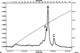
Fig. 2. Elution profile from ion-exchange FPLC. Affinity eluate from yeast glucan loaded. Peak A, fractions 30+31; Peak B, fractions 32+33; Peak C, fractions 34–37; Dashed line, conductivity.
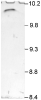
Fig. 3. Native isoelectric focussing of fraction A from FPLC. Range of pH=8–10; 5% polyacrylamide gel, Coomassie Brilliant Blue stained.

Fig. 4. Binding of DCG-04 (biotinylated analogue of the irreversible cysteine protease inhibitor E-64) to Diplostomum pseudospathaceum proteins on a blot. (1) Binding of DCG-04 to the 24 kDa lectin/protease; (2) inhibition of DCG-04 binding by E-64; M, molecular weight markers.

Fig. 5. Dot analysis of proteolytic activity of particular samples during purification of Diplostomum pseudospathaceum lectin (polyacrylamide gel copolymerized with 0·1% gelatin, pH 7·5). (1) Cercarial homogenate; (2) eluate from yeast glucan affinity chromatography; (3) peak A from ion-exchange FPLC; (4) peak B from ion-exchange FPLC; (5) peak C from ion-exchange FPLC. S+E-64, protease sample (S) inhibition with the irreversible inhibitor of cysteine proteases (E-64). S+E-64+TLCK, inhibition of protease sample (S) with irreversible inhibitors of cysteine (E-64) and serine (TLCK) proteases. Trypsin, positive control.
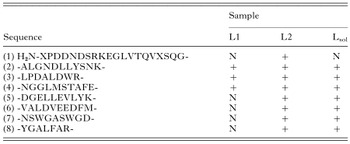
Table 1. List of peptide sequences obtained from Diplostomum lectin/protease by Edman degradation and tandem mass spectrometry, associated with particular samples in which they were detected
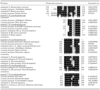
Table 2. Peptide sequences arisen from the isolated D. pseudospathaceum protein (lectin/protease) giving alignments with sequences of selected cysteine proteases found in databases
 You have
Access
- 24
- Cited by