Generalised lymphangiomatosis is a rare disease that is characterised by a diffuse proliferation of lymphatic vessels.Reference Brown, Reiman, Rosenow, Gloviczki and Divertie1 It is believed that the disorder is due to the defective development of the lymphatic system during the intrauterine period.
Its clinical course is different, whereas half of the children were asymptomatic. The others presented with marked respiratory symptoms including cough, dyspnoea, fever, stridor, haemoptysis, dysphagia, or symptoms secondary to infection.
Generalised lymphangiomatosis can involve any organ except the central nervous system, and the prognosis depends on the involved organ and the extent of the disease.Reference Brown, Reiman, Rosenow, Gloviczki and Divertie1–Reference Levey, MacCormac, Sartoris, Haghighi, Resnick and Thorne3 Radiological examination, including computed tomography and magnetic resonance imaging, is useful for evaluating the morphologic features as well as the extent of disease. We report here a case that was referred to us for investigation of marked cardiomegaly.
Case report
A previously healthy 8-year-old girl was referred to our hospital as an outpatient because of incidentally detected cardiomegaly on her chest X-ray. She had no history of fever, night sweats, anorexia, significant chronic cough, wheezing, or shortness of breath. Physical examination revealed no evidence of tachypnoea, increased work of breathing or acute distress. The cardiovascular, abdominal, and neurological examinations were essentially normal.
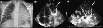
Chest X-ray (Fig 1a) showed cardiomegaly with superior mediastinal widening. Electrocardiography was normal. In echocardiographic examination (Fig 1b and c), there was a cystic formation with septation localised at the left lateral border and just behind the heart extending to the anterior region of the pulmonary artery.
(a) Asymmetric mediastinal enlargement and a well-defined paracardiac lesion (arrow; silhouette sign) are seen in the left hemithorax; (b) a multicystic lesion localised left lateral border of the left ventricle (arrow); (c) lesion extended to the anterior of the pulmonary artery (arrow).

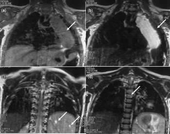
The differential diagnosis of such lesions and their extensive thoracic neurovascular structures and the neighbourhood that offers valuable information to assess the patient's thoracic magnetic resonance imaging was decided upon. Thoracic magnetic resonance imaging (Fig 2a and b) showed a huge cystic lesion extending from the thoracic inlet to the cardiac basis in the left hemithorax. In addition, cystic lesions were visible in the spleen and the dorsal spine in the sections involved (Fig 2c and d).
Coronal T1- and T2-weighted sequences (a and b, respectively), a huge cystic lesion extending from the thoracic inlet to the cardiac basis in the left hemithorax. The lesion is isohyperintense on the T1 W image (arrow), marked hyperintense on the T2 W image (arrow). Entering the upper section of the spleen in the abdominal cross-sectional area (c), millimetric cystic lesions are seen (arrows). (d) At the dorsal spine (arrow), there are a few cystic lesions of which the largest one is 1centimetre (T2 section).

Aspiration biopsy was performed in the mediastinum for differential diagnosis with lymphoma. The biopsy histology was consistent with lymphangioma.
Due to the size of the lesion and probable surgical complications, we planned to give Bleomycin (an intralesional sclerosing agent) and then if it became resectable, surgical removal would be considered.
Discussion
Generalised lymphangiomatosis is a rare congenital disorder mainly affecting children. It was first described by Rodenber in 1828; 65% of lesions are found in children and they do not exhibit any gender predilection.Reference Wunderbaldinger, Paya and Patrik2, Reference Nagar, Bhatgadde, Raut and Narlawar4 These constitute 0.7%–4.5% of mediastinal tumours;Reference Shenoy, Barua, Patel, Culver and Jennings5 90% of these tumours manifest as a mass in the neck or axilla in children, and only 10% had internal or visceral locations such as the abdomen or thorax.
The presence of multiple lymphangiomas is termed lymphangiomatosis. Generalised lymphangiomatosis is reported to frequently involve the bones, mediastinum, spleen, liver, lungs, neck, and pleura.Reference Brown, Reiman, Rosenow, Gloviczki and Divertie1–Reference Levey, MacCormac, Sartoris, Haghighi, Resnick and Thorne3, Reference Singh, Baboo and Pathak6 Our patient had cystic lesions in the mediastinum, spleen, and dorsal spine. The differential diagnosis of a cystic mass in the mediastinum includes necrotic malignant tumours and lymph nodes, pericardial cyst, teratoma, hydatid cyst, mediastinal abscess, haemangioma, and lymphangioma.
The diagnosis of generalised lymphangiomatosis should be made in association with the radiological findings and the results of histological examinations because the histological findings are not always specific.Reference Levey, MacCormac, Sartoris, Haghighi, Resnick and Thorne3 In many cases, the lesions of lymphangiomatosis do not differentiate histological from cavernous or capillary haemangioma.Reference Levey, MacCormac, Sartoris, Haghighi, Resnick and Thorne3 The radiological findings of generalised lymphangiomatosis are reported by several authors.Reference Brown, Reiman, Rosenow, Gloviczki and Divertie1–Reference Levey, MacCormac, Sartoris, Haghighi, Resnick and Thorne3, Reference Singh, Baboo and Pathak6 The lesions showed as sharply defined, non-enhanced cystic lesion. In the skeletal system, multiple osteolytic lesions were noted. These radiological findings were consistent with those of the previous reports.
Since the site and extent of lymphangioma are important prognostic factors, performing radiological evaluation for generalised lymphangiomatosis is crucial for planning the patient's management. Ultrasonography, computed tomography, and magnetic resonance imaging are all used to establish a pre-operative diagnosis.Reference Daniel, Misra, Karak, Mukhopadhyaya and Sharma7, Reference Thomas, Leung and Lynn8 Magnetic resonance may be the best technique to assess the complete extent of thoracic lesions and their relationship to neurovascular structures. Magnetic resonance may also show details of the tubular structures of a cavernous lymphangioma better than does computed tomography.
Treatment is usually conservative. Surgical excision's results are often unsatisfactory because of complications including damage to surrounding structures, particularly nerves, and blood vessels, scarring, and recurrence owing to incomplete excision.Reference Ravitch and Rush9, Reference Emery, Bailey and Evans10 For these reasons, we preferred intralesional sclerosing agent injection at first.