Introduction
Owing to their unique physical, chemical, and structural properties,Reference Wang, Li, Wang, Li and Lin1 graphene and its derivatives show outstanding potential in many fields, such as energy storage and conversion,Reference Sun, Wu and Shi2 high-frequency electronics,Reference Wu, Pisula and Mullen3 and bioscience/biotechnologies.Reference Wang, Li, Wang, Li and Lin1 (See the introductory article in this issue for more background on the history and properties of graphene.)
Of the techniques developed to produce graphene (detailed in the articles in this issue by Nyakiti et al., Bartelt and McCarty, and Jaber-Ansari and Hersam), one of the most economical for mass production is chemical synthesis from graphite by oxidation to graphene oxide (GO) and subsequent reduction.Reference Park and Ruoff4, Reference Fan, Peng, Li, Li, Wang, Zhang and Zhang5 Different functional groups and other nanomaterials can be linked to the graphene surface during the reduction procedure because GO consists of sp 2-bonded carbon atoms with easily functionalized six-atom rings and is abundant in reactive carboxyl and epoxy groups.
In addition to its ease of functionalization, graphene exhibits distinct properties that are very attractive for biosensing and bioimaging, such as a high electrical conductivity (up to 6000 S/cm),Reference Du, Skachko, Barker and Andrei6 a large planar area (theoretically 2630 m2/g for single-layer graphene),Reference Stoller, Park, Zhu, An and Ruoff7 and an excellent ability to quench fluorescence.Reference Chang, Tang, Wang, Jiang and Li8, Reference Tang, Wu, Cort, Buchko, Zhang, Shao, Aksay, Liu and Lin9
In this article, we discuss the immobilization of enzymes, deoxyribonucleic acid (DNA), and proteins on graphene materials and review selected electrochemical and fluorescence biosensors based on graphene and its derivatives for detecting small molecules, DNA, and cells. Graphene-based magnetic resonance and fluorescence for cell imaging are also discussed.
Graphene-based enzyme biosensors
Enzymes are biological molecules (typically proteins) that catalyze chemical reactions. The reaction takes place in a small part of the enzyme called the active site, with the rest of the molecule acting as “scaffolding.” The amino acids around the active site attach to the target molecule (called the “substrate”) and hold it in position while the reaction takes place. This makes each enzyme specific for a single reaction or for a particular type of chemical bond or functional group. A key goal of modern analytical science is to monitor biomolecular content with methods that are highly sensitive, have a short turnaround time, and are inexpensive. Because enzymes often have high efficiency and target specificity, their reactions are suitable for detecting single analytes at low concentration. For example, a number of biosensors have been reported, including sensors using the enzyme horseradish peroxidase (HRP) to detect hydrogen peroxide, using glucose oxidase to detect glucose, and using acetylcholinesterase (AChE) to detect organophosphates.
In the construction of an enzyme biosensor, fabrication of the sensing interface is extremely important but also extremely challenging. For instance, to improve enzyme survival (maintenance of the activity of the immobilized enzyme in the maximum range) and stability (immobility within the biosensor structure),Reference Yang, Jia, Qian, Zhou and Xia10 a biosensing interface should allow analytes to interact quickly and selectively.Reference Gooding11, Reference Patolsky, Zheng and Lieber12 Multiplexing biosensors are also desirable to detect multiple analytes with one device.Reference Tang, Tang, Niessner, Chen and Knopp13, Reference Strömberg, Zardán Gómez de la Torre, Göransson, Gunnarson, Nilsson, Svedlindh and Stromme14
Novel nanomaterials are helping to solve these problems.Reference Wang15 In particular, with its good conductivity, high carrier mobility, and high quenching efficiency, graphene is an excellent material for modifying the surface of an enzyme biosensor. In this section, we describe a few selected examples of typical graphene-based electrochemical biosensors. More information on this topic can be found in recent reviews.Reference Shao, Wang, Wu, Liu, Aksay and Lin16, Reference Ratinac, Yang, Gooding, Thordarson and Brael17
Biofunctionalization with enzymes
Because their two-dimensional structure provides a large area for the immobilization of enzymes and because enriched oxygen-containing groups are apt to react with amino-acid residues, graphene derivatives are ideal substrates for the immobilization of certain biomolecules. Enzymes can be immobilized on graphene surfaces through the following three means: (1) electrostatic interaction between positively charged graphene and a negatively charged enzyme, (2) covalent linkage of an enzyme molecule to the graphene surface, and (3) entrapment of the enzyme using the polysaccharide chitosan or another polymer.
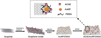
In an example of the first approach, GO is first synthesized by Hummer’s method. During the subsequent reduction of GO, the positive polymer poly(diallyldimethylammonium chloride) (PDDA) and gold nanoparticles (AuNPs) are introduced onto the surface of the chemically reduced GO (CRGO). As shown in Figure 1, the enzyme acetylcholinesterase (AChE) can interact with PDDA-enwrapped AuNP/CRGO under neutral conditions.Reference Wang, Zhang, Du, Shao, Li, Wang, Engelhard, Li and Lin18 The electrostatic interaction between GO and the long polyelectrolyte chains of PDDA prevents AChE molecules from aggregating on the carbon support. The presence of PDDA not only improves the dispersion of the AuNPs but also stabilizes AChE with high activity and loading efficiency.
Schematic illustration of gold nanoparticle (AuNP)/chemically reduced graphene oxide (CRGO) hybrid synthesis and acetylcholinesterase (AChE)/AuNP/CRGO nanoassembly generation using poly(diallyldimethylammonium chloride) (PDDA). Negatively charged AChE was immobilized on the positively charged AuNP/CRGO strongly through electrostatic interactions, which prevented the aggregation of AChE. Red balls represent the defect sites. Reproduced with permission from Reference Reference Wang, Zhang, Du, Shao, Li, Wang, Engelhard, Li and Lin18. ©2011, Royal Society of Chemistry.

Following the second approach, Zhang et al. reported an enzyme-immobilization matrix based on GO.Reference Zhang, Zhang, Yang, Huang, Liu, Zhang and Guo19 They demonstrated that enzyme immobilization on GO sheets can take place readily without use of any cross-linking reagents or additional surface modification. Because GO has a large specific surface area and is abundant in oxygen-containing groups, an enzyme can be bound to its surface through a condensation reaction between the terminal amino groups of the enzyme and the carboxyl groups on GO. More importantly, enzymes retain their structural integrity and biological activity when they are bonded to GO.Reference Pumera, Scipioni, Iwai, Ohno, Miyahara and Boero20
As an example of the third approach, an electrode for direct electrochemistry and glucose sensing was built using the enzyme glucose oxidase immobilized on graphene with the help of chitosan.Reference Kang, Wang, Wu, Aksay, Liu and Lin21 Specifically, wrinkled graphene sheets modified with hydroxyl and carboxylic groups were obtained by thermal reduction of GO, and then a mixed solution of reduced GO and chitosan was dropped onto the surface of a glassy carbon electrode (GCE). Glucose oxidase solution was further coated on the graphene/chitosan-film-modified GCE. The resulting electrode was found to be highly sensitive to glucose, exhibiting a detection limit of 0.02 mM. In this research, biocompatible chitosan was employed to disperse water-insoluble graphene and immobilize the enzyme molecules.
Electrochemical detection
The excellent electrochemical behaviors of graphene indicate that graphene is a promising electrode material in electroanalysis. Several electrochemical sensors based on graphene for bioanalysis have been developed.Reference Yang, Ratinac, Ringer, Thordarson, Gooding and Braet22 Graphene-based materials enhance the direct electron transfer that is characteristic of some redox enzymes and retain bioactivity because of their favorable electronic properties and biocompatibility. As described in the preceding section, Lin’s group constructed a glucose oxidase/graphene/chitosan nanocomposite for glucose detection,Reference Kang, Wang, Wu, Aksay, Liu and Lin21 with which a pair of well-defined redox peaks, characteristic of the reversible electron-transfer process of the redox-active center in the oxidase, could be observed. More graphene-based redox enzyme biosensors have also been developed.Reference Shan, Yang, Song, Han, Ivaska and Niu23, Reference Wang, Shao, Matson, Li and Lin24
Wang et al. reported an electrochemical sensor based on AChE/AuNP/CRGO, whose synthesis was described in the preceding section.Reference Wang, Zhang, Du, Shao, Li, Wang, Engelhard, Li and Lin18 AChE hydrolyzes its target acetylthiocholine, and the production of thiocholine can be detected directly. A well-defined oxidation peak was observed for AChE/AuNP/CRGO/GCE at –0.38 V with respect to GCE. The combination of CRGO and AuNPs promotes electron transfer and thus catalyzes the electro-oxidation of thiocholine at a lower potential. Furthermore, the current peak was 20 times higher than that of AChE/CRGO/GCE. The AChE/AuNP/CRGO/GCE biosensor has been used for the highly sensitive detection of OPs (based on the enzyme-inhibition principle), with a detection limit of 0.1 pM.
DNA-based biosensors
Biofunctionalization with DNA
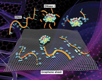
Tang et al. systematically explored the interactions between DNA and carboxyl-functionalized graphene.Reference Tang, Wu, Cort, Buchko, Zhang, Shao, Aksay, Liu and Lin9 Compared to free single-stranded DNA (ssDNA), no detectable hydrolysis by a DNase (deoxyribonuclease, a common enzyme that promotes DNA degradation) was observed after the ssDNA had been adsorbed on the graphene surface and thereby protected by the graphene. The protection occurs because ssDNA is promptly adsorbed onto functionalized graphene, forming strong molecular interactions that prevent DNase from approaching the constrained DNA (Figure 2).
Schematic illustration of the constraint of DNA molecules on functionalized graphene and its effects. Single-stranded DNA can be effectively constrained on the surface of graphene through adsorption. The enzyme DNase I can digest free DNA but not graphene-bound DNA. Reproduced with permission from Reference Reference Tang, Wu, Cort, Buchko, Zhang, Shao, Aksay, Liu and Lin9. ©2010, Wiley-VCH.

Another group reported that the solubility of graphene improved markedly after interaction with ssDNA.Reference Tang, Wang and Loh25 This phenomenon can be attributed to the high loading capacity of graphene for DNA through effective π–π cooperative stacking. The adsorption capacity corresponds to 87 mg/g of graphene and 30 mg/g of GO at a concentration of 20 μM ssDNA. The binding of GO and ssDNA occurs mainly through hydrogen bonding or electrostatic interactions. However, at a neutral pH of 7, GO is negatively charged, which promotes electrostatic repulsion by the negatively charged ssDNA.
As a result of its good solubility and biocompatibility, GO is a favorable carrier for gene delivery. Feng et al. employed a cationic polymer, poly(ethyleneimine) (PEI), to solve the negative-charge problem of GO.Reference Feng, Zhang and Liu26 GO was noncovalently functionalized with PEI, forming positively charged GO/PEI complexes. Then, negatively charged plasmid (ring-shaped) DNA was loaded onto the GO/PEI complexes by electrostatic interactions. The resulting GO/PEI/DNA complex was effective for intracellular gene delivery into the widely used HeLa cell cultures. (HeLa is a human cell line whose cells can proliferate indefinitely.)
Optical detection
GO has interesting optical properties: It can fluoresce over a wide range of wavelengthsReference Luo, Vora, Mele, Johnson and Kikkawa27 and also effectively quench the fluorescence of other fluorescent dyes.Reference Loh, Bao, Eda and Chhowalla28 These characteristics make it a useful fluorescence label for optical imaging. It is also a useful quenching material in a fluorescence resonance energy transfer (FRET) sensor, in which an excitation on another molecule transfers nonradiatively to the GO, or vice versa. Because the fluorescence quenching drops rapidly with distance, FRET can measure distances and interactions between domains in a single protein or between proteins, for example.
Liu and colleagues used GO as the energy donor and AuNPs as the energy acceptor in a FRET sensor for detecting hybridization of DNA with DNA having the complementary sequence.Reference Liu, Choi and Seo29 With excitation at 400 nm, GO modified with probe DNA displayed a strong fluorescence emission band centered at 547 nm. After hybridization with the AuNP-labeled target ssDNA, the GO fluorescence emission was reduced by more than 95% because of effective quenching by the AuNPs.
GO can also bind and quench a dye-labeled ssDNA probe. Upon addition of the target, the target DNA binds with the probe ssDNA and forms double-stranded DNA (dsDNA). The resulting dsDNA desorbs from the GO surface, because the hybridized dsDNA nucleobases are efficiently shielded within the negatively charged dsDNA phosphate backbone.Reference Lu, Yang, Zhu, Chen and Chen30 Therefore, the dye is no longer in contact with GO, and its fluorescence is restored.Reference He, Song, Li, Zhu, Qi, Wen, Wang, Song, Fang and Fan31, Reference Wu, Kempaiah, Huang, Maheshwari and Liu32
Zhao et al. presented a convenient assay based on catalytic DNA called a DNAzyme for selectively detecting Pb2+.Reference Zhao, Kong, Zhang, Meng, Liu, Tan, Shen and Yu33 They hybridized a substrate labeled with the fluorophore carboxyfluorescein (FAM) with a DNAzyme strand to form a DNAzyme/substrate hybrid containing a large ssDNA loop to bind with GO. Upon the addition of Pb2+, the DNAzyme was activated, so that it cleaved the substrate strand, thereby releasing a short FAM-linked oligonucleotide fragment and recovering the fluorescence. The proposed biosensor exhibited a high sensitivity, with a detection limit of 300 pM for Pb2+.
He et al.Reference He, Song, Li, Zhu, Qi, Wen, Wang, Song, Fang and Fan31 reported a GO-based multicolor fluorescent DNA nanoprobe that allows rapid, sensitive, and selective detection of DNA targets in homogeneous solution by exploiting interactions between GO and DNA molecules. The large planar surface of GO allows simultaneous quenching of multiple DNA probes labeled with different dyes, leading to a multicolor sensor for the detection of multiple DNA targets in the same solution.
Quantum dots (QDs) are excellent fluorescent labels because of their high quantum yield, symmetric and stable fluorescence, low photobleaching, and size-dependent and tunable absorption and emission.Reference Wu, Liu, Liu, Haley, Treadway, Larson, Ge, Peale and Bruchez34 Dong et al. modified CdTe QDs with a molecular beacon (MB), a combination of fluorophore with a chosen oligonucleotide, as a probe to recognize target DNA.Reference Dong, Gao, Yan, Ji and Ju35 They then introduced GO into the system, and the strong interaction between the MB and GO led to fluorescent quenching of the QDs. Upon recognition of the target, the increased distance between the QDs and the GO and the weaker interaction between the target-bound MB and the GO significantly hindered the FRET and thus increased the fluorescence of the QDs. This strategy can also be developed to detect other biomolecules,Reference Dong, Gao, Yan, Ji and Ju35 thus promising wide-ranging applications.
Electrochemical detection
The effects of GO-modified binding can be detected electrochemically. A chemically reduced graphene oxide-modified glassy carbon electrode (CRGO/GCE) was constructed to directly detect DNA.Reference Zhou, Zhai and Dong36 The oxidation currents of the four nucleobases (A, adenine; T, thymine; C, cytosine; and G, guanine) can be separated because of the different oxidation potentials of the nucleobases. In the case of the CRGO/GCE, the anodic potentials of the four free bases move negatively, and their peak currents increase because of the higher electrocatalytic activity for the oxidation of the bases on CRGO/GCE. The high density of edge-plane-like defective sites on CRGO could provide many active sites and accelerate electron transfer between the electrode and species in solution.Reference Banks, Davies, Wildgoose and Compton37 Such an electrocatalytic action enables the simultaneous detection of all four free bases at the CRGO/GCE. The current signals of G, A, T, and C from ssDNA and dsDNA at the CRGO/GCE are all separated efficiently without requiring a prehydrolysis step. Based on this principle, more sensitive graphene-based DNA sensors have been established.Reference Lim, Hoh, Ang and Loh38, Reference Muti, Sharma, Erdem and Papakonstantinou39
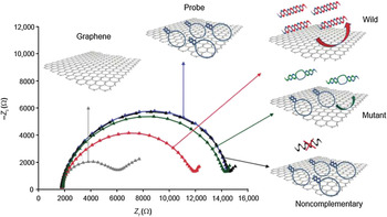
Electrochemical impedance spectroscopy, which distinguishes the resistive and capacitive contributions of the electrochemical double layer, is another efficient label-free method for biosensing. Bonanni et al. employed an electrochemical impedance spectroscopy technique for the rapid detection of a single-nucleotide polymorphism correlated with the development of Alzheimer’s disease.Reference Bonanni and Pumera40 The extracted charge-transfer resistance (R ct), which characterizes the roughness of an electrode surface, increased after hairpin-folded DNA (hpDNA) probes (shown in blue in Figure 3) were immobilized onto the graphene-modified electrode surface (Figure 3). Negatively charged probe DNA repelled the negatively charged redox couple, which increased the R ct value. The formation of the DNA probe film on the electrode surface sterically hindered the reaction, increasing R ct. When a wild-type target (the naturally occurring form) was hybridized with probe DNA, the R ct value decreased markedly (red). The R ct decrease was less significant in the case of hybridization with a mutant (green), and no average R ctvariation was observed with a noncomplementary sequence (black). The researchers also studied the influence of graphene thickness and found that few-layer graphene provides the best sensitivity.
Wang et al. fabricated a CRGO/GCE for monitoring methicillin-resistant Staphylococcus aureus DNA,Reference Wang, Zhang, Chen, Zhou, Yang, Wu, Niu, Han, Wang, Chen, Boey, Zhanga, Liedberg and Zhang41 and Hu et al. built a sensor for the pol gene sequence of human immunodeficiency virus type 1 by etching graphene with 3,4,9,10-perylenetetracarboxylic acid.Reference Hu, Li, Bai, Li, Hua, Wang and Niu42 Both of these sensors exhibited an increase in the R ctvalue with increasing concentration of target DNA.
Schematic of the electrochemical impedance spectroscopy protocol and Nyquist plots of the negative imaginary part of the impedance –Z i versus the real part Z r. Changing the measurement frequency moves the impedance along the circular contours: Graphene surface (gray), hairpin DNA (hpDNA, blue), perfectly complementary target (red), target with a single mismatch (green), and negative control with a noncomplementary sequence (black). In an idealized model, the diameter of the circle indicates the charge-transfer resistance R ct, which quantifies the rate of the reaction. Reproduced with permission from Reference Reference Bonanni and Pumera40. ©2011, American Chemical Society.

Graphene-based immunosensors
The clinical measurement of biomarkers can indicate normal biological processes, pathogenic processes, or pharmacologic responses to a therapeutic intervention. Recently, the “sandwich” immunoassay, which is based on detection of the binding event between an antibody and an antigen, has become one of the most sensitive strategies for the quantification of biomarkers.
Biofunctionalization with antibodies and proteins
The first step in creating graphene-based immunosensors is immobilizing antibodies or other proteins. To this end, our group successfully synthesized carboxylic graphene. Secondary antibody and horseradish peroxidase (HRP, an enzyme that catalyzes the oxidation of hydrogen peroxide and is often used as a label to amplify weak biochemical signals) were efficiently immobilized on the carboxylic graphene using a standard amidization protocol.Reference Du, Wang, Shao, Wang, Engelhard and Lin43 This method is also suitable for immobilizing antibodies and proteins on the surface of GO or graphene/chitosan composite.Reference Wan, Wang, Wu and Zhang44
In another demonstration, (3-glycidyloxypropyl)trimethoxysilane (GOPS) molecules were conjugated onto Fe3O4-nanoparticle-coated graphene nanosheets (magnetic graphene oxide, MGO) through the reaction between –OH groups on MGO and –OCH3groups on GOPS. Antibody and protein were then combined with the resulting active epoxy groups to establish an immunosensor.Reference Tang, Tang, Niessner, Chen and Knopp45
Another approach to anchoring proteins to graphene uses glutaraldehyde, an amine-reactive homobifunctional cross-linker (meaning that it has identical reactive groups at opposite ends of its spacer arm) that can lead to cross-linkages between the oligomeric state of proteins and amino groups. Wei et al.Reference Wei, Xin, Du, Wu, Han, Zhao, Cai, Li, Yang and Li46 and Yang et al.Reference Yang, Javadi, Li and Gong47 mixed thionine (TH) and graphene sheets (GSs) as the base and immobilized an antibody onto the surfaces of GS/TH through linkages with glutaraldehyde. The prepared GS/TH/antibody system was found to be very stable because thionine contains several amino groups.
Immunosensors for detection of proteins
Construction of a useful immunosensor depends on the successful binding of a primary antibody (Ab1), which directly binds the protein, and a number of labeled secondary antibodies (Ab2), which bind to Ab1. At present, two typical functions of graphene can be applied in the design of electrochemical immunosensors. The first involves use of graphene as a modification material to enhance the conductivity and surface area of electrodes. As an example, our group reported an electrochemical immunosensor for sensitive detection of the cancer biomarker α-fetoprotein (AFP).Reference Du, Zou, Shin, Wang, Wu, Engelhard, Liu, Aksay and Lin48 To fabricate this immunosensor, a GCE modified with graphene/chitosan composite was used as a sensing platform for capturing Ab1. The bioconjugate was created by co-linking HRP and AFP secondary antibodies (Ab2) to functionalized carbon nanospheres (CNSs). Upon complete sandwich immunoreaction, the AFP/Ab2/HRP/CNS conjugate was attached to the electrode surface. HRP can catalyze the oxidation of H2O2, so the concentration of AFP can be confirmed by calculating the increased oxidation response of a H2O2 solution. The electrochemical signal from the redox reaction of H2O2 was increased 7-fold compared to that from an HRP/Ab2/AFP/Ab1/chitosan/GCE. This improved response can be attributed to the superior electrochemical and electrical properties of graphene, the ability of the large surface area of graphene to capture a large amount of primary antibodies, and the ability of CNSs to carry multiple HRP molecules.
As another example, GO has been used as a carrier to load a large amount of probes or biomolecular recognition elements for signal amplification. We developed a new electrochemical immunosensor for ultrasensitive detection of phosphorylated protein that used GO as a carrier in a multienzyme amplification strategy.Reference Du, Wang, Shao, Wang, Engelhard and Lin43 Phosphorylated p53 molecules were bound to primary antibodies that had been attached to a AuNP-modified screen-printed electrode. This structure was then incubated with GO/Ab2/HRP complex to complete the immunological reaction. The resulting immunosensor showed excellent analytical performance for the detection of phosphorylated p53 with a range of 0.02–2 nM and a detection limit of 0.01 nM.
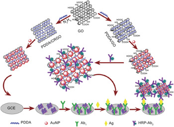
Liu et al. used graphene as both a modification material and a carrier for establishing a better immunosensor (Figure 4).Reference Liu, Zhang, Wang and Zhu49 A AuNP/PDDA/GO hybrid was constructed through simple sonication-induced assembly. On one hand, the synthesized hybrid is a favorable material with outstanding electrical conductivity. On the other hand, the architecture thus formed provides a large-surface-area matrix for antibody immobilization with good stability and bioactivity. Antigens were recognized after primary antibodies were incubated with a AuNP/PDDA/CRGO-modified GCE. HRP-labeled secondary antibodies were attached to the surfaces of AuNP/PDDA/GO through the interaction between AuNPs and mercapto or primary amine groups of biomolecules. The immunosensor fabricated according to this method was found to be extremely sensitive, with a detection limit of 0.05 ng ml–1.
Schematic illustration of the preparation of graphene-based hybrids and the construction of a sandwich-type electrochemical immunosensor. Graphene is used in two ways. First, as illustrated on the left branch, AuNP/PDDA/CRGO hybrids modify a glass carbon electrode (GCE) to immobilize the primary antibodies (Ab1) in preparation for reaction with the antigen to be detected (Ag). Second, as illustrated on the right branch, AuNP/PDDA/GO is bound to a horseradish peroxidase (HRP) labeled secondary antibody (Ab2). Reproduced with permission from Reference Reference Liu, Zhang, Wang and Zhu49. ©2011, Elsevier.

Jung and coworkers developed a fluorescence immunosensor for pathogen detection.Reference Jung, Cheon, Liu, Lee and Seo50 The energy acceptors, AuNPs, were linked to a secondary antibody through a 100-mer ssDNA molecule. The primary antibodies for rotavirus were immobilized on the GO array through a carbodiimide-assisted amidation reaction, and the rotavirus was captured through a specific antigen–antibody interaction. In conjunction with the immunoreaction between the antigen and the secondary antibody, the AuNPs on the GO surface quenched the fluorescence.
Graphene-based biotechnology for cell imaging
In recent years, researchers have begun to explore the feasibility of using GO for bioimaging with optical and magnetic modalities. In one recent example, graphene-based FRET for cellular imaging and gene delivery was reported.Reference Wang, Li, Hu, Lin, Li and Lin51 FAM-labeled adenosine-5′-triphosphate (ATP) aptamer (i.e., an oligonucleic acid that can bind to an ATP molecule) was adsorbed onto the surface of a graphene oxide nanosheet (GONS) through the particular interaction between GO and DNA molecules. The ATP aptamer could target ATP when the aptamer/FAM/GONS was transported into living cells. The formed duplex configuration was then released from the surface of the GONS as a result of weak adsorption, so that the FAM-labeled aptamer was no longer in contact with GONS and the fluorescence was recovered. After 8 h of incubation with aptamer/FAM/GONS, fluorescence derived from the FAM-labeled aptamer/ATP duplex was seen only in the middle of the cell, indicating that the aptamer/FAM/GONS nanocomplex was successfully imported into cells by endocytosis.
Another example of imaging exploits the photoluminescence (PL) from graphene quantum dotsReference Mueller, Yan, McGuire and Li52–Reference Shen, Zhu, Chen, Yang and Li55 (GQDs), which can be obtained from GO by chemical or electrochemical synthesis. GQDs are effective fluorescent probes for bioimaging because of their chemical inertness and low toxicity under physiological conditions. Zhu et al. reported the synthesis of strongly green photoluminescent GQDs and their use in cell imaging.Reference Zhu, Zhang, Qiao, Tang, Li, Yuan, Li, Tian, Liu, Hu, Gao, Wei, Zhang, Sun and Yang56 The synthesized GQDs showed high solubility, excellent biocompatibility, and favorable optical properties and could be used directly for intracellular imaging without any surface processing or functionalization.
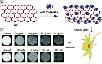
Magnetic resonance imaging (MRI) is a powerful technique that is widely used in clinical diagnosis. Zhang et al.Reference Chen, Yi, Zhang, Zhang, Deng and Zhang57 synthesized dextran-coated Fe3O4 nanoparticle/graphene oxide composites (Fe3O4/GO) as a contrast agent for efficient cellular MRI (Figure 5).Reference Chen, Yi, Zhang, Zhang, Deng and Zhang57 Fe3O4 nanoparticles in a particular region induced local inhomogeneity of the magnetic field and, therefore, caused a decrease in regional signal intensity because of more rapid dephasing of the precessing spins (shorter T 2 relaxation time). The composites exhibited good physiological stability and low cytotoxicity and can be internalized efficiently by HeLa cells. Furthermore, compared with isolated Fe3O4 NPs, the Fe3O4/GO composites showed significantly enhanced cellular MRI.
(a) Schematic illustration of the preparation of Fe3O4 nanoparticle/graphene oxide (Fe3O4/GO) composites and (b) T 2-weighted cellular magnetic resonance images (which map dephasing time): (top) HeLa cells (2 × 10⁵ cells ml–1) incubated with the Fe3O4/GO composites at different concentrations and (bottom) Fe3O4/GO composites (20 μg of Fe ml–1) incubated with HeLa cells at different cell densities, wherein Fe3O4 nanoparticles (NPs) form aggregates on the GO sheets, resulting in a considerably enhanced dephasing (shorter T 2). AMD, amino-modified dextran; HeLa, human cell line whose cells can proliferate indefinitely; MRI, magnetic resonance imaging. Reproduced with permission from Reference Reference Chen, Yi, Zhang, Zhang, Deng and Zhang57. ©2011, American Chemical Society.

Summary
The use of graphene-based materials for biosensing and bioimaging has achieved great success within a short time. Graphene-based biosensors show high sensitivity and can be employed for the detection of both small molecules and biological macromolecules. In this review, we have described graphene-based enzymatic and DNA biosensors based on distinct transduction approaches, notably fluorescence and electrochemistry. Moreover, we discussed the successful application of such sensors in cell imaging because of their particular optical properties.
Although the advances are encouraging, some technical challenges still need to be conquered before use of graphene-based materials for biosensing and bioimaging becomes widespread: (1) The graphene used in electrochemical biosensors is often obtained by chemical or thermal reduction from graphene oxide (GO). This reduced GO has low conductivity. Thus, developing new, simple, and low-cost methods for synthesizing highly conductive graphene is crucial. (2) Graphene materials are used as carriers for antibody, DNA, protein, and drug delivery. Controlling the number of layers and minimizing folding would be advantageous for immobilizing biomolecules on graphene-based structures. Moreover, irregular graphene particle sizes cause variations in the concentrations of probes and secondary antibodies. Synthesis of uniform graphene can help increase the repeatability needed for accurate biosensing and bioimaging. (3) Better understanding of the physics and chemistry at the surface of graphene and of the interactions of chemicals and biomolecules at the graphene interface will play an important role in applying graphene as a nanoscaffold in biosensing, bioimaging, and drug delivery. For example, studying the adsorption mechanism and orientation of biomolecules on graphene will provide further understanding of graphene and its interactions with molecules, which could, in turn, lead to advances in graphene science and applications. (4) The cytotoxicity characteristics, cellular-uptake mechanism, and intracellular metabolic pathway of graphene and its derivatives are still not well-understood (for more on this topic, see the article in this issue by Jachak et al.). These critical research areas are essential for the future use of graphene in living-cell studies and in vivo applications.
In summary, graphene is an excellent new material for biosensing and biological imaging. With a focus on innovative applications, the development of new graphene-based materials will certainly lead to numerous future advances in bioscience and technology.
Acknowledgments
This work was supported by the National Natural Science Foundation of China (21075047) and the Special Fund for Basic Scientific Research of Central Colleges (CCNU11C01002, CCNU10A02005). Y. Lin acknowledges the financial support of an NIH grant (U54 ES16015) from the National Institute of Environmental Health Sciences (NIEHS) and the U.S. Department of Energy (DOE) Laboratory Directed Research and Development program. Pacific Northwest National Laboratory is operated by Battelle for the DOE under Contract DE-AC05-76RL01830.