INTRODUCTION
The African trypanosome, Trypanosoma brucei, the causative agent of human African trypanosomiasis (Barrett et al. Reference Barrett, Burchmore, Stich, Lazzari, Frasch, Cazzulo and Krishna2003), has in recent years emerged as a front runner in systems biology analysis. Reproducible cultivation methods exist and quantitative analysis is relatively well developed. For example, a quantitative mathematical model of energy metabolism in the long slender form of the trypanosome (i.e. the form that replicates in the mammalian bloodstream) has been developed (Bakker et al. Reference Bakker, Michels, Opperdoes and Westerhoff1997) and iteratively updated after experimental testing (Bakker et al. Reference Bakker, Michels, Opperdoes and Westerhoff1999a, Reference Bakker, Walsh, ter Kuile, Mensonides, Michels, Opperdoes and Westerhoffb; Albert et al. Reference Albert, Haanstra, Hannaert, Van Roy, Opperdoes, Bakker and Michels2005; Haanstra et al. 2008 a). Moreover, a model of the gene expression cascade, based on quantitative knowledge of transcription, RNA precursor degradation, trans-splicing and mRNA degradation for phosphoglycerate kinase (PGK) has recently been generated (Haanstra et al. Reference Haanstra, Stewart, Luu, van Tuijl, Westerhoff, Clayton and Bakker2008b). It is therefore possible to envisage the derivation of a fully comprehensive molecular model of trypanosome physiology. Another paper in this volume details the birth of the “silicon trypanosome” project and applications of dynamic modelling to understand function in the cells. Here we focus on the experimental approaches to metabolome analysis that are required to sustain this ambitious endeavour.
ACQUISITION OF METABOLOMICS DATA
In recent years, methods have become available for the rapid acquisition of whole genome data, from which it is possible to infer the entire repertoire of metabolically active enzymes present within a cell. Transcriptome analysis can reveal which genes are expressed at a given time, under a given set of environmental conditions. Proteomic techniques, although less able to achieve comprehensive coverage of expressed proteins, can reveal if specific proteins are indeed present under given conditions. Metabolomics, which aspires to the simultaneous measurement of all low molecular weight chemicals within a cell, has been less easy to achieve, mainly because of the wide range of physicochemical properties characteristic of cellular metabolites and the huge dynamic range over which these are present (Van der Werf et al. Reference van der Werf, Jellema and Hankemeier2005; Breitling et al. Reference Breitling, Ritchie, Goodenowe, Stewart and Barrett2006a). However, a variety of techniques are available to measure cellular metabolites. These include nuclear magnetic resonance (NMR) and a variety of mass spectrometry-based approaches, such as mass spectrometry coupled to liquid chromatography (LC-MS), gas chromatography (GC-MS), or capillary electrophoresis (CE-MS). Different types of mass spectrometer can be used to identify the masses of metabolites separated through the different chromatography platforms.
Alternatively, nuclear magnetic resonance spectrometry (NMR) is used in highly quantitative analysis and also for identifying metabolites that are difficult to ionise or which are difficult to analyse by MS, due to their idiosyncratic behaviour on different chromatographic platforms. The most promising recent development is the combination of MS and NMR technology in hybrid instruments that unite the sensitivity of MS and the superior quantitative capacity and specificity of NMR (Yang, Reference Yang2006).
Relatively little previous work on identification and quantification of metabolites in T. brucei has been performed. The glycolytic intermediates have been identified as part of the programme investigating energy metabolism using HPLC (Visser and Opperdoes, Reference Visser and Opperdoes1980) or enzymatic analysis (Visser et al. Reference Visser and Opperdoes1980; Albert et al. Reference Albert, Haanstra, Hannaert, Van Roy, Opperdoes, Bakker and Michels2005, Haanstra et al. Reference Haanstra, van Tuijl, Kessler, Reijnders, Michels, Westerhoff, Parsons and Bakker2008a). Polyamines and the thiol intermediates of the trypanothione pathway have also been characterized using HPLC-based methodology (Fairlamb et al. Reference Fairlamb, Henderson, Bacchi and Cerami1987; Shim and Fairlamb, Reference Shim and Fairlamb1988; Xiao et al. Reference Xiao, McCloskey and Phillips2009). NMR has been used to trace steady-state concentrations of abundant phosphorylated compounds within T. brucei (Moreno et al. Reference Moreno, Urbina, Oldfield, Bailey, Rodrigues and Docampo2000) and also to assess the abundant end products of metabolism in procyclic trypanosomes from which key enzymes have been deleted in order to assess likely pathways of metabolism in this form of the parasite (Rivière et al. Reference Rivière, van Weelden, Glass, Vegh, Coustou, Biran, van Hellemond, Bringaud, Tielens and Boshart2004; Coustou et al. Reference Coustou, Besteiro, Rivière, Biran, Biteau, Franconi, Boshart, Baltz and Bringaud2005, Reference Coustou, Biran, Breton, Guegan, Rivière, Plazolles, Nolan, Barrett, Franconi and Bringaud2008). Similar studies were also used to assess end products of glucose metabolism in bloodstream forms (Mackenzie et al. Reference Mackenzie, Hall, Flynn and Scott1983).
It is imperative that metabolism is quenched as rapidly as possible prior to extracting metabolites. Directly applying trypanosomes in suspension to boiling ethanol achieves this (Kamleh et al. Reference Kamleh, Barrett, Wildridge, Burchmore, Scheltema and Watson2008a), although it is necessary also to measure medium without cells to distinguish intracellular and extracellular metabolite content. Rapidly chilling cells in culture in a dry ice-ethanol bath, followed by centrifugation, washing and extraction in chloroform-water-methanol, as used for Leishmania (Robinson et al. Reference Robinson, De Souza, Keen, Saunders, McConville, Speed and Likić2007; Ruben T'Kind, personal communication) offers advantages in terms of allowing removal of extracellular medium and gentler extraction conditions than boiling ethanol. Continued development of novel extraction methods will enhance metabolite coverage.
ADVANTAGES OF ULTRA-HIGH RESOLUTION, ULTRA-HIGH MASS ACCURACY MASS SPECTROMETRY
We have recently adopted a platform of ultra-high resolution, ultra-high mass accuracy mass spectrometry to systematically acquire metabolite data in T. brucei (Breitling et al. Reference Breitling, Ritchie, Goodenowe, Stewart and Barrett2006a, Reference Breitling, Pitt and Barrettb, Reference Breitling, Vitkup and Barrett2008). Modern Fourier transform instruments can measure the mass of analytes to within 1 ppm, so a metabolite like glucose (or any of its structural isomers; Mw=180·0634) can be distinguished from a metabolite of similar mass such as theophylline (Mw=180·0647) in a manner that less accurate machines could not achieve. The problem of structural isomers, however, requires further analysis to resolve: e.g. fructose, mannose, galactose, inositol have the same mass as glucose. Orthogonal approaches such as chromatographic behaviour or analysis of metabolite fragments in GC-MS or tandem mass spectrometry are required to resolve this issue.
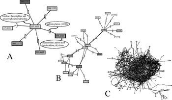
In a first study, trypanosome extracts were generated from parasites isolated from rats and separated as a buffy coat. The extracts were directly injected into a Fourier transform ion cyclotron resonance mass spectrometer. On the order of 1,000 peaks were detected (Breitling et al. Reference Breitling, Ritchie, Goodenowe, Stewart and Barrett2006a). Many of these were associated with the T. brucei lipidome, and it is possible that the highly ionisable nature of these metabolites suppressed the ionisation of many non-lipid metabolites (in spite of using a range of different extraction solvents to recover a balanced set of metabolites). Notwithstanding these problems, the initial datasets were rich enough to enable the inference of large networks of metabolites that could be linked through potential metabolic or chemical links (Breitling et al. Reference Breitling, Ritchie, Goodenowe, Stewart and Barrett2006a). Such ab initio networks reveal the advantage of using high accuracy–high resolution approaches in which each metabolite can be assigned an exact mass: since every possible biochemical transformation (phosphate addition, hydroxylation, methylation, prenylation etc.) will alter the size of a metabolite by a unique exact mass, each metabolite measured in a dataset can be related to others through the simple addition or subtraction of those characteristic metabolic masses (Breitling et al. Reference Breitling, Ritchie, Goodenowe, Stewart and Barrett2006a, Reference Breitling, Vitkup and Barrett2008; Rogers et al. Reference Rogers, Scheltema, Girolami and Breitling2009; Fig. 1). This ab initio network building technique has a major role in the inference of novel metabolic pathways (Jourdan et al. Reference Jourdan, Breitling, Barrett and Gilbert2008).
Building ab initio networks of potential chemical connectivity within metabolomic datasets. This figure demonstrates the principle of the construction of ab initio metabolic networks as described by Breitling et al. (Reference Breitling, Ritchie, Goodenowe, Stewart and Barrett2006a). (A) shows how one measured mass (257.1028) can be connected to others within a dataset by comparing all masses for potential connectivity through a list of the mass differences associated with known biochemical, metabolic, transformations. In (B) the central panel, the network is extended by identifying further metabolites that can be linked to the connected masses by a second metabolic transformation (second level network), and by (C) the network has been extended by six of these iterations to identify potential connectivity between the observed masses.

Magnetic sector ion cyclotron resonance instruments, as used in that first study, are relatively expensive and complex to operate. The development of the Orbitrap mass spectrometer, which can achieve similar levels of accuracy and resolution, represented an important advance in metabolomics research (Breitling et al. Reference Breitling, Pitt and Barrett2006; Lim et al. Reference Lim, Chen, Sensenhauser, Cook and Subrahmanyam2007; Ding et al. Reference Ding, Sorensen, Zhang, Jiang, Jaitly, Livesay, Shen, Smith and Metz2007; Dunn et al. Reference Dunn, Broadhurst, Brown, Baker, Redman, Kenny and Kell2008; Kamleh et al. Reference Kamleh, Barrett, Wildridge, Burchmore, Scheltema and Watson2008 a, Reference Kamleh, Hobani, Dow and Watsonb; Kiefer et al. Reference Kiefer, Portais and Vorholt2008). Furthermore, coupling Orbitrap mass spectrometry to a range of chromatographic systems, most importantly HILIC columns that resolve hydrophilic compounds, has greatly enhanced our ability to identify many polar cellular metabolites (Kamleh et al. Reference Kamleh, Barrett, Wildridge, Burchmore, Scheltema and Watson2008a; Cubbon et al. Reference Cubbon, Antonio, Wilson and Thomas-Oates2009).
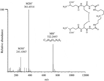
HILIC chromatography linked to an Orbitrap has now become our standard approach to trypanosome metabolomics, and early runs have shown that hundreds of metabolites can be successfully identified through this route, including signature trypanosome metabolites such as trypanothione (Fig. 2). Using these data it has been possible to develop algorithms to enhance the accuracy of mass spectra by calibration based on background ions constantly detected in the chromatograms (Scheltema et al. Reference Scheltema, Kamleh, Wildridge, Ebikeme, Watson, Barrett, Jansen and Breitling2008). Furthermore, software that can remove mass spectrometry artefacts including common adducts (Na+, K+ NH4+, Cl–, etc.), identify isotopomers (based on 13C abundance) and compare spectra along time series, have been applied to identify those peaks that represent true metabolites (Scheltema et al. Reference Scheltema, Decuypere, Dujardin, Watson, Jansen and Breitling2009). This approach allows the identification of several hundred metabolites. However, this coverage is significantly lower than the thousand or so expected for most cells. Given the chemical diversity of the small chemical species that comprise the metabolome, comprehensive coverage can only be achieved if the same sample is exposed to a range of different analytical platforms, including different types of liquid chromatography and solvents, as well as gas chromatography-MS approaches. The latter platform has underpinned much of the pioneering work in metabolomics of Leishmania (Doyle et al. Reference Doyle, MacRae, De Souza, Saunders, McConville and Likić2009), and also offers a valuable tool for trypanosome research.
Extracted mass chromatogram for trypanothione.

ANALYSIS OF METABOLIC FLUX AND DYNAMICS
The ability to generate static views of the metabolome is now available. However, much of what we wish to understand about the system must come from dynamic views of how metabolites flow through the metabolic network. In the case of the glycolytic pathway, a combination of modelling and experimentation has shown that the control of flux through the pathway is distributed between several steps including the glucose transporter (THT1), which exerts some 40% of the flux control (Bakker et al. Reference Bakker, Walsh, ter Kuile, Mensonides, Michels, Opperdoes and Westerhoff1999b), and most likely glyceraldehyde 3-phosphate dehydrogenase, glycerol phosphate dehydrogenase, phosphoglycerate mutase and the mitochondrial glycerol-3-phosphate oxidase (Albert et al. Reference Albert, Haanstra, Hannaert, Van Roy, Opperdoes, Bakker and Michels2005). In contrast, in mammals, hexokinase and phosphofructokinase assume most control over the pathway. A great deal of further work is required to refine even these current relatively simple models. For example, kinetic data are usually obtained from purified enzymes in relatively simple buffer systems, that by definition lack much of the complexity that will govern kinetic behaviour in vivo. Teusink et al. (Reference Teusink, Passarge, Reijenga, Esgalhado, Van der Weijden, Schepper, Walsh, Bakker, Van Dam, Westerhoff and Snoep2000) have shown in yeast that such differences between in vivo and in vitro parameters currently limit our capability to explain pathway behaviour ab initio from enzyme kinetic properties. Time-dependent metabolite patterns have a much richer information content than steady-state data and will be vital to bridge the gap between in vitro biochemistry and the in vivo behaviour of metabolic pathways (Hynne et al. Reference Hynne, Danø and Sørensen2001). Metabolomics approaches, particularly those using mass spectrometry, now offer the possibility to measure flux through multiple pathways simultaneously (Weichert et al. Reference Wiechert, Schweissgut, Takanaga and Frommer2007; Tang et al. Reference Tang, Martin, Myers, Rodriguez, Baidoo and Keasling2009; Niittylae et al. Reference Niittylae, Chaudhuri, Sauer and Frommer2009). In addition to providing a means of testing mathematical models of metabolism, these approaches can provide data that might even allow the prediction of kinetic parameters (or at least a range of parameters that could account for observed behaviour) (Kümmel et al. Reference Kümmel, Panke and Heinemann2006; Hynne et al. Reference Hynne, Danø and Sørensen2001). Such model improvement will be important to make better predictions of the adaptive capacity of metabolism towards environmental changes, such as the addition of drugs and/or the signals that trigger parasite differentiation.
Metabolomics approaches will also reveal the relative importance of parallel routes in the network. Pulse-chase experiments can follow the distribution of a precursor metabolite through the metabolome in time. For example, by adding a pulse of heavy atom-labelled glucose (with one or more of the carbon atoms in glucose labelled with 13C) the accumulation of the heavy isotope in the different metabolites of the glycolytic and pentose phosphate pathways can be measured. In our rapid-stop protocol (Kamleh et al. Reference Kamleh, Barrett, Wildridge, Burchmore, Scheltema and Watson2008a), where trypanosome metabolism is quenched instantaneously by squirting trypanosomes in suspension into boiling ethanol, we can, in principle, make repeated measurements at very short intervals (down to seconds), to measure the dynamic distribution of label over time. The same principle can be applied to other pathways; for example, the flux of labelled cysteine, glutamate and glycine into glutathione and trypanothione pathway intermediates. This ability to actually measure fluxes will play an invaluable role in learning about those parameters that control dynamic flux through the metabolome.
GENOME-WIDE MODELS
Existing kinetic models of trypanosome metabolism describe relatively small parts of the complete metabolic network, while the metabolomics techniques discussed in this paper aim at covering a large part of the metabolic network. They are usually interpreted by mapping the results onto genome-based reconstructions of the metabolic potential of the organism of interest. This is, for instance, possible using the Pathway Tools software (Karp et al. Reference Karp, Paley and Romero2002) that allows interactive interrogation of the results and has already proven useful for many organisms. A genome-wide reconstruction of the Leishmania metabolic network (LeishCyc) has recently been published (Doyle et al. Reference Doyle, MacRae, De Souza, Saunders, McConville and Likić2009). A T. brucei reconstruction (TrypanoCyc) is also being made (Chukualim et al. Reference Chukualim, Peters, Hertz-Fowler and Berriman2008; Jourdan et al. unpublished). The KEGG environment (Kanehisa et al. Reference Kanehisa, Araki, Goto, Hattori, Hirakawa, Itoh, Katayama, Kawashima, Okuda, Tokimatsu and Yamanishi2008) also represents a good background in which to represent measurements. Software that allows direct projection of high accuracy metabolomics datasets onto the KEGG contextual overview is available (Suhre and Schmitt-Kopplin, Reference Suhre and Schmitt-Kopplin2008).
In addition to the simple, static views of the metabolome provided by the MetaCyc and KEGG environments, other computational techniques, that have been developed to study the metabolic potential at a genome scale in a more dynamic fashion, are available (Breitling et al. Reference Breitling, Vitkup and Barrett2008; Feist et al. Reference Feist, Herrgård, Thiele, Reed and Palsson2009). Constraints-based models based on stoichiometric matrices have become widely used (Edwards et al. Reference Edwards, Ibarra and Palsson2001) and have recently been generated for Leishmania (Chavali et al. Reference Chavali, Whittemore, Eddy, Williams and Papin2008) and T. cruzi (Roberts et al. Reference Roberts, Robichaux, Chavali, Manque, Lee, Lara, Papin and Buck2009). These can assist in drug target discovery by identifying essential enzymes whose absence is catastrophic to cellular functioning (Jamshidi and Palsson, Reference Jamshidi and Palsson2007).
In the long run, the ambitious aim of this work is to help identify those parts of metabolism most amenable to targeting by novel drugs.
ACKNOWLEDGMENTS
MPB gratefully acknowledges funding from the BBSRC in the “Systryp” consortium funded jointly by the BBSRC and the ANR. RB is supported by a NWO-Vidi fellowship. BMB is supported by a Rosalind Franklin Fellowship at the University of Groningen.