




No CrossRef data available.
Article contents
Impairments of procedures for implementing complex language are due to disruption of frontal attention processes
Published online by Cambridge University Press: 22 March 2006
Abstract
Production of complex discourse—lengthy, open-ended utterances and narratives—requires intact basic language operations, but it also requires a series of learned procedures for construction of complex, goal-directed communications. The progression of clinical disorders from transcortical motor aphasia to dynamic aphasia to discourse impairments represents a progression of procedural deficits from basic morpho-syntax to complex grammatical structures to narrative and a progression of lesions from posterior frontal to polar and/or lateral frontal to medial frontal. Two cases of impaired utilization of language exemplify the range of impairments from clearly aphasic agrammatic, nonfluency to less and less “aphasic” and more and more executive impairments from transcortical motor aphasia to dynamic aphasia to narrative discourse disorder. The clinical phenomenology of these disorders gradually comes to be more accurately defined in the terminology of executive deficits than that of aphasia. The executive deficits are, in turn, based on impairments in various components of attention. Specific impairments in energizing attention and setting response criteria associated, respectively, with lesions in superior medial and left ventrolateral frontal regions may cause defective recruitment of the procedures of complex language assembly. (JINS, 2006, 12, 236–247.)
Information
- Type
- SYMPOSIUM
- Information
- Journal of the International Neuropsychological Society , Volume 12 , Issue 2 , March 2006 , pp. 236 - 247
- Copyright
- © 2006 The International Neuropsychological Society
INTRODUCTION
Investigation of disorders of language has traditionally been the domain of aphasia studies. Within those studies the focus has been on impairments of basic linguistic functions and their common patterns of cluster after damage to various brain regions in—typically—the left hemisphere. The basic operations of language can be described at several levels of resolution but might include phonology, semantics, perhaps a lexicon or two, and grammar (excluding for this discussion the cognitive, perceptual and motor processes specifically required for reading, writing and speaking). Each operation can be measured on the receptive/comprehension dimension and on the production dimension. The common patterns of impairment are the well-known aphasia syndromes.
Fully developed human language is, however, put to many complex purposes. It is a medium for learning, recollection, and communication. Language is utilized to relate vacation tales, tell jokes, give directions, summarize medical histories (as a patient or as a physician), prepare scientific reports, and so on. Although each of these uses of language is dependent upon recruitment of the basic operations of language, as well as elements from other cognitive and emotional domains, to accomplish a communication goal each requires additional capacities. If a communication requires more than a sentence or two or a list of a few key elements, then the speaker must set an overall communication goal, sustain activity to reach the goal, monitor progress to the goal, inhibit intrusions that are not relevant to the goal, and be attentive to the listener's expectations and reactions. For the purposes of this paper, the capacity to communicate lengthy, complex verbal messages will be labeled “narrative discourse.”
Deficits in discourse are more closely related to disorders of action planning than they are to aphasia. There is now a substantial literature on disorders of action planning due to brain injury: from eating breakfast (Schwartz et al., 1991) to running errands (Shallice & Burgess, 1991) to financial planning (Goel et al., 1997). In common with other examples of action planning, discourse presumes a goal and perhaps one or more subgoals, presumes the normal function of the cognitive and motor functions necessary for implementation of the plan, and presumes that there is not a fixed, invariant route to the goal. Different people might solve the path to a goal in different manners that reflect different procedural experience or skills, different contextual constraints, or simply equally plausible alternative paths to completion. Some steps are contingent on others, but there should be many possible approaches to the goal. Action planning is the setting of one approach with preparation to shift as subgoals are attained or as context might require. Definitions of the cognitive mechanism that might be damaged in patients with discourse impairments parallel those of other action planning deficits: establishing (narrative) intent (Luria & Tsevtkova, 1967), assembling a (verbal) plan (Costello & Warrington, 1989); generating a (narrative) procedure (Robinson et al., 1998).
A Hierarchy of Procedures of Language Utilization
The ability to produce complex communications develops in orderly manner with brain development as a set of learned skills sequentially evolving from attention-dependent to automatic. As language develops in young children, a capacity to produce longer utterances emerges: initially often simply tabulating single concepts but gradually containing rudimentary grammatical properties: action words, such as infinitive verb forms, and attributes and modifiers of objects and actions, such as size, color, order. From this essentially agrammatic beginning, the complexity of language structure grows through listening exposure, direct teaching in school, and trial and error, sometimes with explicit correcting by listeners. Increasing proficiency with complexity means that the cognitive bases for production of complex language forms must have become increasingly proceduralized; that is, increasingly automatic and decreasingly dependent on attention. This review will identify three levels of language use procedures: (1) grammar and simple syntax; (2) complex syntax; and (3) narrative discourse. The boundaries between these levels are not entirely distinct.
Basic grammar and syntax: Learning the use of articles, the meaning of prepositions, the varieties of verb forms for number and tense, subject-verb and noun-pronoun agreement rules, and inflection and/or word order rules. Some of this is lexicalized, that is, separate grammatical forms of a word may be independently represented in lexical-semantic association cortex—irregular past tense forms, gerunds, or the specific locative and chronologic meanings of prepositions, for example—but most appear to be represented as rules or procedures of application (Pinker, 1991). The procedures for grammar begin to develop very early in language use and are essentially universally acquired. They appear to become automatic (non–attention dependent) and instantiated in frontal operculum (and probably in inferior parietal structures) by age 10, as before that age lesions anywhere in language competent left brain produce agrammatism and after that age a clinico-anatomical relationship similar to adults is seen (Van Donegan et al., 1985).
Complex syntax: Learning the procedures to implement more complex forms of language structure to capture more complicated or subtle meanings, such as added precision in conditionality and chronology in verb forms, lengthier and often embedded modifying clauses, and more complicated forms of reference. These procedures of communication are learned at a later developmental age, probably late childhood through adolescence, and their use emerges as the simpler grammatical procedures become more automatic (Reilly et al., 1998). The completeness of acquisition (procedural learning) of these more complex forms of language is more variable than the simpler forms, and acquisition may depend upon education and cultural contexts.
Narrative discourse: Learning to utilize the full range of semantic and lexical knowledge and syntactic skills to achieve specific communication goals (Chapman et al., 1998). The properties of discourse can vary. Procedural discourse tends to have relatively constrained options for content and order: fixing a flat tire, making a hamburger, and so on. Procedural discourse requires recall of the necessary experience or knowledge, but it is not much affected by context: Intent is obvious and it can be presented in virtually tabular forms. Narrative discourse, on the other hand, does not have the same restrictions on content, though it is still bound by intent and context. Narratives may require event recollection, judgment about inclusion of detail, working memory for just related details, or, for those intended further into the discourse, attention to the effect on the listener and many other factors (Mackenzie, 2000). Some forms of discourse are entirely dependent upon education or vocation: relating a medical history, arguing a legal case, writing a scientific report, and so on. The developmental neural time frame for discourse is probably life long beginning in late childhood and maturing through adolescence and early adulthood (Mackenzie, 2000). Goal-directed behavior utilizing language requires considerable executive and attention resources (Chapman et al., 2003; Chapman & Ulatowska, 1989). At this level discourse is no different than any other complex, goal-directed activity. It requires intent, planning, memory and sustained effort across time—the elements of executive function.
It would be possible to reverse the review above, that is, summarize the development of executive functions and then analyze language development as an example of emerging cognition dependent upon executive functions. It is beyond the scope of this paper to review development of executive functions, but there is ample evidence that supports the claim that maturation of prefrontal cortex (and its connections to caudate and cerebellum) is required before children develop the capacity for complex cognitive operations. Frontal cortex gray matter volumes and prefrontal synaptic density both decrease in size until adult levels are reached in adolescence (Huttenlocher, 1979; Sowell et al., 1999). The decrease in frontal cortical volume is associated with an increase in source memory and recognition memory (Sowell et al., 2001). It may not be intuitive that these relationships should be inverse—volume decreasing as capacity increases—but they are consistent with higher efficiency of network connections with experience. In an excellent review, Diamond describes the maturation of prefrontal function as a stepwise increase working memory and attention/inhibition capacities (Diamond, 2002). The characterization of the role of frontal cortex in cognition as dependent on an expansion of attention capacity that relies on frontal-subcortical networks will return below as the general hypothesis of this article.
Damage to That Hierarchy: Clinical Syndromes and Associated Lesion Sites
The different levels of language proceduralization develop sequentially but somewhat independently, and brain injuries demonstrate that each level can be impaired somewhat independently. Because the levels of language proceduralization are surely represented in overlapping brain regions, most brain lesions cannot cause isolated impairments in one level, but there are prototypical clinical forms for damage to each level. These clinical prototypes define the hierarchy of procedures as they unravel just as development defines them as they emerge.
Patients with Broca's aphasia have spontaneous language (and repetition) reduced to the agrammatical level of production: single word or only short noun-verb utterances, preferential use of infinitive or simple verb forms, lack of tense and number consistency, and loss of articles and modifiers (Mohr et al., 1978). Broca's aphasia is caused by large lesions that damage the posterior lateral frontal lobe, including operculum, the anterior, superior insula, the anterior parietal lobe, and the white matter deep to those structures.
Patients with transcortical motor aphasia (TCMA) in the classical aphasia taxonomy, have “nonfluent” spontaneous output (Freedman et al., 1984) with grammatically intact repetition and oral reading. “Nonfluency” has encompassed agrammatism as described above. Most commonly, however, output is terse and unelaborated but basically grammatical with subject-verb agreement, correct use of prepositions and articles but with few modifying words and little output beyond simple sentences. (Nonfluency has also included mutism, discussed below.) Clinical-anatomical studies of TCMA have demonstrated lesions in the left lateral frontal lobe or structures deep to it (Freedman et al., 1984). Each of the uses of “nonfluency” above likely represents a slightly different lesion location. The most common lesion site is lateral frontal lobe, areas 45, 46, 9, and 6. Patients with “Broca's area aphasia” have overtly agrammatical spontaneous output acutely but much better, even fully grammatical repetition; lesions are restricted to frontal operculum (areas 44, 45, and 4) and anterior insular structures (Alexander et al., 1990; Mohr et al., 1978). These patients typically evolve to grammatical but terse spontaneous output with essentially no output more complex than simple sentences. Depending on dorsal vs. ventral lesion site, there may be slightly more or less overt agrammatism, and depending on anterior vs. posterior lesion site, there may be slightly more or less overt phonemic paraphasia. There is, however, no fixed line separating “Broca's area aphasia” from TCMA. At its most prototypical, TCMA represents preservation of only the simpler forms of morpho-syntax in spontaneous output.
At this point in consideration of the clinical syndromes associated with impaired language use, we leave the domain of traditional aphasia studies. The term dynamic aphasia was first proposed for the entire range of TCMA (Luria & Tsevtkova, 1967). It has come to have a more restricted definition: normal word use, sentence structure, and grammar for most output but variable utterance length and syntax and a reduced capacity for expanded output that is directly dependent on the complexity of the goal of the output and inversely related to the degree of external cues. Clinical-anatomical studies of patients characterized as dynamic aphasia are few, but lesions have been in left DL frontal regions, areas 46, 9v and 10, and critically, extensively in deep white matter of the left prefrontal lobe (Costello & Warrington, 1989; Robinson et al., 1998). Dynamic aphasia represents preservation of grammar and most aspects of syntax in spontaneous output. As long as interactions are on familiar topics or responses are straightforward, patients with dynamic aphasia may not even appear to have any language problem. This is one form of “aphasia without aphasia” (Von Stockert, 1974). Only when they have to construct an open-ended utterance without structure provided do they become impaired.
In the other form of “aphasia without aphasia,” patients have initial mutism that evolves to very short but grammatical utterances, with few modifying words (Rubens, 1976) and little output beyond simple sentences, often with very long latencies to speak; lesions involve left superior or medial regions (Freedman et al., 1984; Masdeu et al., 1978; Rubens, 1976). These patients also evolve to terse but fully grammatical spontaneous output, such that they are not overtly aphasic, although they are generally very laconic. Because similar mutism evolving to terse but normal output has been described with lesions of the right superior medial regions (Gelmers, 1983), and because bilateral superior medial lesions produce long-lasting akinetic mutism (Freeman, 1971), it is possible that this is not a disorder of utilization of language but a disorder of activation of language (and speech and motor) functions. Depending on lesion site (superomedial vs. dorsolateral), there may be more or less overall reduction in language output, but there is no fixed line separating these two “aphasias without aphasia.” It has been suggested from functional imaging studies that presupplemental motor area (preSMA) is particularly critical for activation of internally generated language, that is, language that is not constrained or cued by external factors (Crosson et al., 2001).
Narrative discourse deficits represent the final level of impaired use of language procedures to assemble complex propositional utterances, often serial utterances, and always determined by context and by a specific communication goal. The nature of the goal—telling a story, giving directions, giving instructions, and so forth—will affect the structure of the discourse form. Discourse deficits have been reported with damage to either the left or the right frontal lobe, in patients with diffuse brain injury and even in patients in confusional states. Some coarse differences in discourse deficits after right or left frontal injuries (Chatterjee et al., 1997) have been described, but there is little anatomically or functionally detailed study. A broad clinical picture of the effects of left frontal injuries can be identified. Grammatical and syntactic structures are normal. The patient's output is typically not heard as aphasic but is described as vague or confused. The patient will be characterized as a bad historian, not as an aphasic historian. A narrative that cannot be reduced to a simple tabular production will seem poorly planned: chronologically disorganized, repetitious, and under-referenced. Left frontal lesions associated with discourse deficits have rarely been specified although when it is possible to determine lesion sites, they appear to be distinctly prefrontal: 9, 10, and 46 (Costello & Warrington, 1989; Robinson et al., 1998). Depending on lesion site (polar vs. anterior dorsolateral), there may be differences in the level of complexity at which language implementation to communicate becomes poorly organized, but there is no fixed line separating dynamic aphasia from narrative discourse impairment.
Lesions in the more anterior portions of the frontal lobe are increasingly likely to affect cortical regions or projections on both the medial and lateral surfaces. A dorsolateral lesion that extends deep into the middle frontal white matter will disrupt projections from polar frontal cortex to posterior language zones. It is no wonder that the clinical syndromes of classical TCMA, dynamic aphasia, medial frontal laconic output and discourse impairment have a large amount of overlap or that one may evolve to another during progression of or recovery from disease. Review of relevant publications suggests that most reports of TCMA are “contaminated” by cases perhaps better considered dynamic aphasia and include patients with both medial and lateral versions of “aphasia without aphasia.”
Without trying to parcel out various reports to one diagnosis, from the most impaired to the most subtle, all of these disorders are defined with very similar vocabularies. Initiation is delayed, and there are pauses at transition points (Mega & Alexander, 1994). Productions are under elaborated (Freedman et al., 1984; Novoa & Ardila, 1987). Fragmentary sentences are common (Kaczmarek, 1984; Luria & Tsevtkova, 1967). Grammar and syntax are normal, but few multisentence utterances are produced (Nadeau, 1988). Syntactic and sentential structures tend to be repeated (Mega & Alexander, 1994; Novoa & Ardila, 1987). There is a tendency to utilize structures made available by the examiner, from frank echolalia to incorporation of portions of a question into the response (Mega & Alexander, 1994). When an open-ended response is required, there is difficulty selecting a sequence (Luria & Tsevtkova, 1967) or establishing a plan to proceed. Shifting topics from the initial proposition is difficult (Kaczmarek, 1984).
Attempts to define the nature of these deficits have moved beyond typical aphasia vocabulary to the types of accounts mentioned in the introduction: an inability to transform narrative intent into language procedures (Luria & Tsevtkova, 1967), an inability to assemble a verbal plan for the full narrative before the step of actual sentence construction (Costello & Warrington, 1989), or an inability to generate a procedure for narrative in the absence of a concrete context (Robinson et al., 1998).
Patients with discourse deficits or dynamic aphasia due to left prefrontal injury may, paradoxically, be less adept at getting the key overall point of a narrative across, than more overtly aphasic patients. Patients with Broca's area lesions (centered on areas 44 and 45) are unable to assemble scrambled words into sentences, but they are able to assemble scrambled sentences into coherent stories. Patients with more anterior lesions (centered on areas 9 and 46) can perform sentence assembly but not narrative assembly (Crozier et al., 1999; Sirigu et al., 1998). Aphasic patients, in general, have retained capacity to transmit the gist of a narrative approximately proportional to preservation of basic language operations—word finding, paraphasias, and comprehension (Chapman & Ulatowska, 1989; Reilly et al., 1998; Ulatowska et al., 1983).
Other patient groups may have more significant impairment in the capacity to transmit the critical gist of a narrative. Numerous studies of patients with right brain injuries of various etiologies and locations have demonstrated impaired logical coherence and incomplete specification of the essential core of the intended narrative (Brownell et al., 1990; Davis et al., 1997; Galski et al., 1998; Grindrod & Baum, 2005), but precise lesion analyses have not been reported. Adults with severe traumatic brain injury show impairment in many aspects of discourse (Galski et al., 1998): fewer units of content and overall incoherence. The trauma patients have executive cognitive deficits, but studies thus far have made little attempt to establish specific regional effects or even to differentiate between diffuse and focal mechanisms for discourse impairments. Deficits in discourse years after traumatic injury are very pronounced in children, particularly children who were initially aphasic (Chapman et al., 1998; Ewing-Cobbs et al., 1998). Frontal lesions are particularly critical.
Summary (see Table 1): Patients with TCMA cannot easily (automatically) recruit procedures for constructing sentences despite largely intact grammatical and other basic language operations. Patients with dynamic aphasia cannot easily (automatically) recruit procedures for constructing lengthier, more complex sentence constructions, especially when they are not constrained by context, despite largely intact basic syntactic forms. Patients with discourse impairment cannot easily (automatically) manage the procedures required to assemble a narrative despite intact language capacity. One interpretation might be that with maturation, experience, and development of language the frontal and then the prefrontal regions progressively master the procedures necessary for grammar, simple sentence structures, complex sentence structures, and finally, assembly of sentences into coherent narratives. These procedures become centered in progressively more anterior regions of the left frontal lobe from operculum to polar. Language productions that are initially attention-demanding and slow become progressively more automatic and rapid, but open-ended communications will always make some demands upon attention, and those demands must be met quickly or the stream of the communication will be disrupted. The medial frontal regions bilaterally serve to activate language (as they activate every other motor and cognitive function), and the more complex or less constrained the language utterance the more activation that is required. Complex language thus requires setting a communication plan, maintaining activation of the basic language operations that are going to be recruited for the lexical, phonological and essential grammatical and syntactical structure of any utterance, and monitoring the progress of the communication. Right hemisphere integrity appears required for much of monitoring the coherence and the response of the listener, but not for language structure.
Levels of impaired language assembly and clinico-anatomical correlates

Case Examples
Patient 1
At age 52 this right handed woman had an embolic infarction. She was a native English speaker, educated through high school and employed in an unskilled labor position. In the emergency department, a few hours after onset, she was mute, but within the first day of admission was producing single words. Formal testing demonstrated global aphasia. Within a week comprehension had rapidly improved, and she was speaking in longer phrases. There was frequent echolalia and perseveration of sentence forms. Repetition was normal.
At one month after onset she was speaking more, although her family noted that she could only easily respond to questions that could be answered with yes/no or 1–2 words. When asked about her activities at home, she replied, “I have a lot of stuff I have to get most bearing with.” When asked if she was doing any housekeeping, she replied, “I'm pretty much taking it easy because no one wants me to do anything.” All other utterances were limited, often perseverative and rarely carried any information. Repetition was normal, including lengthy functor-loaded targets: “She is the one who brought it to me.” On the Boston Naming Test-Short Form (BNT-SF) she named only 2/15 items but she had unusual semantic errors: “dinosaur” for octopus and “colonial” for house. Given initial phonemic cues, she produced the correct name or completion for all but one of the other items. She could name no animals on a fluency task until she said, after a long pause, “I have … Ruby, I have … and cat.”
At three months after onset, language was much improved although she could not describe her activities in any clear detail. Latencies to initiate responses were prolonged. Grammar and syntax were good. There were word-finding pauses that could stretch for several seconds, but the target was usually eventually produced. She named 7 animals in 60s. The Boston Naming Test (BNT) score (Kaplan et al., 1983) was 31/60. Comprehension of the syntax sections of the BDAE (Goodglass & Kaplan, 1983) was at the 2nd percentile, although word, command, and conversational comprehension were normal.
At six months, there was more improvement although any attempt at lengthy description of activities was slow to start, hesitant, fragmentary, and simplified. Naming score was 44/60. She named 5 animals in 60s. When asked to construct a sentence using a specified verb, her productions were delayed. Some responses were odd: using “distribute,” she said, “[pause of 17s] Some people distribute the shopping.” Using “applaud,” she said, “I applaud … [pause of 5] when I went to the concert, I applauded.” Other responses showed reuse of sentence structure: using “take,” she said, “[pause of 5] I take off my stuff in the bathroom” followed by using “give,” “[pause of 4] He gives me … uh … he gives me … things to take off in the shower.”
An MRI scan performed six weeks after onset (Figure 1) showed a left middle frontal gyrus infarct with moderate deep extension.

T1 axial MRI at six weeks after infarction; note edge of lesion in superior operculum (arrow) reaching to white matter above anterior limb of internal capsule, but center of lesion is in middle frontal gyrus.
Summary: Initial mutism quickly evolved to TCMA with minimal control of responses to external stimuli: echolalia, perseveration at every level of response, and remarkable phonemic completion. With additional recovery, naming neared normal, and she could respond coherently at sentence length: dynamic aphasia. She could not, however, carry the conversational burden when an unconstrained response was required.
Patient 2
At age 61 this right handed woman had a spontaneous left frontal intracerebral hemorrhage that was emergently evacuated. She is a native English speaker, educated through a master's degree, very literate, with work experience in editing. She was not initially evaluated locally, but according to her husband, she did not speak at all for several days. At one month after injury, she had fluent output but with many pauses and fragmentary breakdowns in formulation of spontaneous speech. There was mild incorporation: Examiner: “You went to China?” Response: “I went to … went to….” “What kind of work have you been doing most recently?” Response: “I … I don't know … most recently I worked for myself.” When she attempted to describe a picture, she resorted to perseverative use of the same sentence structure repeatedly: “Someone is taking cookies and look, he's, his … looks like he's handling, handing … uh … to the girl and the chair is teetering and he's about to fall. The … uh … woman is drying the dishes and the sink is overflowing and the water is … running and she's stepping back casually in the water … uh….” BNT (Kaplan et al., 1983) was 57/60. Comprehension was normal including the syntax portions of the BDAE (Goodglass & Kaplan, 1983). Repetition was normal.
At three months after onset, output was somewhat better formed, but still slow. She described a recent social encounter that included three major topics. Initiation was delayed and appeared to have a false start. Each topic was related in adequate order but details were lacking. The examiner had to probe to discover what had actually transpired. Transitions between the three topics were very slow, and there were no transitional phrases. BNT-SF was 15/15. She named 12 animals in 60s.
At six months after onset, there was continued improvement, but narrative was still labored with prolonged pauses (2–5s) at transitions between subthemes of her narrative. Asked if she had seen the “debate,” she said, “The debate … the vice-president debate I felt was even though the … I didn't … wasn't … perturbed by it but the reaction of a good friend's … by … he [Cheney] … didn't see … didn't meet … [Edwards] at all and the first time he met him was during the debate. He used it to show that he was absent a lot but it's misleading. He … was absent but not as much as indicated.” She named 19 items of produce in 60s, but the examiner had to suggest subcategory shifts. She could generate synonyms but very slowly and rarely more than one for each target.
An MRI performed six weeks after onset (Figure 2) demonstrated a residual lesion in the left middle and superior dorsolateral frontal regions undercutting superior medial regions.

MRI at six weeks after hemorrhage and surgical evacuation of hemorrhage; upper row axial T2; note lesion extension medially to edge of cingulated gyrus (arrow); lower row T1 parasaggital; center of lesion in SMA (arrow) but deep extension involves middle frontal white matter. Cortical lesion extent does not capture the consequences of subcortical extension for local and distant disconnection of frontal pathways.
Summary: Initial mutism again evolved to TCMA with poor control of responses to external stimuli—incorporation—and perseveration that was mostly at the phrase level. With further improvement she had normal naming and adequate output at the phrase level—dynamic aphasia. Later, she was able to carry some of the conversational burden at the sentence level, but this very intelligent, verbal person had great difficulty unfolding any narrative descriptions—discourse impairment.
From Aphasic Disorder to Executive Disorder to Attentional Disorder
When these patients are most overtly aphasic, their deficits are easily captured by standard terminology of aphasia, but with recovery it is less apparent that aphasia models adequately characterize their difficulties. Shifting the description of their impairments from the language of aphasia to the vocabulary of executive function may illuminate the deficits more accurately. In the context of language utilization, TCMA, DA and discourse impairments reflect deficits in: (1) response selection: a limited and repetitious repertoire; (2) sustained performance: incomplete and fragmentary responses; (3) response set: long latencies to respond and poor shift of topic; and (4) response inhibition: perseveration and utilization behaviors such as echolalia and incorporation. These are executive deficits in language use. A precisely similar analysis (although perhaps not the lesion-behavior relationships) holds for all goal-directed behaviors (Shallice & Burgess, 1991). Discourse has the advantages for experimentation that everyone can do it to some extent and that the subcomponents are readily identified.
Conversion of the model for impairments in complex language use from aphasia to executive disorder allows analysis of a different type and begins to isolate some of the fundamental processes essential for complex behaviors to unfold.
For complex language, the left lateral frontal region executes the procedures of language use and the left (and perhaps the right) medial frontal region provides the activation of language. In this context, execution means setting appropriate output procedures and content for communication and inhibiting or suppressing any inappropriate or collateral procedures or content that might have been activated internally or from external stimuli. Execution also requires monitoring the evolution of the intended scripts and sustaining activation of procedures until completion, and at the level of discourse monitoring appears to require actions of the right lateral frontal region as well. In summary, execution of the procedures requires activating, setting, inhibiting or suppressing, sustaining, and monitoring, over very short time scales as a communication unfolds—and all of these must occur.
We proposed a model of executive deficits based on these attention-dependent processes (Stuss et al., 1995). (Stuss reviews in this volume the model, the methodology and some of the findings of a series of investigations.) By 2003, we had completed several studies investigating the effects of focal frontal lesions on various aspects of attention. The patients in these studies had focal frontal injuries that were mapped on to increasingly specific cortical maps. All of the cases were more than three months after injury. None was clinically aphasic, although in retrospect, some certainly fit in the profiles described here. (Both of the patients described above have been seen since testing for these studies was completed and neither was tested in these protocols.) There were no complicating neurological factors, for, for example, epilepsy, hydrocephalus or unlocalizeable injury such as diffuse traumatic damage or whole brain radiation.
Regarding activating response behaviors, on every reaction time task that we have utilized, over three different patient test populations, patients with superior medial lesions, left or right, have been significantly slower than all other frontal lesion groups. (1) When given a cue to prepare for a response, patients with superior medial lesions, left or right, could demonstrate improved activation, but the improvement was lost within 3 seconds, unlike all other frontal lesion groups (Stuss et al., 2005). (2) On a task of sustained concentration (Alexander et al., 2005) that required a rapid response to push a button in front of whichever of five lights blinked with 200 ms latency from one response to the next stimulus over 500 trials, the patients with superior medial lesions, left or right, started slow and stayed slow; even 6 minutes of repeated stimulus presentation was insufficient to improve the level of activation. (3) On a Stroop-like reaction time task (unpublished data) patients with superior medial lesions, left or right, had very prolonged reaction times, and they were the only patients with a high rate of nonresponses, suggesting a complete failure of activation. On these tasks, when specific lesion sites were associated with poor activation, the right superior medial lesions—areas 24, 32, and 9—were most commonly significant. This asymmetry may only be apparent as the superior medial lesion group included more right than left cases. In our earlier studies of various traditional neuropsychological tests, the distribution of left- and right-sided lesions was more equal, and a lateralized effect of medial lesions was not demonstrated on any task. As noted above, clinical literature supports the conclusion that both left and right superior medial lesions reduce activation. Even if the right medial lesion is more critical than the left, a left medial lesion may produce greater loss of activation because it is situated to disrupt the projections of both medial regions to the left lateral frontal lobe where language procedures are instantiated.
What does this have to do with discourse? Energization of response to a stimulus, whether external or internal, whether simple or complex, is reduced by superior medial lesions. Delay of a few seconds or serial delays of hundreds of milliseconds can disrupt the entire process of recruitment. Within a narrative, the content of the narrative provides a rolling prompt to sustain a response state: Thus, with loss of energization and preparation, there would be delayed initiation and hesitations and pauses at shift points. When shifts in topic occur, the entire activation, setting and recruitment must occur again without contamination by previously activated programs or inappropriate collateral programs.
Regarding setting response behaviors, five studies are illuminating. (1) On a word list generation task, patients with left lateral frontal lesions had particularly poor production in the first 15 seconds of generation (Stuss et al., 1998). (2) On standard administration of the Stroop interference task, only patients with left frontal lesions had difficulty setting responses for color naming (Stuss et al., 2001). (3) On a difficult version of a Stroop-like task performed as a reaction time test (unpublished data), only patients with left lateral lesions could not set response criteria and had excessive false positive responses. (4) On a reaction time task that required discriminating between targets and foils that shared up to a maximum of two (out of three) features with the target, only the patients with left lesions made false positive errors, suggesting bias in setting response criteria (Stuss et al., 2002). (5) On the task of sustained concentration (Alexander et al., 2005), there was no overall increase in errors in any of the groups, but, when analyzed by blocks of 100 trials, the left frontal group, specifically those with ventrolateral lesions—areas 44, 45, and 47/12—had significantly increased errors in the first block. Over three different test populations of patients with frontal lesions, on five different tasks that required setting response behaviors, only patients with left ventrolateral frontal showed impairments, and these were particularly striking when setting response had to be done quickly. The boundary between activating behaviors to respond and setting the initial stimulus-response criteria is not always distinct. A threshold for activation is probably more easily met when a response is already partly set, and whatever stimulus-response set is established relies on activation to be implemented.
What does this have to do with discourse? Within a narrative, there are multiple points of setting a response plan, some constrained and others contingent: if this segment is chosen first, then that one (or one of those) must follow next—a process that is impaired by lesions of the left ventrolateral region. The precise consequences of a response setting impairment on language output are not transparent and have not been precisely tested. To reason backwards from the observed behaviors of dynamic aphasia, it appears that the effect is to delay formation of a clear action plan for narrative, leaving semantic elements that provide specificity and reference insufficiently activated. It seems possible that a deficit in response setting could also cause the hesitations, incompletions, and corrections that may accompany dynamic aphasia.
Regarding monitoring response behaviors, our studies provide less data. On a list-learning task, patients with right frontal lesions had an abnormal number of repetitions (Stuss et al., 1994). On the reaction time tests only patients with right lateral frontal lesions failed to show the normal foreperiod effect—decreasing reaction time with longer interstimulus intervals—and this was observed with passage of as little as 5 seconds (Stuss et al., 2005). Observations from the discourse literature support the possibility that monitoring, even of verbal tasks, is impaired after right lateral lesions, as patients with right frontal lesions are more likely to include irrelevant material and produce incoherent narratives, including frankly confabulated ones.
Frontal-Subcortical Networks
Although the focus here is on frontal lesions and procedures of discourse, each of the frontal regions has important parallel connections to ipsilateral striatum and contralateral neocerebellum, and it is through these networks that frontal regions establish the procedures for sentence and narrative assembly. For complete illustration of the proposed neural basis for language procedures, the subcortical components deserve brief review.
The projections from frontal regions maintain anatomical separation (Alexander et al., 1986; Cummings, 1993). Medial frontal structures (ACG/SMA/preSMA) project over the edge of the ventricle through the subcallosal fasciculus to the posterior head of the caudate (Yakovlev & Locke, 1961). Lateral frontal structures project directly downward onto the more anterior dorsal portion of the head of the caudate (Cummings, 1993). In studies of TCMA collected by clinical criteria, there are cases with lesions in the dorsal caudate and anterior limb or the corona radiata immediately above the anterior limb. In one analysis of cases with infarcts restricted to that deep territory—large infarcts in the lenticulostriate territory—the clinical phenomenology of TCMA was essentially identical to reports of cases involving frontal cortex (Mega & Alexander, 1994). (Although, see Godefrey et al. [1992] for an alternative account.) Study of patients with early HD and PD has demonstrated the same type of deficits of application of language procedures (grammar in this case) as in patients with Broca's aphasia (Ullman et al., 1997).
Regarding the cerebellum, the application of procedural rules is complex. The lateral frontal convexity projects through the anterior limb of the internal capsule, the medial cerebral peduncle, across the pons via medial rostral pontine nuclei and into the neocerebellum, particularly its more dorsal—posterior—parts (Middleton & Strick, 2000). One of the original PET studies of controlled word generation in normal subjects is usually cited for demonstration of activation of left frontal operculum, but activation of right posterior cerebellum was equally prominent (Petersen et al., 1989). Numerous investigations have demonstrated impaired word list generation despite normal direct naming (a common pattern after frontal injury) after focal cerebellar lesions, usually in the right posterior lobe (Marien et al., 2001; Schmahmann & Sherman, 1998). Fiez et al. (1992) performed detailed assessment of language procedures in a patient with a large right posterior cerebellar infarction. Not aphasic, the patient was very impaired on a variety of language tasks that required either rapidly shifting verbal discriminations or else depended on effective verbal learning through repetition. There are several case reports of patients with cerebellar lesions and TCMA-like impairments—short and simplified utterances with normal repetition. Every case has had lesion in the right posterior cerebellum. Most of the reports have been of Italian speakers, and the patients are actually mildly agrammatic—that is, they make errors in morphosyntactic selection and production (Gasparini et al., 1999; Silveri et al., 1994). Marien et al. (1996) demonstrated a similar profile in a Dutch speaker—also with a right cerebellar infarct—and concluded that the deficits constituted dynamic aphasia based on “defective temporal modulation … of cognitive operations”. Perhaps inflected languages make greater demands upon active procedures for grammar than uninflected languages. In English speakers impairments at the level of grammar have been more subtle or absent (Fiez et al., 1992; Schmahmann & Sherman, 1998). It has been suggested that the primary function of the cerebellum is to “learn to predict and prepare for imminent information acquisition, analysis or action” (Allen et al., 1997), including, presumably, speaking. This terminology—“defective temporal modulation” and “predict and prepare for … action”—will return in the conclusion.
Summary: he implementation of procedures for cognitive operations can be damaged by lesions in frontal and prefrontal cortex or by lesions in critical subcortical structures or connections between them. The rough parallels between more anterior frontal lesions and a hierarchy of more complex procedures cannot be demonstrated as yet in subcortical structures, but left dorsal caudate and right posterior cerebellum are critical nodes for the implementation of attention-dependent language-related procedures.
An Attentional Basis for Complex Language Use
It is customary to think of narratives over long time scales—minutes to hours—but the important time scales may be very short: less than a second to energize a response and set the response program, less than 3 seconds to maintain activation after a response prompt, less than 5 seconds to monitor the response, and 15 to 60 seconds to set response contingencies. The time scale of executive deficits is short even when that of the behavior is not. All executive functions require the continuous modulation of attention across brief time spans. Modulation is determined by immediate context or by a goal retained either in working memory or—in an abstract form—in episodic memory. So, telling the story may take 20 minutes, but the actual time scale of essential executive functions is a moving window of a few seconds duration. Across a narrative or with transient complexity within a narrative, different mixes of attention capacities will come and go continuously. These attention capacities are distributed across the lateral, superior medial and polar frontal lobes in a nonhomogeneous manner drawn together functionally by contextual requirements. Different regional frontal injuries affect modulation of different aspects of attention. Burgess et al. have proposed a “gateway hypothesis” of prefrontal cortex (specifically area 10) function that quite elegantly describes the type of attentional shifts and modulations that are proposed here although without any laterality hypotheses (Burgess et al., 2005). Their hypothesis makes very broad claims about rostral frontal capacities to shift on-going cognitive operations between stimulus-oriented and stimulus-independent cognition and behavior, characterized as “an important component of the ‘supervisory attentional system.’ ” The real-time unfolding of complex language use modulated by the several attentional processes of frontal cortex also fits their model.
The prefrontal cortex through ipsilateral anterior, dorsal caudate, and contralateral posterior neocerebellum drives a neural system that executes complex, time-constrained, attention-based recruitment of procedures for language execution. With modest variation in the exact site and extent of frontal (or caudate or cerebellar) lesions, the relative mix of damage to the control of the various levels of language procedures may be quite different, from morphological elements (at least in inflected languages) all the way up to skilled discourse structures.
References
REFERENCES
Alexander, G.E.,
DeLong, M.R., &
Strick, P.L.
(1986).
Parallel organization of functionally segregated circuits linking
basal ganaglia and cortex.
Annual Review of Neuroscience,
9,
357–381.CrossRefGoogle Scholar
Alexander, M.P.,
Naeser, M.A., &
Palumbo, C.
(1990).
Broca's area aphasia.
Neurology,
40,
353–362.CrossRefGoogle Scholar
Alexander, M.P.,
Stuss, D.T.,
Shallice, T.,
Picton, T., &
Gillingham, S.
(2005).
Impaired concentration due to frontal lobe damage from two distinct
lesion sites.
Neurology,
65,
572–579.CrossRefGoogle Scholar
Allen, G.,
Buxton, R.B.,
Wong, E.C., &
Courchesne, E.
(1997).
Attentional activation of the cerebellum independent of motor
involvement.
Science,
275(5308),
1940–1943.CrossRefGoogle Scholar
Brownell, H.H.,
Simpson, T.L.,
Bihrle, A.M.,
Potter, H.H., &
Gardner, H.
(1990).
Appreciation of metaphoric alternative word meanings by left and
right brain-damaged patients.
Neuropsychologia,
28(4),
375–383.CrossRefGoogle Scholar
Burgess, P.W.,
Simons, J.S.,
Dumontheil, I., &
Gilbert, S.J.
(2005).
The gateway hypothesis of rostral prefrontal cortex (Area 10)
function. In
J. Duncan,
L. Phillips, &
P. McLeod (Eds.),
Speed, control and age: In honour of Patrick Rabbitt
(pp. in press).
Oxford:
Oxford University Press.
Chapman, S.B.,
Levin, H.S.,
Wanek, A.,
Weyrauch, J., &
Kufera, J.
(1998).
Discourse after closed head injury in young children.
Brain and Language,
61,
420–449.CrossRefGoogle Scholar
Chapman, S.B.,
Max, J.E.,
Gamino, J.F.,
McGlothlin, J.H., &
Cliff, S.N.
(2003).
Discourse plasticity in children after stroke: Age at injury and
lesion effects.
Pediatric Neurology,
29,
34–41.CrossRefGoogle Scholar
Chapman, S.B. &
Ulatowska, H.K.
(1989).
Discourse in aphasia: Integration deficits in processing
reference.
Brain and Language,
36,
651–668.CrossRefGoogle Scholar
Chatterjee, A.,
Yapundich, R.,
Mennemeier, M.,
Mountz, J.M.,
Inampudi, C.,
Pan, J.W., &
Mitchell, G.W.
(1997).
Thalamic thought disorder: On being “a bit
addled.”
Cortex,
33,
419–440.Google Scholar
Costello, A.L. &
Warrington, E.K.
(1989).
Dynamic aphasia. The selective impairment of verbal planning.
Cortex,
25,
103–114.Google Scholar
Crosson, B.,
Sadek, J.R.,
Maron, L.,
Gokçay, D.,
Mohr, C.M.,
Auerbach, E.J.,
Freeman, A.J.,
Leonard, C.M., &
Briggs, R.W.
(2001).
Relative shift in activity from medial to lateral frontal cortex
during internally versus externally guided word generation.
Journal of Cognitive Neuroscience,
13,
272–283.CrossRefGoogle Scholar
Crozier, S.,
Sirigu, A.,
Lehericy, S.,
van de Moortele, P.F.,
Pillon, B.,
Grafman, J.,
Agid, Y.,
Dubois, B., &
LeBihan, D.
(1999).
Distinct prefrontal activations in processing sequence at the
sentence and script level: An fMRI study.
Neuropsychologia,
37,
1469–1476.CrossRefGoogle Scholar
Cummings, J.L.
(1993).
Frontal-subcortical circuits and human behavior.
Archives of Neurology,
50,
873–880.CrossRefGoogle Scholar
Davis, G.A.,
O'Neil-Pirozzi, T.M., &
Coon, M.
(1997).
Referential cohesion and logical coherence of narration after right
hemisphere stroke.
Brain and Language,
56,
183–210.CrossRefGoogle Scholar
Diamond, A.
(2002).
Normal development of prefrontal cortex from birth to young
adulthood: Cognitive functions, anatomy and biochemistry. In
D.T. Stuss &
R.T. Knight (Eds.),
Principles of frontal lobe function, (pp.
466–503).
Oxford:
Oxford University Press.
Ewing-Cobbs, L.,
Brookshire, B.,
Scott, M.A., &
Fletcher, J.M.
(1998).
Children's narratives following traumatic brain injury:
Linguistic structure, cohesion, and thematic recall.
Brain and Language,
61,
395–419.CrossRefGoogle Scholar
Fiez, J.A.,
Raife, E.A.,
Balota, D.A.,
Schwarz, J.P.,
Raichle, M.E., &
Petersen, S.E.
(1992).
Impaired non-motor learning and error detection associated with
cerebellar damage.
Brain,
115,
155–178.CrossRefGoogle Scholar
Freedman, M.,
Alexander, M.P., &
Naeser, M.A.
(1984).
Anatomic basis of transcortical motor aphasia.
Neurology,
34,
409–417.CrossRefGoogle Scholar
Freeman, F.R.
(1971).
Akinetic mutism and bilateral anterior cerebral artery
occlusion.
Journal of Neurology, Neurosurgery and Psychiatry,
34,
693–698.CrossRefGoogle Scholar
Galski, T.,
Tompkins, C., &
Johnston, M.V.
(1998).
Competence in discourse as a measure of social integration and
quality of life in persons with traumatic brain injury.
Brain Injury,
12,
769–782.CrossRefGoogle Scholar
Gasparini, M.,
Di Piero, V.,
Ciccarelli, O.,
Cacioppo, M.M.,
Pantano, P., &
Lenzi, G.L.
(1999).
Linguistic impairment after right cerebellar stroke: A case
report.
European Journal of Neurology,
6,
353–356.CrossRefGoogle Scholar
Gelmers, H.J.
(1983).
Non-paralytic motor disturbances and speech disorders: The role of
the supplementary motor area.
Journal of Neurology, Neurosurgery and Psychiatry,
46,
1052–1054.CrossRefGoogle Scholar
Godefrey, O.,
Rousseaux, M.,
Leys, D.,
Desteé, A.,
Scheltens, P., &
Pruvo, J.P.
(1992).
Frontal lobe dysfunction in unilateral lenticulostriate infarcts:
Prominent role of cortical lesions.
Archives of Neurology,
49,
1285–1289.CrossRefGoogle Scholar
Goel, V.,
Grafman, J.,
Tajik, J.,
Gana, S., &
Danto, D.
(1997).
A study of the performance of patients with frontal lobe lesions in
a financial planning task.
Brain,
120,
1805–1822.CrossRefGoogle Scholar
Goodglass, H. &
Kaplan, E.
(1983).
The assessment of aphasia and related disorders.
Philadelphia:
Lea & Febiger.
Grindrod, C.M. &
Baum, S.R.
(2005).
Hemispheric contributions to lexical ambiguity resolution in a
discourse context: Evidence from individuals with unilateral left and
right hemisphere lesions.
Brain and Cognition,
57,
70–83.CrossRefGoogle Scholar
Huttenlocher, P.R.
(1979).
Synaptic density in human frontal cortex—developmental changes
and effects of aging.
Brain Research,
163,
195–205.Google Scholar
Kaczmarek, B.L.J.
(1984).
Neurolinguistic analysis of verbal utterances in patients with focal
lesions of frontal lobes.
Brain and Language,
21,
52–58.CrossRefGoogle Scholar
Kaplan, E.,
Goodglass, H., &
Weintraub, S.
(1983).
The Boston Naming Test.
Philadelphia:
Lea & Febiger.
Luria, A.R. &
Tsevtkova, L.S.
(1967).
Towards the mechanism of “dynamic aphasia.”
Acta Neurologica Psychiatrica Belgica,
67,
1045–1067.Google Scholar
Mackenzie, C.
(2000).
Adult spoken discourse: The influences of age and education.
International Journal of Language and Communication
Disorders,
35,
269–285.CrossRefGoogle Scholar
Marien, P.,
Engelborghs, S.,
Fabbro, F., &
De Deyn, P.P.
(2001).
The lateralized linguistic cerebellum: A review and a new
hypothesis.
Brain and Language,
79,
580–600.CrossRefGoogle Scholar
Marien, P.,
Saerens, J.,
Nanhoe, R.,
Moens, E.,
Nagels, G.,
Pickut, B.A.,
Dierckx, R.A., &
De Deyn, P.P.
(1996).
Cerebellar induced aphasia: Case report of cerebellar induced
prefrontal aphasic language phenomena supported by spect findings.
Journal of Neurological Sciences,
144,
34–43.CrossRefGoogle Scholar
Masdeu, J.C.,
Schoene, W.C., &
Funkenstein, H.H.
(1978).
Aphasia following infarction of the left supplementary motor
area.
Neurology,
28,
1220–1223.CrossRefGoogle Scholar
Mega, M.S. &
Alexander, M.P.
(1994).
The core profile of subcortical aphasia.
Neurology,
44,
1824–1829.CrossRefGoogle Scholar
Middleton, F.A. &
Strick, P.L.
(2000).
Basal ganglia and cerebellar loops: Motor and cognitive
circuits.
Brain Research Review,
31,
236–250.CrossRefGoogle Scholar
Mohr, J.P.,
Pessin, M.S.,
Finkelstein, S.,
Funkenstein, H.H.,
Duncan, G.W., &
Davis, K.R.
(1978).
Broca's aphasia: Pathologic and clinical.
Neurology,
28,
311–324.CrossRefGoogle Scholar
Nadeau, S.
(1988).
Impaired grammar with normal fluency and phonology.
Brain,
111,
1111–1137.CrossRefGoogle Scholar
Novoa, O.P. &
Ardila, A.
(1987).
Linguistic abilities in patients with prefrontal damage.
Brain and Language,
30,
206–225.CrossRefGoogle Scholar
Petersen, S.E.,
Fox, P.T.,
Posner, M.I.,
Mintum, M.A., &
Raichle, M.E.
(1989).
Positron emission tomograpic studies of the processing of single
words.
Journal of Cognitive Neuroscience,
1,
153–170.CrossRefGoogle Scholar
Reilly, J.S.,
Bates, E.A., &
Marchman, V.A.
(1998).
Narrative discourse in children with early focal brain
injury.
Brain and Language,
61,
335–375.CrossRefGoogle Scholar
Robinson, G.,
Blair, J., &
Cipolotti, L.
(1998).
Dynamic aphasia: An inability to select between competing verbal
responses?
Brain,
121,
77–89.Google Scholar
Rubens, A.B.
(1976).
Transcortical motor aphasia.
Studies in Neurolinguistics,
1,
293–306.Google Scholar
Schmahmann, J.D. &
Sherman, J.C.
(1998).
The cerebellar cognitive affective syndrome.
Brain,
121,
561–579.CrossRefGoogle Scholar
Schwartz, M.F.,
Reed, E.S.,
Montgomery, M.W.,
Palmer, C., &
Mayer, N.H.
(1991).
The quantitative description of action disorganization after brain
damage: A case study.
Cognitive Neuropsychology,
8,
381–414.CrossRefGoogle Scholar
Shallice, T. &
Burgess, P.W.
(1991).
Deficits in strategy application following frontal lobe damage in
man.
Brain,
114,
727–741.CrossRefGoogle Scholar
Silveri, M.C.,
Leggio, M.G., &
Molinari, M.
(1994).
The cerebellum contributes to linguistic production: A case of
agrammatic speech following a right cerebellar lesion.
Neurology,
44,
2047–2050.CrossRefGoogle Scholar
Sirigu, A.,
Cohen, L.,
Zalla, T.,
Pradat-Diehl, P.,
Van Eeckhout, P.,
Grafman, J., &
Agid, Y.
(1998).
Distinct frontal regions for processing sentence syntax and story
grammar.
Cortex,
34,
771–778.CrossRefGoogle Scholar
Sowell, E.R.,
Delis, D.,
Stile, J., &
Jernigan, T.L.
(2001).
Improved memory functioning and frontal lobe maturation childhood
and adolescence: A structural MRI study.
Journal of the International Neuropsychology Society,
7,
312–322.CrossRefGoogle Scholar
Sowell, E.R.,
Thompson, P.M.,
Holmes, C.J.,
Batth, R.,
Jernigan, T.L., &
Toga, A.W.
(1999).
Localizing changes in brain structure between childhood and
adolescence using statistical parametric mapping.
NeuroImage,
9,
587–597.CrossRefGoogle Scholar
Stuss, D.T.,
Binns, M.,
Murphy, K.J., &
Alexander, M.P.
(2002).
Dissociations within the anterior attentional system: Effects of
task complexity and irrelevant information on reaction time and
accuracy.
Neuropsychology,
16,
500–513.CrossRefGoogle Scholar
Stuss, D.T.,
Alexander, M.P.,
Hamer, L.,
Palumbo, C.,
Dempster, R.,
Binns, M.,
Levine, B., &
Izukawa, D.
(1998).
The effects of focal anterior and posterior brain lesions on verbal
fluency.
Journal of the International Neuropsychology Society,
4,
265–278.Google Scholar
Stuss, D.T.,
Alexander, M.P.,
Palumbo, C.,
Buckle, L., &
Pogue, J.
(1994).
Organizational strategies of patients with unilateral and bilateral
frontal lobe injury in word list learning tasks.
Neuropsychology,
8,
355–373.CrossRefGoogle Scholar
Stuss, D.T.,
Alexander, M.P.,
Shallice, T.,
Picton, T.,
Binns, M.,
Macdonald, R.,
Borowiec, A., &
Katz, D.I.
(2005).
Multiple frontal systems controlling response speed.
Neuropsychologia,
43,
396–417.CrossRefGoogle Scholar
Stuss, D.T.,
Floden, D.,
Alexander, M.P.,
Levine, B., &
Katz, D.I.
(2001).
Stroop performance in focal lesion patients: Dissociation of
processes and frontal lesion location.
Neuropsychologia,
39,
771–786.CrossRefGoogle Scholar
Stuss, D.T.,
Shallice, T.,
Alexander, M.P., &
Picton, T.
(1995).
A multidisciplinary approach to anterior attentional
functions.
Annals of the New York Academy of Sciences,
769,
191–212.CrossRefGoogle Scholar
Ulatowska, H.K.,
Freedman-Stern, R.,
Doyel, A.W.,
Macaluso-Haynes, S., &
North, A.J.
(1983).
Production of narrative discourse in aphasia.
Brain and Language,
19,
317–334.CrossRefGoogle Scholar
Ullman, M.T.,
Corkin, S.,
Coppola, M.,
Hickok, G.,
Growdon, J.H.,
Koroshetz, W.J.,
et al.
(1997).
A neural dissociation within language: Evidence that the mental
dictionary is part of declarative memory, and the grammatical rules are
processed by the procedural system.
Journal of Cognitive Neuroscience,
9,
266–276.CrossRefGoogle Scholar
Van Donegan, H.R.,
Loonen, M.C.B., &
Van Donegan, K.J.
(1985).
Anatomical basis for acquired fluent aphasia in children.
Annals of Neurology,
17,
306–309.Google Scholar
Von Stockert, T.R.
(1974).
Aphasia sine aphasie.
Brain and Language,
1,
277–282.CrossRefGoogle Scholar
Yakovlev, P.I. &
Locke, S.
(1961).
Limbic nuclei of the thalamus and connections of limbic cortex. iii.
Corticocortical connections of the anterior cingulate gyrus, the cingulum
and the subcallosal bundle in monkey.
Archives of Neurology,
5,
364–400.CrossRefGoogle Scholar

Levels of impaired language assembly and clinico-anatomical correlates

T1 axial MRI at six weeks after infarction; note edge of lesion in superior operculum (arrow) reaching to white matter above anterior limb of internal capsule, but center of lesion is in middle frontal gyrus.
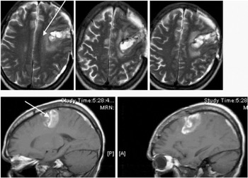
MRI at six weeks after hemorrhage and surgical evacuation of hemorrhage; upper row axial T2; note lesion extension medially to edge of cingulated gyrus (arrow); lower row T1 parasaggital; center of lesion in SMA (arrow) but deep extension involves middle frontal white matter. Cortical lesion extent does not capture the consequences of subcortical extension for local and distant disconnection of frontal pathways.
 You have
Access
- 97
- Cited by