INTRODUCTION
Moniezia expansa (M. expansa), also known as sheep tapeworm, is a very common parasite in ruminants and cattle. It is spread all around the world and is considered to be one of the most common cestode species in sheep. Even though the losses caused by M. expansa are not so severe compared to the nematode parasites, the moniesiasis is still a concern in farm breeding of sheep and goats (Mahin et al. Reference Mahin, Chadli and Id Sidi Yahya1991; Southworth et al. Reference Southworth, Harvey and Larson1996).
Various anticestodal drugs are used to lessen the burden of tapeworm infections. Niclosamide and praziquantel are drugs of choice for the treatment of cestodoses (Elliott, Reference Elliott1984; Mahin et al. Reference Mahin, Chadli and Id Sidi Yahya1991; McKellar and Jackson, Reference McKellar and Jackson2004; Akkaya et al. Reference Akkaya, Deniz, Veren and Esatgil2006). Also benzimidazole drugs, e.g. albendazole and oxfendazole, are effective against M. expansa (Bauer, Reference Bauer1990). However, the treatment can be ineffective because of a development of resistance to anthelmintics. Mamani-Linares (Mamani-Linares, Reference Mamani-Linares2009) described Moniezia spp. resistant to albendazole and fenbendazole. Resistance development to praziquantel in Moniezia spp. was described in New Zealand (Mason et al. Reference Mason, Moffat and Cole2002). The resistance can be caused by various factors. Metabolic detoxification and excretion of anthelmintic drugs both by host and parasite are among discussed ways of resistance development (Alvarez et al. Reference Alvarez, Solana, Mottier, Virkel, Fairweather and Lanusse2005; Cvilink et al. Reference Cvilink, Lamka and Skálová2009a ; Brophy et al. Reference Brophy, MacKintosh and Morphew2012).
There are several ways of drug modification via metabolism. Oxidation is not very common way to modify xenobiotics in helminths. The activities of cytochromes P450 (CYP) are low even if none in most of the helminths, even though the genes were found (Barrett, Reference Barrett1998, Reference Barrett1997; Cvilink et al. Reference Cvilink, Lamka and Skálová2009a ). Solana et al. (Reference Solana, Rodriguez and Lanusse2001) proved the oxidation of ABZ in M. expansa to albendazole sulphoxide (ABZ.SO), but no other information about drug oxidases have been available. On the other hand, the presence and/or activity of antioxidant enzymes superoxide dismutase, glutathione reductase and cytochrome c peroxidase were found (Paul and Barrett, Reference Paul and Barrett1980; McCallum and Barrett, Reference McCallum and Barrett1995). Drug reductases, hydrolases and conjugation enzymes are considered to be more important for helminths (Barrett, Reference Barrett1997; Cvilink et al. Reference Cvilink, Lamka and Skálová2009a ). In M. expansa arylsulphatase, phosphatase and esterase activities as well as deacetylation reactions were detected (Douch and Blair, Reference Douch and Blair1975). Among all types of drug conjugation enzymes, only glutathione S-transferases were found in M. expansa (Douch and Buchanan, Reference Douch and Buchanan1978; Brophy et al. Reference Brophy, Southant and Barrett1989; Sanchez-Moreno et al. Reference Sanchez-Moreno, Orteoa, Sanchez-Navas, Salas-Peregrin and Monteoliva1989). As is obvious from abovementioned facts, the knowledge of drug-metabolizing enzymes and drug biotransformation in M. expansa is not sufficient. Therefore, our aim was to examine activities of common drug metabolizing and antioxidant enzymes and to investigate the metabolism of benzimidazole drugs flubendazole, mebendazole and albendazole.
MATERIALS AND METHODS
Chemicals
Albendazole was purchased from Sigma-Aldrich (Prague, Czech Republic). Albendazole sulphoxide and albendazole sulphone (ABZ.SO2) were obtained from Toronto Research Chemicals Inc. (North York, ON, Canada). Flubendazole, reduced flubendazole (FLU-R), mebendazole and reduced mebendazole (MBZ-R) were provided by Janssen Pharmaceutica (Prague, Czech Republic). SOD Assay Kit-WST (Dojindo, Tabaru, Japan) was purchased from Probior (München, Germany). Liquid sterile-filtered RPMI 1640 medium (Roswell Park Memorial Institute medium) and all other chemicals (high performance liquid chromatography, HPLC or analytical grade) were obtained from Sigma-Aldrich (Prague, Czech Republic).
Biological material
Individuals of M. expansa were obtained from Suffolk lambs (6–7 months old) undergoing natural infection with M. expansa. The occurrence of infection in lambs was determined by coprological examination. Six lambs in total were culled (stunning, exsanguination) in agreement with Czech slaughtering rules for farm animals. Adult tapeworms were rinsed out from small intestine with 0·9% NaCl solution. Obtained tapeworms (only front parts of tapeworms with scolexes, approx. 1 m) were washed 3 times with 0·9% NaCl and 3 times with phosphate buffer saline (0·01 m, pH 7·4) and were used immediately for ex vivo experiments or kept in a freezer (−80 °C) until the subcellular fractions were prepared.
Preparation of subcellular fractions
Microsomes-like, cytosol-like and mitochondria-like fractions were obtained from tapeworms’ homogenate using fractional ultracentrifugation. The tapeworms were homogenized in sodium phosphate buffer (0·1 m, pH 7·4) using a Potter-Elvehjem homogenizer. The 5000 g supernatant was further centrifuged (20 000 g, 60 min, 4 °C). The pellet represents the mitochondria-like fraction. The 20 000 g supernatant was again centrifuged (105 000 g, 65 min, 4 °C) to obtain the cytosol-like and microsomes-like fraction. The pellets with microsomes- and mitochondria-like fractions were resuspended in the phosphate buffer containing 20% (v/v) glycerol. All fractions were stored at −80 °C. Protein concentrations in subcellular fractions were assayed using the bicinchoninic acid method according to Smith et al. (Reference Smith, Krohn, Hermanson, Mallia, Gartner, Provenzano, Fujimoto, Goeke, Olson and Klenk1985).
Enzyme assays
Enzyme activities were assayed in the cytosol-like, mitochondria-like and microsomes-like fractions of M. expansa homogenate. Enzyme assays were performed in 3 measurements for each sample. The amount of organic solvents in the final reaction mixtures did not exceed 1% (v/v).
Peroxidase (Px) activity was determined by measuring o-phenylenediamine dihydrochloride oxidation in the presence of H2O2 spectrophotometrically with a microplate reader (Tecan Infinite M 200) at 490 nm (ε = 1·1 mm −1 × cm−1) (Pérez, Reference Pérez2002).
Catalase (CAT) activity was assayed according to the method described by Góth (Reference Góth1991), which is based on the detection of remaining H2O2 in reaction mixture. H2O2 forms a yellow complex with ammonium molybdate, which was measured spectrophotometrically at 405 nm using a microplate reader (Tecan Infinite M 200).
The assay of superoxide dismutase (SOD) was performed using SOD Assay Kit-WST according to the general protocol. The inhibition activity of SOD on O2 •− formation leads to lower formazan production, which can be determined by a colorimetric method (Tecan Infinite M 200). The activity of SOD is expressed as the decrease (nmol min−1 mg−1) in formazan production.
The other important antioxidant enzyme in Platyhelminthes is thioredoxin glutathione reductase (TGR), which combines both thioredoxin reductase (TrxR) and glutathione reductase (GR) activities. TrxR activity was assayed by the method using 5,5′-dithiobis(2-nitrobenzoic acid) (DTNB). TrxR reduces DTNB by NADPH into yellow 5′-thionitrobenzoic acid, whose formation was followed for 5 min at 412 nm using the Tecan Infinite M 200 (Kuntz et al. Reference Kuntz, Davioud-Charvet, Sayed, Califf, Dessolin, Arnér and Williams2007; Bonilla et al. Reference Bonilla, Denicola, Novoselov, Turanov, Protasio, Izmendi, Gladyshev and Salinas2008). The method for assay of GR activity is based on the NADPH-dependent reduction of oxidized glutathione. Resulting decrease of absorbance due to the consumption of NADPH was observed for 6 min at 340 nm on a Tecan Infinite M 200 (Bonilla et al. Reference Bonilla, Denicola, Novoselov, Turanov, Protasio, Izmendi, Gladyshev and Salinas2008).
The activity of flavin monooxygenases (FMO) towards thiobenzamide was assayed according to Cashman and Hanzlik (Reference Cashman and Hanzlik1981). The final concentration of substrate (dissolved in acetonitrile) was 1 mm. The formation of S-oxide of thiobenzamide was measured spectrophotometrically at 370 nm.
The activities of several isoforms of cytochromes P450 were quantified. The activities of 7-ethoxyresorufin (specific for human⁄rat CYP1A1), 7-methoxyresorufin (CYP1A2), 7-pentoxyresorufin O-dealkylases (CYP2B) and 7-benzyloxyresorufin O-dearylase (CYP3A) were determined at 37 °C using fluorimetric determination of resorufin (Burke et al. Reference Burke, Thompson, Weaver, Wolf and Mayers1994). Each substrate dissolved in dimethylsulphoxide (DMSO) was added at a final concentration of 5 μ m. Assays were conducted using the Perkin-Elmer luminescence spectrophotometer LS 50-B, with the excitation and emission wavelengths of 530 and 585 nm, respectively.
The activities of carbonyl reducing enzymes were tested using the following substrates dissolved in redistilled water: 1 mm metyrapone, 1 mm 4-pyridinecarboxaldehyde and 0·01 mm daunorubicin (at pH 8·5). Spectrophotometric determination (detection wavelength 340 nm, 25 °C) of NADPH consumption in the reaction mixture served for the assessment of reductase activities (Maser, Reference Maser1995; Ohara et al. Reference Ohara, Miyabe, Deyashiki, Matsuura and Kara1995; Palackal et al. Reference Palackal, Burczynski, Harvey and Penning2001). Enzymatic activities for the model substrate acenaphthenol (1·5 mm in DMSO) and menadione (0·5 mm in ethanol) were assayed spectrofluorimetrically using Perkin-Elmer LS 50 B luminescence spectrophotometer. For the acenaphthenol dehydrogenases, the excitation wavelength 340 nm and emission wavelength 480 nm were used. The velocity of substrate dehydrogenation was measured as an increase in fluorescence caused by the reduction of cofactor (NADP+) (Palackal et al. Reference Palackal, Burczynski, Harvey and Penning2001). The assay with substrate menadione was carried out at 380 nm (excitation) and 460 nm (emission). Biotransformation of menadione is coupled with NADPH oxidation which is recorded by the decrease of fluorescence (Maté et al. Reference Maté, Virkel, Lifschitz, Ballent and Lanusse2008).
The UDP-glucuronosyl transferase (UGT) and UDP-glucosyl transferase (UGlcT) was assayed according to Mizuma et al. (Reference Mizuma, Machida, Hayashi and Awazu1982). The final concentration of p-nitrophenol (in redistilled water) was 3·3 μ m. Absorbance was measured using a microplate reader Tecan Infinite M 200 (detection wavelength 415 nm).
Cytosolic glutathione S-transferase (GST) activity was determined using 1 mm 1-chloro-2,4-dinitrobenzene as a substrate (in ethanol). The increase of absorbance at 340 nm was measured spectrophotometrically (Tecan Infinite M 200) (Habig and Jakoby, Reference Habig and Jakoby1981).
In vitro biotransformation of ABZ, FLU and MBZ and drug extraction
The subcellular fractions were incubated with ABZ, FLU or MBZ and coenzyme NADPH. The total volume of the reaction mixture (0·3 mL) contained 100 μL of fractions (0·3–0·9 mg of proteins), anthelmintics (10·0 μ m) pre-dissolved in DMSO (FLU, MBZ) or methanol (ABZ), 1·0 mm NADPH and 0·1 m sodium phosphate buffer (pH 7·4). The first type of blank samples contained 100 μL of 0·1 m sodium phosphate buffer (pH 7·4) instead of fractions, in second type, the fractions were incubated only with DMSO (or methanol) without drugs. The concentration of solvents in all reaction mixtures and blank samples was 1% (v/v). In kinetics study concentration ranges 0·5–15 μ m FLU and 0·5–20 μ m MBZ were used. The incubations were carried out at 37 °C for 30 min. At the end of the incubation, 30 μL of ammonium solution (concentrated, 25% v/v) and 700 μL of ethyl acetate were added. After shaking (3 min, vortex) the mixture and centrifugation (3 min, 5000 g), the supernatants were removed and subsequently evaporated using Eppendorf concentrator 5301. Samples were stored at −20 °C until HPLC analyses.
Ex vivo cultivation of M. expansa with anthelmintics and drug extraction
Only front part of the tapeworm's body with scolex (approximately 1 m) was subject to ex vivo experiments. Prior to the incubation with anthelmintics, separated individuals were kept in phosphate buffer saline (0·01 m, pH 7·4) with penicillin (60 μg mL−1) and streptomycin (100 μg mL−1) for 15 min at 38 °C in a humid aerobic atmosphere with 5% CO2. Cultivation itself was thereafter performed in plastic flasks with 20 mL of RPMI 1640 medium (pH 7·4, with 60 μg mL−1 penicillin and 100 μg mL−1 streptomycin) containing one of the drugs (10 μ m ABZ, FLU or MBZ) at 38 °C in a humid aerobic atmosphere with 5% CO2. FLU and MBZ were pre-dissolved in DMSO, ABZ in methanol. Final concentration of DMSO (or methanol) in RPMI medium was only 0·1% (v/v) to prevent any harmful effects on living tapeworms. Two types of blank samples, medium with anthelmintics but without parasites, and control tapeworms cultivated in drug-free RPMI medium with DMSO (or methanol), were prepared. Tapeworms with drugs and blank samples were incubated for 24 h. To reveal time dependence of metabolite formation, 1 mL of medium was removed from the incubation flasks at time intervals 2, 4 and 8 h after the start of incubation. Medium obtained at each time interval including 24 h was placed into the microtubes. Tapeworms, rinsed thoroughly 3 times with phosphate buffered saline, were weighed and transferred to the tubes. The samples were frozen and stored at −80 °C until processed for HPLC or ultra-high performance liquid chromatography/tandem mass spectrometry (UHPLC/MS/MS) analyses.
Prior to the HPLC analysis liquid/liquid extraction was performed. Tapeworms were quickly homogenized (up to 20 s) in 0·1 m sodium phosphate buffer (pH 7·4) using Sonopuls (Bandelin, Germany). The medium or homogenate (0·3 mL) were alkalized with 30 μL of ammonium solution (concentrated, 25% v/v). After the addition of 700 μL of ethyl acetate, the mixture was shaken (5 min, vortex) and centrifuged (3 min, 5000 g). Supernatants were evaporated and dry samples stored under −20 °C until HPLC analysis. Description of sample preparation for UHPLC/MS/MS is given below (UHPLC/MS/MS conditions).
HPLC analysis
The HPLC analysis was carried out using a Shimadzu chromatograph. Assay of ABZ and its metabolites was performed according to Cvilink et al. (Reference Cvilink, Szotáková, Krízová, Lamka and Skálová2009b ). Briefly, a reverse-phase column LiChroCART 250-3, LiChrospher 60 RP-select B, 250 mm × 3 mm, 5 μm (Merck, Darmstadt, Germany) was used, the mobile phase consisted of acetonitrile − 25 mm, pH 3·0 potassium phosphate buffer (ratio 35:65, v/v), and flow rate was 0·5 mL min−1 in isocratic mode. Parent drug and its metabolites were detected by the fluorescence detector (Shimadzu RF-10AXL, λ EX = 290 nm, λ Em = 320 nm). The retention times for ABZ and metabolites were 17·2 min (ABZ), 5·1 min (ABZ.SO) and 7·1 min (ABZ.SO2). The limit of quantification for ABZ, ABZ.SO and ABZ.SO2 was 120·0, 2·0, and 0·07 pmol mL−1, respectively.
The analysis of FLU, MBZ and their metabolites, previously described by Cvilink et al. (Reference Cvilink, Skálová, Szotáková, Lamka, Kostiainen and Ketola2008b ), was slightly modified. The column Ascentis Express, 100 mm × 3 mm, 2·7 μm (Supelco – Sigma-Aldrich, Prague, Czech Republic) was used. The parent drugs FLU and MBZ were detected by ultra-violet/VIS photodiode array detector SPD-MD10Avp at 246 nm. The fluorescence detector (Shimadzu RF-10AXL, λ EX = 286 nm, λ Em = 306 nm) served for detection of FLU-R and MBZ-R. Mobile phase and flow rate were the same as in the case of ABZ and its metabolites. Under these chromatographic conditions, the retention times were 5·6 min (FLU), 2·0 min (FLU-R), 4·4 min (MBZ) and 1·75 min (MBZ-R).
UHPLC/MS/MS conditions
The samples for UHPLC/MS/MS analyses were prepared from ex vivo cultivation of M. expansa. The samples were initially extracted by liquid/liquid method as described above, and then using SPE extraction method previously performed by Stuchlíková et al. (Reference Stuchlíková, Jirásko, Vokřál, Lamka, Špulák, Holčapek, Szotáková, Bártíková, Pour and Skálová2013).
UHPLC/MS/MS chromatograms of samples were measured using electrospray ionization (ESI) on a hybrid quadrupole-time-of-flight mass analyser (micrOTOF-Q, Bruker Daltonics, Germany). UHPLC was performed on an Agilent 1290 Infinity liquid chromatograph (Agilent Technologies, Waldbronn, Germany) using Zorbax Eclipse C18 column 150 mm × 2·1 mm, 1·8 μm (Agilent Technologies, Waldbronn, Germany), temperature 25 °C, flow rate 0·4 mL min−1 and the injection volume 3 μL. The mobile phase consisted of 0·5 mm ammonium acetate adjusted to pH = 4·0 (A) and acetonitrile (B). The linear gradient was as follows: 0 min – 15% B, 10 min – 50% B, 12 min – 95% B for ABZ; 0 min – 15% B, 8 min – 40% B, 10 min – 95% B for FLU; 0 min – 15% B, 6 min – 40% B, 10 min – 95% B for MBZ. The washing and reconditioning of the column were carried out after linear gradient for all separations. The QqTOF mass spectrometer with average resolving power higher than 13 000 was used with the following setting of tuning parameters: capillary voltage 4·5 kV, drying temperature 220 °C, the flow rate and pressure of nitrogen were 8 L min−1 and 1·3 bar, respectively. The external calibration was performed with sodium formate clusters before individual measurements. ESI mass spectra were recorded in the range of m/z 50–1000 in the positive-ion mode. The isolation width ∆m/z 4 and the collision energy 20 eV (found as optimal energy for fragmentation of studied metabolite ions) using argon as the collision gas were employed for MS/MS experiments.
Statistical analysis
The reported data are expressed as mean ± s.d. Statistical comparisons were carried out using Student's t-test (Microsoft Office Excel 2007). A probability of P < 0·05 was considered statistically significant.
RESULTS
Enzyme assays
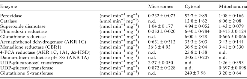
The specific activities of oxidation/antioxidant, reduction and conjugation enzymes which participate in biotransformation of anthelmintic drugs were evaluated in microsomes-like, cytosol-like and mitochondria-like fractions of M. expansa homogenate. The assays are based on spectrophotometric or spectrofluorimetric detection of the rising cofactor/product of biotransformation or the diminishing substrate/cofactor. The antioxidant enzymes (Px, CAT, SOD, GR and TrxR activity of TGR) had the highest activity in cytosol-like fraction; the activities in microsomes- and mitochondria-like fractions were several times lower or were not detected. Oxidation enzymes – CYP and FMO – failed to show any activity in all fractions. Regarding the carbonyl-reducing enzymes, the cytosol-like fraction reduced acenaphthenol, menadione, daunorubicin at pH 8·5 and 4-pyridinecarboxaldehyde. Substrates acenaphthenol and menadione were also metabolized by microsomes- and mitochondria-like fractions. Reduction activity towards metyrapone was not proven. The glucuronidation and glucosylation of p-nitrophenol mediated by UGT and UGlcT were detected in microsomes- and mitochondria-like fractions. High activity of GST was observed in cytosol-like fraction. The values of specific activities are summarized in Table 1.
Specific activities of antioxidant, reduction and conjugation enzymes found in microsomes-like, cytosol-like and mitochondria-like subcellular fractions of M. expansa

The values represent means ± s.d. from 3 to 6 samples, n.d., not detected.
CBR1, carbonyl reductase 1; AKR, aldo-keto reductase; 3α-HSD, 3α-hydroxysteroid dehydrogenase; 4-PCA, 4-pyridine carboxaldehyde.
HPLC analysis of ABZ, FLU and MBZ biotransformation in vitro
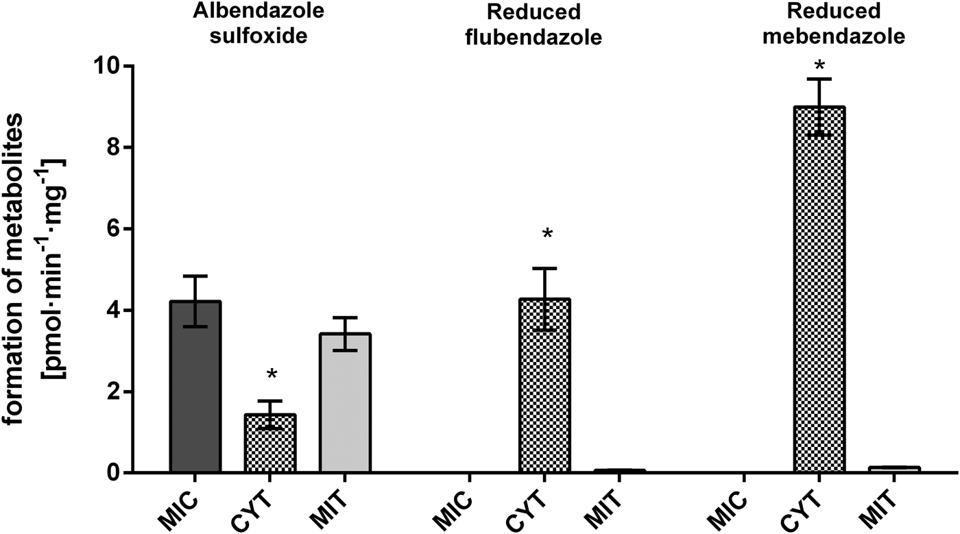
The subcellular fractions of M. expansa homogenate were incubated with one of the anthelmintics (ABZ, FLU and MBZ) at concentration 10 μ m in the presence of coenzyme NADPH. ABZ was metabolized in all subcellular fractions. ABZ.SO was the major product of the reaction. The highest amount of ABZ.SO was detected in microsomes-like fraction, lower amounts in mitochondria- and cytosol-like fractions. In cytosol-like fraction the amount of ABZ.SO was 2·4–3 times lower than in mitochondria- and microsomes-like fractions, respectively. The second step of oxidation, i.e. formation of ABZ.SO2, occurred in very low extent with the amount of metabolite being under the limit of quantification. Considering the FLU and MBZ reactions only carbonyl-reduced metabolites (FLU-R, MBZ-R) were found. Cytosol-like fraction was the only fraction, where the measurable amounts of FLU-R and MBZ-R were detected. In vitro reduction of MBZ was approximately twice more intensive than the reduction of FLU. The results of in vitro incubation are shown in Fig. 1.
Concentration of ABZ, FLU and MBZ metabolites analysed by HPLC after 30 min in vitro incubation of microsomes-like, cytosol-like and mitochondria-like subcellular fractions of M. expansa homogenate with 10 μ m parent drugs. Results represent means ± s.d., n = 3. *Significantly different compared with microsomes- and mitochondria-like fractions, P < 0·05.

Kinetics of FLU and MBZ reduction in vitro
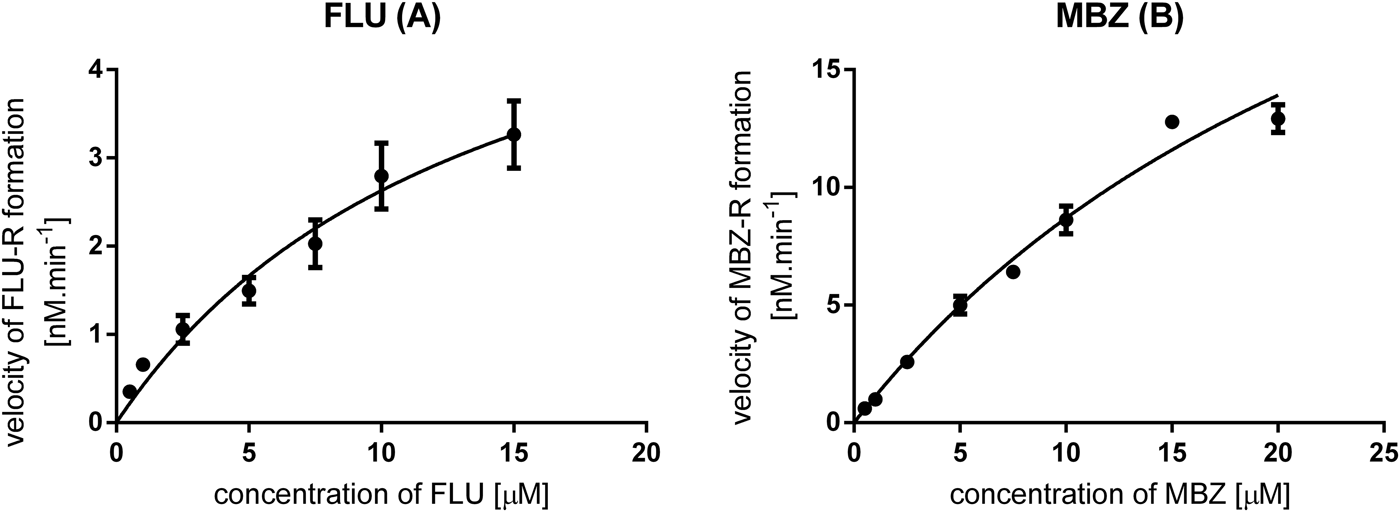
The cytosol-like fraction of M. expansa was incubated with various concentrations of FLU (0·5–15·0 μ
m) and MBZ (0·5–20 μ
m) and amount of reduced metabolites (FLU-R and MBZ-R) was measured by HPLC and the velocity of reaction was calculated. The direct plot of the reaction velocity vs substrate concentration fit well the Michaelis–Menten equation, as it is shown in Fig. 2. Using GraphPad Prism 6 software, basic kinetic parameters, apparent Michaelis constant and apparent maximal velocity (
 $K_{\rm m}^{\rm ^{\prime}} $
and
 $V_{\max} ^{\rm ^{\prime}} $
), of reactions were calculated. The ratio of and
 $K_{\rm m} ^{\rm ^{\prime}} $
is called intrinsic clearance (Clint) and represents the efficiency of enzymatic transformation. The values of
 $K_{\rm m} ^{\rm ^{\prime}} $
,
 $V_{\max} ^{\rm ^{\prime}} $
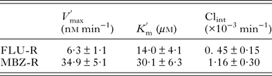
and Clint are presented in Table 2.
 $V_{\max} ^{\rm ^{\prime}} $
for MBZ is about five times higher than for FLU and
 $K_{\rm m} ^{\rm ^{\prime}} $
of MBZ is twice the
 $K_{\rm m} ^{\rm ^{\prime}} $
of FLU making the Clint of MBZ reduction three times higher than that of FLU reduction.
Kinetics of flubendazole (FLU) reduction (A) and mebendazole (MBZ) reduction (B) in cytosol-like fraction of M. expansa.

The kinetic parameters of mebendazole and flubendazole reduction in M. expansa cytosol-like fraction

The results represent means ± s.d. from 4 samples.
HPLC analysis of ABZ, FLU and MBZ biotransformation ex vivo
The living tapeworms were cultivated individually in plastic flasks in RPMI 1640 medium containing ABZ, FLU or MBZ at concentration 10·0 μ m for 24 h. The analyses of metabolites were carried out in a cultivation medium and homogenate of tapeworms’ bodies. As in the experiments performed in vitro, the certain amount of ABZ.SO was detected in medium and homogenate from living tapeworms. However, the blank samples contained significant concentration of ABZ.SO as well. The amount of ABZ.SO formed by the non-enzymatic conversion was subtracted from the ABZ.SO amount detected in samples. The results obtained by HPLC measurement did not significantly prove that living tapeworms actually transform ABZ by its oxidation.
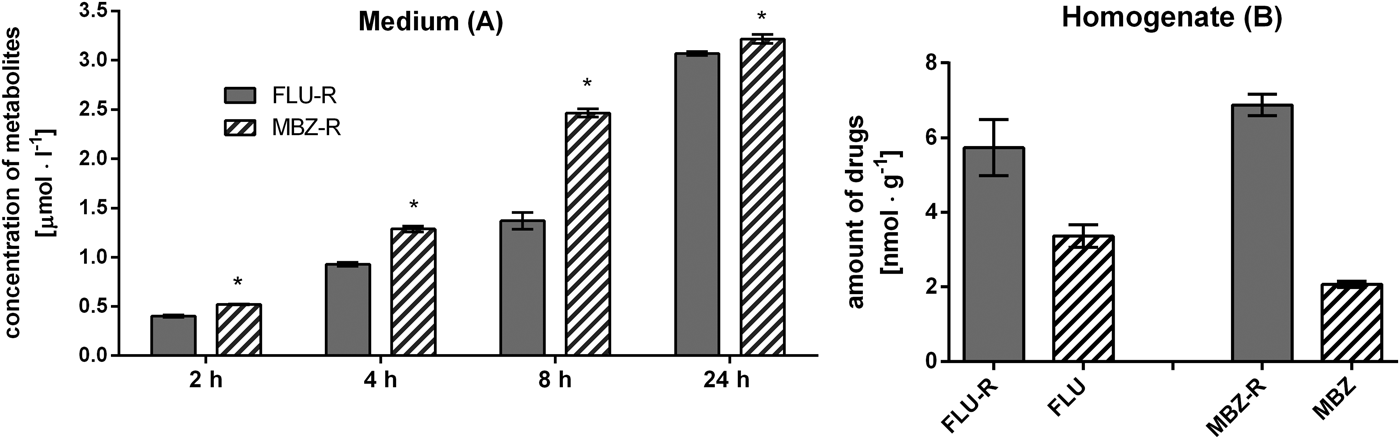
The reduced metabolites FLU-R and MBZ-R were the only phase I metabolites detected after ex vivo incubation of the tapeworms with FLU and MBZ. In the medium, concentrations of metabolites were analysed in the time intervals 2, 4, 8 and 24 h of incubation. The formation of metabolites detected in medium was time-dependent, with significantly higher level of MBZ-R than FLU-R (see Fig. 3A). Amount of metabolites measured in the homogenate of tapeworms confirmed the higher extent of MBZ reduction than FLU reduction, which is recorded in Fig. 3B. These results correspond with the higher concentration of parent drug FLU in comparison to MBZ both in homogenate (Fig. 3B) and medium (data not shown).
Concentration of reduced metabolites (FLU-R, MBZ-R) in cultivation medium analysed by HPLC after 2, 4, 8 and 24 h of ex vivo incubation of living tapeworms with 10 μ m FLU or MBZ (A). Concentration of reduced metabolites (FLU-R, MBZ-R) and parent drugs (FLU, MBZ) in tapeworms homogenate analysed by HPLC after 24 h of ex vivo incubation of living tapeworms with 10 μ m FLU or MBZ (B). Results represent means ± s.d., n = 3. *Significantly different compared with FLU-R, P < 0·05.

UHPLC/MS/MS of studied benzimidazole drugs and their metabolites
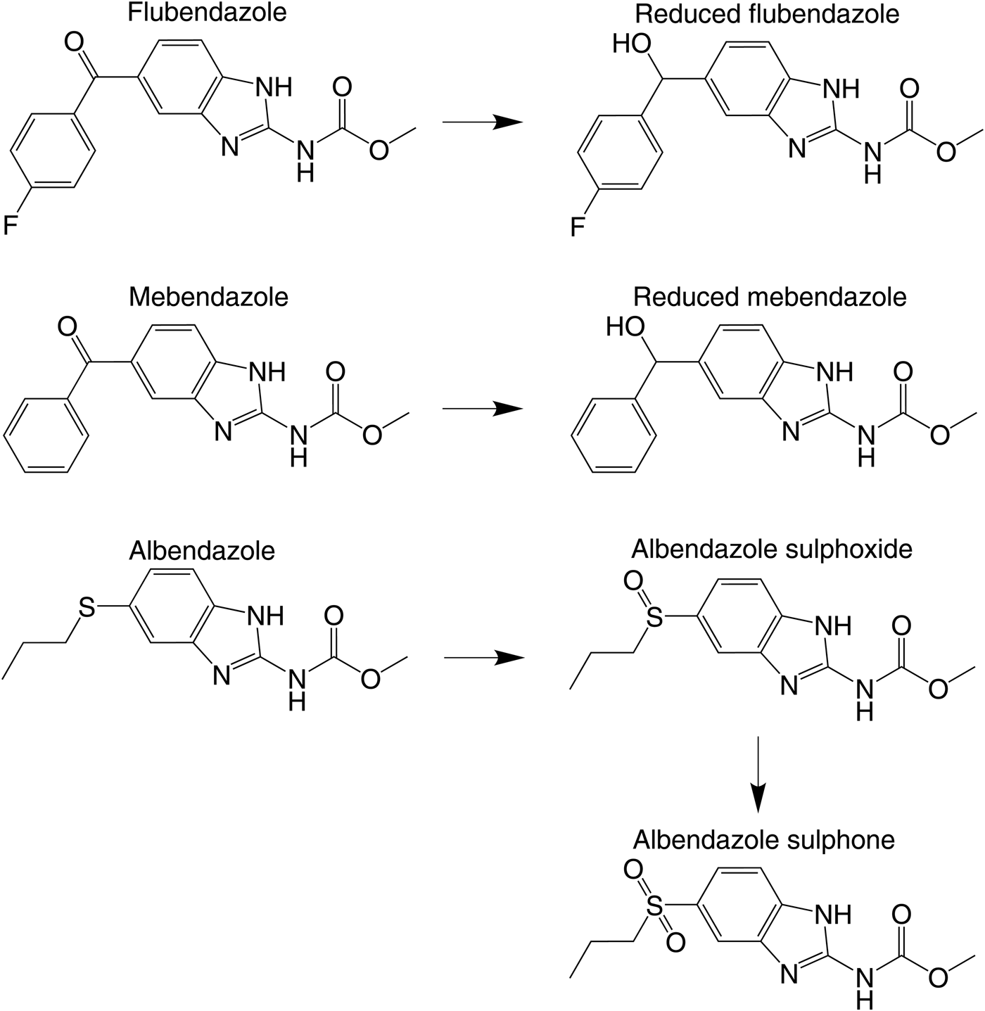
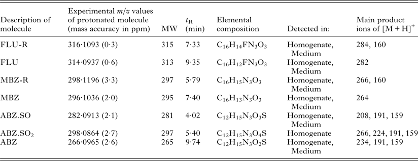
The analytical characterization of benzimidazole drugs and their metabolites in studied samples (see Experimental part) was performed using UHPLC/ESI-MS/MS. Detected ex vivo metabolites were identified based on the presence of protonated molecules [M + H]+ and the interpretation of their product ion spectra. High mass accuracy measurement allowed the confirmation of elemental composition and the type of metabolic reactions according to exact mass defects. Retention times, m/z values, molecular weights, elemental compositions and key fragment ions of benzimidazole metabolites detected in MS/MS experiments are summarized in Table 3. Results were confirmed by the comparison with UHPLC/ESI-MS/MS measurements of available standards. Structures of parent drugs and metabolites are shown in Fig. 4. The neutral loss (NL) ∆m/z 32 (CH3OH), typical fragmentation for methylesters (Holčapek et al. Reference Holčapek, Jirásko and Lísa2010), was in accordance with the present methylcarbamate group and was observed for all benzimidazole drugs and their metabolites. The cleavage of bond between benzimidazole and (phenyl)hydroxymethyl part for reduced MBZ or (4-fluorophenyl)hydroxymethyl part for reduced FLU was among other observed fragmentation mechanisms. This fragmentation combined with NL of methanol yielded product ion at m/z 160. Due to high stability of conjugated system, this fragmentation was not observed for parent drugs MBZ and FLU. On the other hand, the cleavage of bond between propylthio substituent (propylsulphoinyl for ABZ.SO or propylsulphonyl for ABZ.SO 2 ) and benzimidazole part of ABZ related compounds resulted in the formation of radical ion at m/z 191 and m/z 159 in case of additional NL of methanol. The combined NL of methanol and propene yielding ion at m/z 208 and 224 was also observed in case of ABZ.SO and ABZ.SO 2 . The results obtained by UHPLC/MS/MS measurement significantly prove that living tapeworms reduced FLU and MBZ carbonyl group and oxidized ABZ to ABZ.SO and ABZ.SO2.
Chemical structures of FLU, MBZ, ABZ and their metabolites present in ex vivo cultivation of M. expansa.

Benzimidazole drugs and their metabolites in M. expansa detected by UHPLC/MS/MS. Experimental m/z values and main product ions values are necessary for identification of parent drugs and their metabolites. The main product ions give the information about structure of metabolites.

Experimental m/z values of protonated molecules [M + H]+ in ESI full scan mass spectra, mass accuracies; molecular weights (MW); retention time (t R); elemental compositions and MS/MS product ions.
DISCUSSION
Moniezia spp. is one of the most spread tapeworms affecting ruminants, therefore it was intensively studied in many ways including their physiology and biochemistry as well as the methods of treatment. Unfortunately till now only little information about ability of M. expansa to biotransform drugs and other xenobiotics has been available. The aim of our study was to fill in this empty space. The knowledge of helminths biotransformation enzymes and metabolism of selected anthelmintic drugs is important as the biotransformation can contribute to drug resistance development e.g. the metabolite formation and efflux of triclabendazole was greater in resistant strains of Fasciola hepatica than in susceptible ones (Wolstenholme et al. Reference Wolstenholme, Fairweather, Prichard, von Samson-Himmelstjerna and Sangster2004; Alvarez et al. Reference Alvarez, Solana, Mottier, Virkel, Fairweather and Lanusse2005; Brophy et al. Reference Brophy, MacKintosh and Morphew2012).
Reduction is considered to be the main pathway of detoxification of xenobiotics in helminths. The presence of reductive drug metabolism in M. expansa was proven by results from ex vivo cultivations, where the less potent FLU and MBZ metabolites with reduced carbonyl group were found (Dayan, Reference Dayan2003; Ceballos et al.
Reference Ceballos, Elissondo, Sánchez Bruni, Denegri, Lanusse and Alvarez2011). These metabolites were previously identified in other helminths species: Haemonchus contortus, Hymenolepis diminuta and Dicrocoelium dendriticum (Cvilink et al.
Reference Cvilink, Lamka and Skálová2009a
). From in vitro experiments it can be seen that the formation of reduced metabolites took place in cytosol-like fraction of M. expansa. It is the same in other helminth species such as H. contortus and H. diminuta (Bártíková et al.
Reference Bártíková, Vokřál, Skálová, Kubíček, Firbasová, Briestenský, Lamka and Szotáková2012; Vokřál et al.
Reference Vokřál, Bártíková, Prchal, Stuchlíková, Skálová, Szotáková, Lamka, Várady and Kubíček2012). The activities of these enzymes were further studied by determining the basic kinetic parameters, apparent Michaelis constant and apparent maximal velocity. When the parameters were compared to those of H. diminuta (Bártíková et al.
Reference Bártíková, Vokřál, Skálová, Kubíček, Firbasová, Briestenský, Lamka and Szotáková2012),
 $V_{\max} ^{\rm ^{\prime}} $
for the FLU reduction was almost 6 times higher in M. expansa than in the H. diminuta, while the
 $K_{\rm m} ^{\rm ^{\prime}} $
, representing the affinity of the enzymes to the FLU, is very similar in both cestodes. In case of MBZ reduction, the
 $V_{\max} ^{\rm ^{\prime}} $
does not differ from H. diminuta, while the
 $K_{\rm m} ^{\rm ^{\prime}} $
in the M. expansa is twice lower than in H. diminuta, suggesting that MBZ has greater affinity to the enzyme of M. expansa.
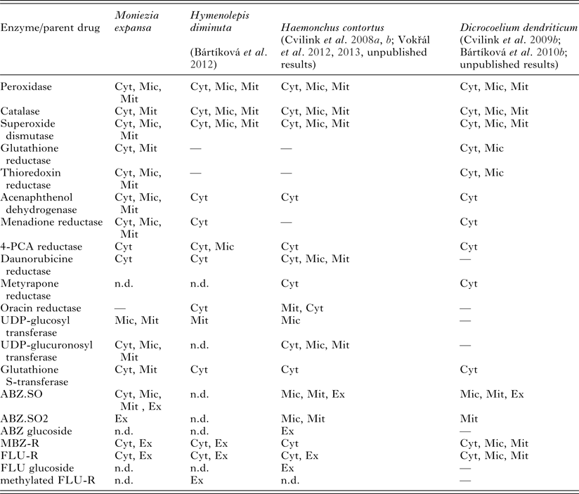
Consequently, reduction of other xenobiotics with carbonyl group in M. expansa was tested using various substrates such as acenaphthenol, menadione, metyrapone, 4-pyridinecarboxaldehyde and daunorubicin. All substrates except metyrapone were metabolized, mainly in cytosol-like fraction. In comparison with previously performed testing of cestode H. diminuta (Bártíková et al. Reference Bártíková, Vokřál, Skálová, Kubíček, Firbasová, Briestenský, Lamka and Szotáková2012), the activities found in M. expansa are lower. In the case of acenaphthenol dehydrogenase the activity is almost ten times lower. However, the localization of the carbonyl reducing enzymes seems to be different in both cestodes. In M. expansa, acenaphthenol dehydrogenase and menadione reductase activities were proven in all three tested fractions, while in the H. diminuta only cytosol-like fraction showed the activity towards acenaphthenol and menadione. On the contrary, H. diminuta possessed the highest transformation capacity of 4-pyridinecarboxylaldehyde reductase in microsome-like fraction, while it was absent in M. expansa. Brief comparison of drug-metabolizing enzymes in several helminth species is shown in Table 4.
Comparison of drug-metabolizing enzymes activities and biotransformation of anthelmintics in different helminths (M. expansa, H. diminuta, H. contortus, D. dendriticum)

Cyt, cytosol-like fraction; Mic, microsomes-like fraction; Mit, mitochondria-like fraction; Ex, ex vivo; n.d., not detected; (—), not tested.
Oxidative metabolism in helminths is not as common and developed as in other species (Barrett, Reference Barrett1998). Cytochrome P450 activities were found only in few species: H. contortus mainly in larvae (Kotze, Reference Kotze1997, Reference Kotze1999; Laing et al. Reference Laing, Kikuchi, Martinelli, Tsai, Beech, Redman, Holroyd, Bartley, Beasley, Britton, Curran, Devaney, Gilabert, Hunt, Jackson, Johnston, Kryukov, Li, Morrison, Reid, Sargison, Saunders, Wasmuth, Wolstenholme, Berriman, Gilleard and Cotton2013), Schistosoma mansoni (Saeed et al. Reference Saeed, Mostafa, O'Connor, Rafferty and Doenhoff2002; Berriman et al. Reference Berriman, Haas, LoVerde, Wilson, Dillon, Cerqueira, Mashiyama, Al-Lazikani, Andrade, Ashton, Aslett, Bartholomeu, Blandin, Caffrey, Coghlan, Coulson, Day, Delcher, DeMarco, Djikeng, Eyre, Gamble, Ghedin, Gu, Hertz-Fowler, Hirai, Hirai, Houston, Ivens and Johnston2009). The cytochrome P450 genes were also found in other helminths: roundworm C. elegans, tapeworms Hymenolepis microstoma, Echinococcus granulosus, Echinococcus multiocularis, Taenia solium and fluke Schistosoma japonicum (C. elegans Sequencing Consortium, 1998; Schistosoma japonicum Genome Sequencing and Functional Analysis Consortium, 2009; Tsai et al. Reference Tsai, Zarowiecki, Holroyd, Garciarrubio, Sanchez-Flores, Brooks, Tracey, Bobes, Fragoso, Sciutto, Aslett, Beasley, Bennett, Cai, Camicia, Clark, Cucher, De Silva, Day, Deplazes, Estrada, Fernández, Holland, Hou, Hu, Huckvale, Hung, Kamenetzky, Keane and Kiss2013). In our assay, there was found no activity in any of the CYP isoforms tested. The oxidative metabolism of xenobiotics can be catalysed by enzymes, which usually serve as protection system against oxygen radicals and other molecules causing oxidative stress in organisms, like peroxidase, catalase, glutathione peroxidase or superoxide dismutase. The other important antioxidant enzyme in Platyhelminthes is TGR combining both thioredoxin reductase and glutathione reductase activities. Activities of Px, CAT, SOD, TrxR and GR were found in M. expansa. Paul and Barrett (Reference Paul and Barrett1980) found only the activity of Px and SOD. Compared to H. diminuta the activities of Px, CAT and SOD are very similar. The only exception is the activity of Px in cytosol-like fraction, which is 10 times higher than in H. diminuta (Bártíková et al. Reference Bártíková, Vokřál, Skálová, Kubíček, Firbasová, Briestenský, Lamka and Szotáková2012). These enzymes may participate in the oxidation of anthelmintics including ABZ.
The oxidation of ABZ has been reported in many helminth species like H. contortus, F. hepatica, Ascaris suum and D. dendriticum (Solana et al. Reference Solana, Rodriguez and Lanusse2001, Reference Solana, Scarcella, Virkel, Ceriani, Rodríguez and Lanusse2009; Cvilink et al. Reference Cvilink, Skálová, Szotáková, Lamka, Kostiainen and Ketola2008b , Reference Cvilink, Szotáková, Krízová, Lamka and Skálová2009b ; Vokřál et al. Reference Vokřál, Jirásko, Stuchlíková, Bártíková, Szotáková, Lamka, Várady and Skálová2013). In vitro experiments revealed that the microsome-like fraction of M. expansa metabolized ABZ to active ABZ.SO, also known as ricobendazole anthelmintic drug. HPLC analysis did not prove sufficiently the ex vivo metabolism of ABZ, therefore more sensitive UHPLC/MS analysis was carried out. ABZ.SO was found in both tapeworm homogenate and cultivation medium based on UHPLC/MS/MS analysis of ex vivo samples. The amounts of ABZ.SO in ex vivo samples were several times higher, than those in blank ones, proving that the ABZ was metabolized and not just spontaneously oxidized. The MS analysis confirmed notable amount of anthelminticly ineffective albendazole sulphone (ABZ.SO2), further oxidation product of ABZ.SO, in homogenate of the tapeworm bodies. On the contrary, no trace of ABZ.SO2 was detected in the medium where the tapeworms were cultivated. It seems that more polar ABZ.SO2 cannot be excreted by diffusion or any other transport mechanism, thus leading to the accumulation of metabolite in the tapeworm's body.
In the second phase of biotransformation, drugs or their metabolites are conjugated with endogenous molecules to further reduce their toxicity or enhance their excretion. One of the very important enzymes of the second phase metabolism is glutathione S-transferase. In M. expansa the activity of GST prevailed in cytosol-like fraction, as in H. diminuta, with specific activity rate also very similar to the H. diminuta and even higher than in their host's intestinal tissue (Bártíková et al. Reference Bártíková, Krízová, Lamka, Kubícek, Skálová and Szotáková2010a , Reference Bártíková, Vokřál, Skálová, Kubíček, Firbasová, Briestenský, Lamka and Szotáková2012). Only low activities were measured for UDP-glucosyl transferase and UDP-glucuronosyl transferase. Glycosylation is very unusual conjugation reaction in animals, but it is a way to deactivate benzimidazole anthelmintics, such as FLU and ABZ in Haemonchus contortus (Cvilink et al. Reference Cvilink, Skálová, Szotáková, Lamka, Kostiainen and Ketola2008b ). This reaction is also performed by other helminth species (Cvilink et al. Reference Cvilink, Lamka and Skálová2009a ). However, in ex vivo biotransformation study with M. expansa no conjugated metabolites were detected. It seems that tested anthelmintic drugs are not suitable substrates for conjugation enzymes of M. expansa.
The results of our ex vivo and in vitro study show that M. expansa has metabolic systems allowing it to biotransform various xenobiotics including benzimidazole drugs. Moniezia expansa biotransforms anthelmintics FLU and MBZ via reduction of carbonyl group to less active metabolites. Moniezia expansa is capable to oxidize ABZ to ABZ.SO and further to ABZ.SO2. This deactivated drug remains in the body of the parasite, because it probably has no transporter system to excrete it. No second phase metabolites of tested anthelmintics were found.
ACKNOWLEDGEMENTS
We would like to thank to technical assistance of Alena Pakostová.
FINANCIAL SUPPORT
The project was supported by the Czech Science Foundation (GA ČR, grant No. P502/10/0217), by the Grant Agency of Charles University (GA UK, grant No. 673612/B-CH/2012) and by the Charles University in Prague (grant number SVV 260 065). The project was co-financed by the European Social Fund and the state budget of the Czech Republic (Project No. CZ.1.07/2.3.00/30.0022).