INTRODUCTION
Over the last century, there have been several reports describing the genus Prymnesium Massart emend. Edvardsen, Eikrem and Probert as toxic. The first report of a Prymnesium parvum bloom in Danish waters noted diseased and dying fish (Liebert & Deerns, Reference Liebert and Deerns1920). Since this initial report, several blooms have occurred in different parts of the world, above all in temperate and subtropical regions and most of them in brackish waters (Manning & La Claire, Reference Manning and La Claire2010; Bertin et al., Reference Bertin, Zimba, Beauchesne, Huncik and Moeller2012a). These blooms cause very large economic losses to aquaculture and fisheries around the world (Moestrup, Reference Moestrup, Green and Leadbeater1994).
The toxicity of Prymnesium includes cytotoxic, ichthyotoxic, neurotoxic and haemolytic activity (Bertin et al., Reference Bertin, Zimba, Beauchesne, Huncik and Moeller2012b), which enables Prymnesium to affect a wide range of organisms, from vertebrates to phytoplankton. Apart from its ichthyotoxicity, Prymnesium has been described as a very effective competitor due to the absence of other eukaryotic or prokaryotic algae when a bloom of P. parvum occurs (Granéli & Johansson, Reference Granéli and Johansson2003) and due to its allelopathic capabilities (Schmidt & Hansen, Reference Schmidt and Hansen2001). The toxicity and ability to outcompete other organisms arises from its production of a diverse group of toxic compounds with a very broad range of biological activity and solubility (Bertin et al., Reference Bertin, Zimba, Beauchesne, Huncik and Moeller2012a). We found among them Haemolysin 1 (Kozakai et al., Reference Kozakai, Oshima and Yasumoto1982), Prymnesin-1 and Prymnesin-2 (Manning & La Claire, Reference Manning and La Claire2010), fatty acids (Henrikson et al., Reference Henrikson, Gharfeh, Easton, Easton, Glenn, Shadfan, Mooberry, Hambright and Cichewicz2010) and fatty acid amides (Bertin et al., Reference Bertin, Zimba, Beauchesne, Huncik and Moeller2012a).
The ichthyotoxicity of Prymnesium species arises from its impact on the permeability mechanism in the gills of fish and aquatic invertebrates (Yariv & Hestrin, Reference Yariv and Hestrin1961; Ulitzer & Shilo, Reference Ulitzer and Shilo1966). Haemolytic activity may be another of its allelopathic mechanisms (Granéli & Hansen, Reference Granéli, Hansen, Granéli and Turner2006). Sometimes the same molecule can have different effects and activities (Yasumoto et al., Reference Yasumoto, Underdal, Aune, Hormazabal, Skulberg, Oshima, Graneli, Sundström, Edler and Anderson1990). Some bloom-forming phytoplankton taxa produce potent toxins that can kill fish, birds and mammals, and also accumulate in shellfish rendering them unsafe for human consumption. These toxins, however, are not linked to allelopathic effects against other phytoplankton (Prince et al., Reference Prince, Poulson, Myers, Sieg and Kubanek2010).
Methods to determine the toxicity or allelopathy of Prymnesium organisms include haemolysis assays (Uronen et al., Reference Uronen, Lehtinen, Legrand, Kuuppo and Tamminen2005), determining mortality rates for the brine shrimp Artemia (Larsen & Bryant, Reference Larsen and Bryant1998; Granéli & Johansson, Reference Granéli and Johansson2003) or quantifying mortality or growth inhibition of surrounding algal cells (Schmidt & Hansen, Reference Schmidt and Hansen2001; Fistarol et al., Reference Fistarol, Legrand and Granéli2003; Freitag et al., Reference Freitag, Beszteri, Vogel and John2011).
The toxicology of this genus is dependent upon growth conditions including light, nutrients, temperature and other parameters (reviewed in Granéli et al., Reference Granéli, Edvardsen, Roelke and Hagström2012), although results are sometimes contradictory. After reviewing the literature, toxin production by P. parvum is a very complex process that depends on several factors.
The extraction method can also affect the results. The toxic substances produced by P. parvum are called prymnesins and consist of glycolipids, galactolipids, polyketides, proteolipids, fatty acids and fatty acid amides (Manning & La Claire, Reference Manning and La Claire2013; Blossom et al., Reference Blossom, Andersen, Rasmussen and Hansen2014). This heterogeneous mixture makes it difficult to find a unique solvent with which to extract all of the components. Among all fractions, the toxic component is most soluble in polar organic solvents (Manning & La Claire, Reference Manning and La Claire2010).
Most studies involving the genus Prymnesium have been conducted with the species P. parvum. One of the reasons for this could be the relatively recent discovery (after the 1980s) of other species in the Prymnesium genus (Billard, Reference Billard1983; Fresnel et al., Reference Fresnel, Probert and Billard2001; Seoane et al., Reference Seoane, Elkrem, Pienaar and Edvardsen2009; Grant et al., Reference Grant, Waller and Wetherbee2011; Sym et al., Reference Sym, Pienaar, Edvardsen and Egge2011). Another reason could be the prevalence of P. parvum in culture collections that usually do not include other Prymnesium species.
In this study, we have measured the haemolytic activity of eight strains from at least seven different species of Prymnesium while taking into account the growth phase (exponential vs stationary). We have also compared the efficacy of methanol and cold acetone as the extraction solvent.
MATERIALS AND METHODS
Strains
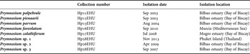
Eight strains from at least seven different species in the genus Prymnesium (Haptophyta) were isolated from Bilbao and Mogro estuaries in the Bay of Biscay, the Mediterranean Sea and the Phuket Islands (Thailand) (Table 1). Five of the strains correspond to known species that have been characterized by genetic analysis and electron microscopy. Two other strains will be submitted as new species (Hp 26 EHU and Hp 28 EHU), and the final strain, Hp 58 EHU, has not been analysed by electron microscopy or other molecular methods, and thus we do not know if it is identical to any of the other species in this study.
Strains of Prymnesium used in the haemolytic assays

Culture conditions
The cultures were grown at 18 ± 1°C with a salinity of 30 psu in f/2 medium (Guillard & Ryther, Reference Guillard and Ryther1962) prepared with filtered and autoclave-sterilized seawater. Cultures were exposed to a 12 L:12 D cycle. To obtain a growth curve, fluorescence was measured daily with a Turner Designs 10-100R fluorimeter (Turner Designs, USA) as a proxy of cell density. Based on these growth curves, samples for haemolytic analyses were obtained from the exponential and stationary phases.
Cell density and biovolume
One millilitre of each culture was extracted prior to filtration in order to ascertain the density, using a Neubauer chamber.
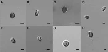
The biovolume (μm3) of each strain was calculated according to the equations provided by Olenina et al. (Reference Olenina, Hajdu, Edler, Andersson, Wasmund, Busch, Göbel, Gromisz, Huseby, Huttunen, Jaanus, Kokkonen, Ledaine and Niemkiewicz2006) for cell shapes and sizes. At least 10 individuals from each strain were measured in order to obtain the average dimensions of cells and these values were used in the corresponding equations. The biovolumes for all of the strains were calculated using the geometric shape of rotational ellipsoid (π/6 × d2 × l), with d being the diameter and l being the length, except for the strain Hp26 EHU, for which biovolume was calculated using the equation for a sphere (π/6 × d3) due to its spherical form. Cells from each strain were photographed live with a Leica DMRB microscope (Leica, Wetzlar, Germany) and an Olympus BX 53 microscope (Olympus, Tokyo, Japan) and analysed using the NIS-Elements viewer software (Nikon, Melville, USA) (Figure 1).
Shapes of the Prymnesium strains used in this study: (A) Prymnesium palpebrale; (B) Prymnesium pienaarii; (C) Prymnesium parvum; (D) Prymnesium faveolatum; (E) Prymnesium calathiferum; (F) Prymnesium sp. 1; (G) Prymnesium sp. 2; (H) Prymnesium sp. 3. Scale bar: 5 µm.

Toxin extraction
When the cultures reached the selected phase of growth, cells were harvested and distributed into two aliquots of ~ 500 ml each. These duplicate samples were filtered through 0.7 µm GF/F glass fibre filters that were 47 mm in diameter. The cells collected on each filter were resuspended, one with methanol and the other with cold acetone, and extracted as follows. First, they were sonicated to homogenize the suspension and then they were centrifuged for 10 min at 3000 rpm at 10°C. Finally, the two supernatants were stored at −20°C prior to analysis. The complete procedure was carried out in the dark to avoid degradation of the prymnesin toxins (Parnas et al., Reference Parnas, Reich and Bergmann1962).
Haemolysis assay
The haemolytic activity of Prymnesium cultures was analysed according to Stolte et al. (Reference Stolte, Panosso, Gisselson and Granéli2002) using sheep's blood in Alsever's solution. Erythrocytes were separated from the plasma by centrifugation for 10 min at 400 × g at 10°C and washed twice with isotonic phosphate buffer (PBS). Then blood was diluted in PBS to a final concentration of 1.7 × 10⁷ red blood cells ml−1. Two millilitre aliquots of the methanol and acetone extracts were dried down and redissolved in 200 µl of methanol and 800 µl of PBS buffer. Toxicity of the extracts was calculated relative to saponin (Sigma S-2149, USA) and haemolytic activity was expressed as μg saponin equivalent per cell. Each assay was carried out in triplicate in a 96 microwell plate by mixing 150 µl of blood with 150 µl of extract diluted in PBS. Haemolysis controls for 0 and 100% lysis were prepared by mixing 150 µl of blood with 150 µl of PBS or distilled water, respectively. Microplates were incubated for 1 h in the dark at room temperature and then the whole plate was centrifuged for 10 min at 400 × g at 10°C. From each well, 200 µl of the supernatant was transferred to a new plate and the absorbance at 405 nm was measured with a microplate reader (BioRad 550, USA). Measurements from three replicates were averaged.
Statistical analysis
All statistical analyses were performed using IBM SPSS Statistics 23.0. The influence of the solvent and the growth phase on toxicity were analysed by a Student t-test. Prior to the study, data were log transformed to fit a normal distribution.
RESULTS
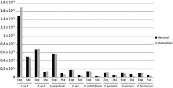
All of the strains exhibited haemolytic activity in both exponential and stationary phases. The values per cell indicated a maximum of 1.7 × 10⁻⁵ μg saponin equivalent per cell in Prymnesium sp. 3 (exponential phase, extracted with cold acetone), and a minimum of 2.8 × 10⁻⁷ μg saponin equivalent per cell in P. parvum (stationary phase, extracted with cold acetone) (Figure 2). There were three strains with haemolytic activity above 5 × 10⁻⁶ μg saponin equivalent per cell: Prymnesium sp. 3, Prymnesium sp. 2 and Prymnesium palpebrale (exponential phase in both solvents for all). The rest did not reach 5 × 10⁻⁶ μg saponin equivalent per cell under any condition. Among samples collected from the stationary phase, there was a large difference between Prymnesium sp. 3 and the rest of the strains. In Prymnesium sp. 3, the haemolytic activity was almost 5 × 10⁻⁶ μg saponin equivalent per cell while the rest of the strains were below 2 × 10⁻⁶.
Haemolytic activity of Prymnesium strains normalized to cell number (μg saponin equivalent per cell). Exp = exponential phase; Sta = stationary phase.

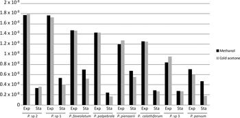
A comparison of haemolytic activity relative to cell number vs biovolume revealed that the former showed larger differences between strains than the latter (Figures 2 & 3). When haemolytic activity was normalized to biovolume, we observed a maximum in Prymnesium sp. 2 with 1.8 × 10⁻⁸ μg saponin equivalent per μm3 (exponential phase, extracted with cold acetone) and a minimum in Prymnesium palpebrale with 1.69 × 10⁻⁹ μg saponin equivalent per μm3 (stationary phase, extracted with cold acetone).
Haemolytic activity of Prymnesium strains normalized to biovolume (μg saponin equivalent per μm3). Exp = exponential phase; Sta = stationary phase.

The large differences in toxicity between strains when normalized to cell number were reduced when toxicity was normalized to biovolume. When data were normalized to cell number, Prymnesium sp. 3 was much more toxic than the rest of the strains. This is due to the larger size of Prymnesium sp. 3 cells, which were 4–24 times bigger than the other strains. When normalized to biovolume, the haemolytic activity of Prymnesium sp. 3 was only higher than P. parvum (Figures 2 & 3). Because normalization of the results relative to biovolume avoids the large size discrepancy between cells from different strains, we made the rest of our comparisons (growth phase and extraction solvent) using data normalized to biovolume.
The growth phase had an impact on haemolytic activity. A comparison of the data from stationary and exponential phases revealed that haemolytic activity was 1.49–8.4 times higher in the exponential phase (Student t-test; P < 0.001) (Figure 3).
However, there were no significant differences between the haemolytic activity in the samples extracted with methanol and those extracted with cold acetone (Student t-test; P = 0.622) (Figure 3).
DISCUSSION
All strains examined in this study exhibited haemolytic activity in both exponential and stationary phases, which agrees with the results observed in other studies performed in strains of this genus over the past few decades (Shilo & Rosenberger, Reference Shilo and Rosenberger1960; Shilo, Reference Shilo1967; Martin & Padilla, Reference Martin and Padilla1971; Kozakai et al., Reference Kozakai, Oshima and Yasumoto1982; Igarashi et al., Reference Igarashi, Satake and Yasumoto1996; Schug et al., Reference Schug, Skingel, Spencer, Serrano, Le, Schug, Valenti, Brooks, Mydlarz and Grover2010).
All strains had clearly higher values of haemolytic activity in the exponential phase relative to the stationary phase. Similar results were obtained by Simonsen & Moestrup (Reference Simonsen and Moestrup1997). They observed that levels of haemolytic compounds within P. parvum cells were higher during the late exponential phase than during the stationary phase. By contrast, the haemolytic activity of the culture medium was higher during the stationary phase, which they attributed to the release of the toxins into the medium (Simonsen & Moestrup, Reference Simonsen and Moestrup1997). It has also been shown that P. parvum accumulated haemolytic toxins during the exponential phase, whereas ichthyotoxin is accumulated during the stationary phase (Padilla, Reference Padilla1970). The different times for maximal accumulation of each type of toxin (haemolytic vs ichthyotoxic) observed in P. parvum could mean that distinct molecules are responsible for different toxic effects and could be produced at different stages of growth and in response to diverse physicochemical conditions. Other phytoplankton groups also exhibit a similar pattern in which the location of toxins on the inside or outside of cells is related to the growth phase. For example, in Nodularia spumigena nodularin is mostly retained inside cells during log phase but is released from cells into the media during stationary phase (Suikkanen et al., Reference Suikkanen, Fistarl and Granéli2004).
The wide range of substances produced by the organisms that affect different targets could lead to a misunderstanding in terminology. Sometimes we only refer to toxicity when animals are involved while at other times toxicity is used when other algae are negatively impacted by these substances, even when these interactions are normally referred to as allelopathy. Some molecules such as the glycoglycerolipids from Prymnesium polylepis and Karenia mikimotoi are both haemolytic and ichthyotoxic (Yasumoto et al., Reference Yasumoto, Underdal, Aune, Hormazabal, Skulberg, Oshima, Graneli, Sundström, Edler and Anderson1990). However, molecules implicated in toxicity to animals do not always have an effect on other phytoplankton organisms. For example, allelopathic dinoflagellates of the genus Alexandrium cause paralytic shellfish poisoning in humans due to their sodium channel blocking agents (Baden et al., Reference Baden, Rein, Gawley and Cooksey1998), but these toxins do not have allelopathic effects (growth inhibition, paralysis and cell lysis) when they are exposed to cultures or natural assemblages of phytoplankton competitors (Tillman et al., Reference Tillman, Alpermann, John and Cembella2008). Another example is the dinoflagellate Prorocentrum lima, which produces okadaic acid, the main cause of diarrhetic shellfish poisoning, but does not exert any allelopathic activity (Sugg & Van Dolah, Reference Sugg and Van Dolah1999).
In this work, we have extracted toxins from the cell filtrate of eight Prymnesium strains with two polar organic solvents, methanol and cold acetone. The data are similar for both solvents indicating that the molecules responsible for the haemolytic activity of the extracts have similar solubility in these two solvents. These solvents have also been used for other extractions such as in chromatographic methods for pigments (Gieskes & Kraay, Reference Gieskes and Kraay1983; Zapata et al., Reference Zapata, Rodríguez and Garrido2000). For extraction of toxins, methanol is the solvent typically used. However, cold acetone is also used in toxin extraction protocols, although normally it is used as a first step to remove pigments and then this fraction is discarded (Manning & Le Claire, Reference Manning and La Claire2013). Our results show that both solvents are able to extract molecules responsible for the haemolytic activity, and thus we felt it was necessary to review the extraction methods for these substances.
When haemolytic activity was normalized to cell number, we clearly observed that cell size can account for the differences in haemolytic activity, with the three biggest species showing the highest values, especially Prymnesium sp. 3 which is notably bigger than the other strains. When the data were normalized to biovolume, we noticed that the last three cultures to be isolated were those with the highest haemolytic activity. In other species, such as Gonyaulax excavate, toxicity was higher in strains found in nature rather than in a unialgal culture (White, Reference White1986), and saxitoxin production in Alexandrium can be lost during routine culture maintenance over a prolonged period of time (Martins et al., Reference Martins, Kulis, Franca and Anderson2004). Thus, we cannot rule out that some strains may have lost toxicity over the years in a culture collection.
One of the gaps we observed in the literature regarding toxicity in the genus Prymnesium was the lack of experiments conducted with species other than P. parvum or P. polylepis. Very few manuscripts can be found that focus on other species in this genus. Sometimes there is a small note in the manuscripts describing a new species (Fresnel et al., Reference Fresnel, Probert and Billard2001), but this does not always occur and we cannot find any studies comparing the efficacy of toxins produced from different Prymnesium species. To the best of our knowledge, this manuscript is the first example of a comparative study, and we encourage others to perform comparative studies between species or strains in this genus because, as we demonstrate, there are different behaviours among species and strains.
Studies regarding the toxicity of phytoplankton species have increased dramatically in recent years due to the great importance of the marine resources industry, not only for food production but also as a source of health or beauty products (Abida et al., Reference Abida, Ruchaud, Rios, Humeau, Probert, De Vargas, Bach and Bowler2013). Understanding the behaviour of different phytoplankton species, especially those that produce blooms leading to significant economic losses, is a challenge for this century. Further work needs to be carried out to better understand these small organisms that sustain all life in the sea and can cause terrible damage to aquaculture or fisheries.
ACKNOWLEDGEMENTS
The authors are grateful to Aitor Laza-Martínez for isolating the strains Hp26EHU and Hp28EHU. We would also like to thank two anonymous referees for the constructive comments regarding this manuscript.
FINANCIAL SUPPORT
This work was supported by the projects EHUA 12/22 and EHUA 15/30 from the University of the Basque Country (UPV/EHU).