Introduction
Nematodirus, one of the most common parasitic nematodes in ruminants (sheep, goats and cattle) worldwide, occurs in the small intestine, causing watery diarrhoea, progressive emaciation and even death in severe cases (Zheng et al., Reference Zheng, Zhao, Gao, Geng and Hu1997; Tariq et al., Reference Tariq, Chishti, Ahmad and Shawl2008; J.F. Wang et al., Reference Wang and Cao2008; G.L. Wang et al., Reference Wang, Liu, Nu and Yi2010; Nissen et al., Reference Nissen, Poulsen, Nejsum, Olsen, Roepstorff, Rubaire-Akiiki and Thamsborg2011; Justine et al., Reference Justine, Briand and Bray2012). Currently, controlling gastrointestinal nematode infections has focused mainly on the development of anthelmintics and vaccines (Ross et al., Reference Ross, Ivanova, Spiridonov, Waeyenberge, Moens, Nicol and Wilson2010). However, long-term administration of anthelmintics has induced host resistance and very few vaccines against nematode infections have been developed. Therefore, diagnostic strategies would be preferable for controlling nematode infections and, in particular, accurate identification of nematode species is essential (Zhao et al., Reference Zhao, Hu, Song, Jia, Li, Wang, Lin, Xu, Yu and Deng2012a, Reference Zhao, Li, Song, Li, Chen, Lin, Yuan, Weng, Hu, Zou and Zhub). Traditional approaches to identify Nematodirus spp. have focused mainly on morphological characteristics of male worms, especially the tips of spicules and the copulatory bursa, including the distribution of bosses on the inner surface of the bursa (Becklund & Walker, Reference Becklund and Walker1967a, Reference Becklund and Walkerb; Stringfellow, Reference Stringfellow1968). In North America, six Nematodirus species, namely N. filicollis, N. davtiani, N. helvetianus, N. oiratianus interruptus ssp. n., N. abnormalis and N. spathiger, in sheep, mountain goats, cattle or gazelles, have been identified by physical characteristics using light and electron microscopy in whole mounts and cross-sections (Lichtenfels & Pilitt, Reference Lichtenfels and Pilitt1983). Although these morphological features could differentiate both male and female worms of Nematodirus spp., a microscopic examination is not suitable for field application in detecting Nematodirus infection.
During the past two decades, molecular detection methods based on polymerase chain reaction (PCR) technologies have provided alternative, convenient and fast approaches for the identification of nematodes, trematodes and ectozoic parasites (Christensen et al., Reference Christensen, Zarlenga and Gasbarre1994; Gasser & Hoste, Reference Gasser and Hoste1995; Gasser et al., Reference Gasser, Stevenson, Chilton, Nansen, Bucknell and Beveridge1996, Reference Gasser, Bott, Chilton, Hunt and Beveridge2008; Chitimia et al., Reference Chitimia, Lin, Cosoroaba, Braila, Song and Zhu2009; Ahmed et al., Reference Ahmed, Singh, Bera, Bandyopadhyay and Bhattacharya2011; Martínez-Ibeas et al., Reference Martínez-Ibeas, Martínez-Valladares, González-Lanza, Miñambres and Manga-González2011; Testini et al., Reference Testini, Papini, Lia, Parisi, Dantas-Torres, Traversa and Otranto2011; Wang et al., Reference Wang, Gao, Zhu and Zhao2012). In 1998, the second internal transcribed spacer ribosomal DNA (ITS2 rDNA) was sequenced extensively for N. spathiger, N. filicollis, N. helvetianus and N. battus, and endonuclease restriction sites in ITS2 regions were also mapped (Newton et al., Reference Newton, Chilton, Beveridge and Gasser1998b). Based on the presence of the NlaIII site, these four Nematodirus species were differentiated using PCR-restriction fragment length polymorphisms (RFLP) (Newton et al., Reference Newton, Chilton, Beveridge, Hoste, Nansen and Gasser1998a). Gasser et al. (Reference Gasser, Rossi and Zhu1999) also used PCR-RFLP of ITS2 rDNA to identify four Nematodirus species (N. rupicaprae, N. oiratianus, N. davtiani alpinus and N. europaeus) from wild ruminants in Italy. The entire ITS rDNA sequence also proved to be a suitable marker to infer phylogenetic relationships of N. spathiger, N. filicollis, N. helvetianus and N. battus (Audebert et al., Reference Audebert, Durette-Desset and Chilton2000). Based on ApoI and BstEII restriction sites in ITS rDNA regions, PCR-RFLP methods were established to differentiate these four Nematodirus species (Nadler et al., Reference Nadler, Hoberg, Hudspeth and Rickard2000). However, these results were obtained with a small number of samples and no sample from China was included. The present study analysed genetic variations in the complete ITS rDNA of the three most common Nematodirus species (N. spathiger, N. helvetianus and N. oiratianus) in goats and sheep in north-western China, and established molecular approaches to identify these three Nematodirus species in sheep and goats.
Materials and methods
Collection of adult worms and DNA extraction
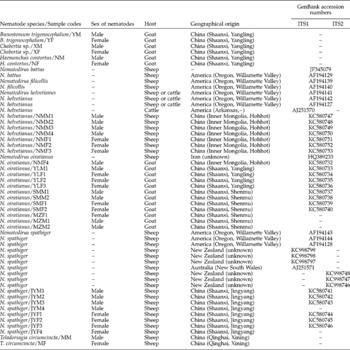
Twenty-two adult nematodes, 9 from goats and 13 from sheep, were collected from the Yangling district and the counties of Jingyang and Shenmu in Shaanxi province, together with Hohhot city in Inner Mongolia Autonomous Region, China. Geographical distances of more than 10 km were selected and distances between these two sites ranged from 20 km between Yangling district and Jingyang county to 1149 km between Yangling county and Hohhot city. Parasite samples with codes, geographical origins, genders and GenBank accession numbers are listed in table 1. Each worm was washed in physiological saline, identified to Nematodirus species based on morphological features, and kept at − 20°C until DNA extraction.
ITS sequences of ribosomal DNA of gastrointestinal nematodes in ruminants, relative to male or female worms, host, geographical origin; accession numbers for ITS1 and ITS2 sequences are the same in the majority of nematodes listed.

Total genomic DNA (gDNA) was extracted from individual worms by proteinase K treatment, column-purified (TIANamp Genomic DNA Purification System, Tiangen, China) and eluted into 40 μl H2O according to the manufacturer's recommendations. Each DNA sample was stored at − 20°C for further use.
PCR amplification and sequencing of ITS rDNA
The rDNA region, comprising ITS1, 5.8S and the ITS2 rDNA and primer flanking sequences, was amplified by PCR using universal primers NC5 and NC2 (Zhu et al., Reference Zhu, Gasser, Podolska and Chilton1998). PCR reactions were carried out using a 25 μl reaction system containing 1 × Taq MasterMix (CWBIO, Beijing, China), 0.4 mm of each primer, and 3 μl of each gDNA, under the following conditions: an initial denaturation at 94°C for 5 min, 35 cycles of 94°C for 2 min (denaturation), 45°C for 45 s (annealing), 72°C for 2 min (extension), and a final extension at 72°C for 10 min. Samples without gDNA and with host DNA (goat and sheep) as negative controls were included in each amplification. Each amplicon (5 μl) was examined by agarose gel (1.0%) electrophoresis to validate amplification efficiency, and positive amplicons were sent to Sangon Company (Shanghai, China) for direct sequencing.
Sequence analysis and phylogenetic reconstruction
ITS1, 5.8S, ITS2 rDNA fragments of N. helvetianus, N. spathiger and N. oiratianus isolated from different geographical regions were determined by comparison with those of N. helvetianus (GenBank accession number AF194141), N. spathiger (AF194144) and N. oiratianus (HQ389233). Completed sequences of ITS rDNA were aligned using the computer program Clustal X 1.83 (Thompson et al., Reference Thompson, Gibson, Plewniak, Jeanmougin and Higgins1997).
Since similar sequences of 5.8S rDNA occur within Nematodirus species (data not shown), sequences of ITS1 and ITS2 were used for further study. Levels of sequence differences (D) in two rDNA, respectively, within Nematodirus sp. were obtained by pairwise comparison of nucleotides using the formula D= 1 − (M/L), where M is the number of alignment positions where both sequences have a common base, and L is the total number of alignment positions over which the two sequences are compared (Chilton et al., Reference Chilton, Gasser and Beveridge1995). Inter-species genetic differences of two rDNA fragments within the genus Nematodirus were evaluated separately using the Megalign procedure in DNAStar 5.0 (Burland, Reference Burland2000), and base composition, transitions and transversions were calculated by Mega 4.0 (Tamura et al., Reference Tamura, Dudley, Nei and Kumar2007). Sequences of Nematodirus nematodes available in GenBank were also downloaded for genetic analysis, including 7, 1, 1 and 6 sequences from sheep or cattle in America, Australia, Iran and New Zealand, respectively.
In order to infer phylogenetic relationships of N. helvetianus, N. spathiger and N. oiratianus, genetic trees were reconstructed based on ITS1 and ITS2 rDNA sequences, respectively, using the maximum likelihood (ML) method, with Ostertagia sp. (HQ844228) as the outgroup. Ambiguously aligned regions for each fragment were excluded using Gblocks online server (http://molevol.cmima.csic.es/castresana/Gblocks_server.html) with the options for a less stringent selection. ML analyses were performed using PhyML 3.0 (Guindon & Gascuel, Reference Guindon and Gascuel2003), and the GTR model with its parameter for the concatenated dataset. The consensus tree was obtained after bootstrap analysis, with 1000 replications and values above 50% reported. Phylograms were drawn using the Tree View program version 1.65 (Page, Reference Page1996).
PCR-RFLP analysis
Based on sequence alignments of ITS rDNA (data not shown), including ITS rDNA sequences of the three species available in GenBank, two restriction endonucleases (BstpI and ApaLI) were selected to differentiate the three Nematodirus species. PCR amplicons of ITS rDNA regions were obtained using the same conditions above, and digested using BstpI and ApaLI (TaKaRa, Dalian, China), respectively, in 20 μl reaction systems containing 2 μl of 10 × H buffer (for BstpI) or 10 × L buffer (for ApaLI), 1 U of BstpI or ApaLI, and 17 μl PCR products, at 60°C (for BstpI) or 37°C (for ApaLI) for 3 h. Each digested product (4 μl) was examined by 1.5% agarose gel electrophoresis under ultraviolet transillumination, and photographed.
As there are no BstpI or ApaLI enzymatic sites in ITS rDNA regions of N. oiratianus, a specific PCR was established to identify N. oiratianus from some common gastrointestinal nematodes. According to ITS rDNA sequence alignments (data not shown), a pair of specific primers was designed as follows: OLQ (forward; 5′-GTACTCGCTGATATGGTGTC-3′) and NC2 (reverse; 5′-TTAGTTTCTTTTCCTCCGCT-3′). The PCR condition was optimized by varying the amounts of DNA template, Mg2+ concentrations and annealing temperatures. Reactions were carried out in 25 μl reaction system containing 1 × Taq MasterMix (CWBIO), 0.1–1 mm of MgCl2, 0.4 mm of each primer and 0.5–1.5 μl of each gDNA template in a thermocycler (Biometra, Gottingen, Germany) under the following conditions: an initial denaturation at 94°C for 5 min, 30 cycles of 94°C for 30 s (denaturation), 45–60°C for 30 s (annealing), 72°C for 30 s (extension), and a final extension at 72°C for 10 min. The specificity of this PCR was determined with amplifications of gDNAs of some common gastrointestinal nematodes (N. spathiger, N. helvetianus, Teladorsagia circumcincta, Bunostomum trigonocephalum, Chabertia spp. and Haemonchus contortus). The sensitivity was assessed with amounts of male N. oiratianus DNA of 32.5 ng, 5.4 ng, 0.9 ng, 0.2 ng, 0.03 ng, 4 pg, 0.69 pg, 0.12 pg, 0.02 pg, respectively. The limit of detection was defined as the least amount of DNA amplifiable.
To validate this specific PCR for identifying N. oiratianus, a total of 22 adult Nematodirus spp., including 12 of N. oiratianus, 5 of N. helvetianus, and 5 of N. spathiger from different geographical regions in China (table 1), were used for amplifications using an optimized reaction system. The PCR-RFLP approach was used to identify 20 N. spathiger and 20 N. helvetianus collected from Jingyang county in Shaanxi province and Hohhot city in Inner Mongolia Autonomous Region, to ascertain its usefulness for the identification and differentiation of two Nematodirus species.
Results and discussion
Genomic DNA samples were isolated from 22 individuals representing three Nematodirus species, namely N. helvetianus, N. spathiger and N. oiratianus, and used for PCR amplifications of ITS rDNA fragments. Each amplicon appeared as a single band of approximately 800 bp for all three species, and there were no detectable length differences between samples.
In order to examine intra- and inter-species sequence differences in the ITS rDNA regions within genus Nematodirus, positive amplicons were sequenced directly. After ambiguous bases at both ends, and partial sequences of 18S and 28S rDNA, were discarded, the lengths of the 5.8S were identical (143 bp) for the three species. The respective lengths of ITS1 and ITS2 rDNA were 383 bp and 240 bp for N. spathiger and 383 bp and 246 bp for N. oiratianus. The respective lengths of the ITS1 and ITS2 rDNA differed in female N. helvetianus (383 bp and 240 bp) compared with males (382 bp and 239 bp), indicating that, combined with the morphological features, such differences are useful in distinguishing female and male N. helvetianus. The A+T contents of ITS1 and ITS2 rDNA sequences for the three species were 49.6–51.7% and 55.7–57.7%, respectively. A pairwise comparison of the 5.8S rDNA sequences showed that no intra-species difference in 5.8S rDNA sequences was detected within N. spathiger and N. oiratianus, and one mutation site was found in samples of male and female N. helvetianus. The respective intra-species sequence variations of ITS1 and ITS2 rDNA were 0–4.4% and 0–6.1% in N. oiratianus, 0.3–1.8% and 0–0.4% in N. spathiger, and 0–6.5% and 0–5.4% in N. helvetianus. The relatively lower intra-specific variations found in N. spathiger, compared with the other two species, could be associated with differences in sampling sites and parasite populations.
The respective inter-specific differences in ITS1 and ITS2 rDNA sequences were 5.2–9.7% and 5.2–12.8% (data not shown), which were comparable with previous studies for ITS1 (2.3–16.5%) and ITS2 (3.9–24.7%) (Newton et al., Reference Newton, Chilton, Beveridge and Gasser1998b). Alignments of ITS rDNA fragments showed 98 mutation sites among different species, consisting of 50 transitions (A ↔ G, n= 27; T ↔ C, n= 23), 32 transversions (T ↔ G, n= 8; C ↔ G, n= 5; A ↔ T, n= 15; A ↔ C, n= 4) and 10 multiple substitutions. Interestingly, a gap of six bases (positions 591–596) was observed in the ITS rDNA sequences, and these sites would be ideal markers to differentiate N. oiratianus from the other two Nematodirus species.
Intra-specific variations of the three species from different countries were determined using ITS1 and ITS2 rDNA sequences available in GenBank. The respective intra-specific variations of ITS1 and ITS2 rDNA were 1.8–4.4% and 1.6–6.1% between N. oiratianus isolates from China and Iran, 5.7–7.1% and 6.3–8.3% between N. helvetianus samples from China and America. For N. spathiger, compared with samples from China, sequence differences in ITS1 rDNA were 0.3–2.4% in isolates from America, 0.3–2.9% in New Zealand, and 2.1–2.4% in Australia. Genetic variations in ITS2 rDNA of N. spathiger were 0–0.4% between samples from China and America, and 0–0.8% between China and New Zealand.
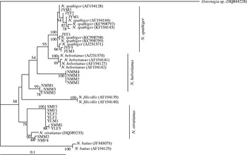
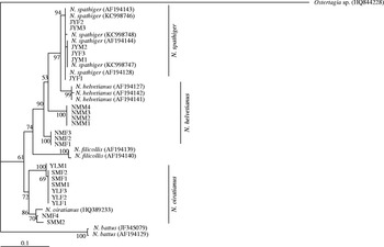
To determine phylogenetic relationships of N. oiratianus, N. helvetianus and N. spathiger, based on ITS1 and ITS2 rDNA sequences, genetic trees of species within genus Nematodirus were reconstructed using ML methods (figs 1 and 2). For Nematodirus isolates in the present study, samples of N. oiratianus were clustered with reference N. oiratianus isolate in sheep (HQ389233) in a solitary clade, and N. spathiger isolates were located with reference N. spathiger isolates in sheep (AF194143, AF194144, AF194128, KC998799, KC998798, KC998797, AJ251571, KC998746, KC998747 and KC998748). However, samples of N. helvetianus in the present study were clustered in two separate clades that were different from the cluster of reference isolates in sheep or cattle (AF194141, AF194142, AF194127 and AJ251570). These results were slightly different from trees reconstructed by Newton et al. (1998b) and Nadler et al. (Reference Nadler, Hoberg, Hudspeth and Rickard2000), who reported that all N. helvetianus samples were positioned in one solid clade. These differences may be associated with the amount of samples, geographical origins and host species.
Phylogenetic relationships of Nematodirus spp. inferred by maximum parsimony (ML) based on ITS1 rDNA sequences, with Ostertagia sp. as the outgroup; bootstrap values (%) above 50% from 1000 pseudo-replicates are shown for the maximum likelihood analyses.

Phylogenetic relationships of Nematodirus spp. inferred by maximum parsimony (ML) based on ITS2 rDNA sequences, with Ostertagia sp. as the outgroup; bootstrap values (%) above 50% from 1000 pseudo-replicates are shown for the maximum likelihood analyses.

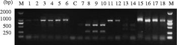
Two restriction endonucleases (ApaLI and BstpI) were selected according to the entire ITS rDNA sequence alignments in the present study and available in GenBank (data not shown) to differentiate the three species. The respective positions of ApaLI in the sequences were 249, 248 and 249 for N. spathiger, male and female N. helvetianus, and the position of BstpI was 304 for N. spathiger samples, with no sites in the other two species. Following PCR-RFLP, both amplicons of N. helvetianus and N. spathiger were digested into two fragments (approximately 600 bp and 300 bp) by ApaLI after 3 h incubation at 37°C, but only amplicons of N. spathiger samples were digested into two fragments (approximately 550 bp and 350 bp) by BstpI after 3 h at 60°C. However, neither of the two restriction endonucleases could digest any amplicons from N. oiratianus isolates (fig. 3).
PCR-based restriction enzyme products of Nematodirus species. Lanes: M, DNA size marker 2000 with ordinate values in bp; C, no-DNA control; 1–6 are PCR products, JMF2, JYM1, NMF3, NMM1, SMF1 and SMM1; 7–12 are ApaLI digestions of JYF2, JYM1, NMF3, NMM1, SMF1 and SMM1; 13–18 are BstpI digestions of JYF2, JYM1, NMF3, NMM1, SMF1 and SMM1.

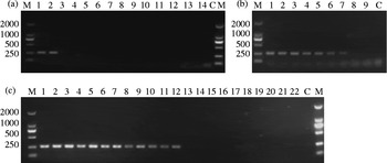
Then, a specific PCR was developed to identify N. oriatianus. Specific PCR protocols were optimized by varying the amounts of DNA template, Mg2+ concentrations and annealing temperatures, for which 1 μl, 60°C and 0.5 mm were the most appropriate. Under the optimized cycling conditions, the expected fragment of 250 bp was examined by 1.0% agarose gel electrophoresis from each N. oiratianus sample, but no products were amplified from other closely related species, such as N. spathiger, N. helvetianus, T. circumcincta, B. trigonocephalum, Chabertia spp. and H. contortus (fig. 4a). The smallest amount of DNA detectable in male N. oiratianus by specific PCR assay was 0.69 pg (fig. 4b). These results indicated a high specificity and sensitivity of the established PCR protocol.
Specific PCR assays for the detection of Nematodirus oiratianus. Lanes: M, DNA size marker 2000 with ordinate values in bp; C, no-DNA as control. (a) Lanes 1–14 are PCR products of SMF1, SMM1, NMF1, NMM1, JYF1, JYM1, MM, MF, YM, YF, XM, XF, NM, NF; (b) lanes 1–9 are DNA in sample SMM1 at 32.5 ng, 5.4 ng, 0.9 ng, 0.2 ng, 0.03 ng, 4 pg, 0.69 pg, 0.12 pg, 0.02 pg; (c) lanes 1–22 are samples of SMM1, SMF1, SMM2, SMF2, MZM1, MZM2, MZF1, YLF1, YLM1, YLF2, YLF3, NMF4, JYM1, JYF1, JYM2, JYF2, JYF3, NMM1, NMF1, NMM2, NMF2, NMM3 from geographical regions in China.

In order to evaluate the application of both molecular approaches, specific PCR and PCR-RFLP, 22 Nematodirus samples originating from sheep or goats in two endemic provinces (Shaanxi province and Inner Mongolia Autonomous Region) of mainland China were analysed (table 1). On agarose gels, there was a visible band only for N. oiratianus (fig. 4c), with no amplicon for N. spathiger or N. helvetianus. All samples representing N. spathiger and N. helvetianus could be further differentiated by PCR-RFLP based on ApaLI and BstpI. Each PCR amplicon of N. spathiger could be digested by two restriction endonucleases, but PCR products of N. helvetianus could only be digested by ApaLI (data not shown).
In summary, the entire ITS rDNA fragments of N. oiratianus, N. spathiger and N. helvetianus from goats and sheep in China were obtained and analysed. Based on alignments of these ITS rDNA sequences, the PCR-RFLP approach could effectively differentiate the three Nematodirus species, whereas specific PCR accurately identified N. oiratianus from some common gastrointestinal nematodes in sheep or goats with high sensitivity. These data provide valuable molecular approaches for epidemiological surveillance and for assisting in the control of Nematodirus infection in sheep and goats.
Financial support
Project support was provided in part by grants from the Funds of Basic Research Key Program (ZD2012010) to G.H.Z. and the Study of controlling parasitic diseases in goats and sheep in Shenmu county (K332021210) to S.K.Y.
Conflict of interest
None.