Device occlusion of patent ductus arteriosus has now become a commonplace and safe procedure at most tertiary paediatric cardiac centres.Reference Yarrabolu and Rao 1 Baruteau et alReference Baruteau, Hascoët and Baruteau 2 offer an excellent review of the history, current practices, and future developments of ductal occlusion. In our practice at the Red Cross War Memorial Children’s Hospital, all patients with significant left-to-right shunting via the patent ductus arteriosus, as evidenced by echocardiographic parameters of left atrial and/or left ventricular volume overload, are considered for device occlusion of their patent ducti. Contraindications to transcutaneous occlusion include those with additional cardiac lesions amenable to surgical repair, ducti with unsuitable anatomy for device occlusion, usually Krichenko Type B,Reference Krichenko, Benson and Burrows 3 and evidence for severe pulmonary hypertension – that is, pulmonary artery pressures or vascular resistance >2/3 of the systemic blood pressure or vascular resistance.Reference Gournay 4 The latter two categories are occasionally referred for surgical ligation only after diagnostic cardiac catheterisation.
Most ductal occlusions are completed without significant complications, but ductal spasm before or during the procedure remains a significant problem when encountered (Fig 1). This phenomenon was highlighted by Batlivala et al.Reference Batlivala, Glatz, Gillespie, Dori and Jonathan Rome 5 In a series of 331 occlusions of patent ducti in 10 years, they described seven cases (2.1%) with ductal spasm, all born prematurely. In our series of 267 ductal occlusions in 12.5 years, we have encountered 10 cases (3.7%), eight of which are included in this case series. Batlivala et alReference Batlivala, Glatz, Gillespie, Dori and Jonathan Rome 5 and Baruteau et alReference Baruteau, Hascoët and Baruteau 2 have very recently thoroughly reviewed the literature in this field; consequently, we have not repeated it here.
A patent ductus arteriosus in complete ductal spasm (left) and (right), 10 minutes after initiating our novel protocol.

We became aware of the problem of ductal spasm due to the following two early cases of attempted percutaneous patent ductus arteriosus closures. The first patient that we encountered was not recognised as suffering from ductal spasm until, after cardiac catheterisation, the spontaneously “closed” (on angiogram) ductus re-opened fully once the patient had returned to the ward; a continuous murmur was again heard on auscultation. The second was recognised with ductal spasm during the catheterisation process, but due to concern regarding correct ductal sizing, at a time when we were using detachable coils only, the patient was referred for surgical ductal ligation. We were therefore concerned that this complication is under-diagnosed, and thereby constitutes a risk to the safe completion of ductal occlusions when partial spasm is not recognised, and that patients are unnecessarily referred for surgery when device occlusion may, in fact, be feasible.
We therefore agree with Batlivala et alReference Batlivala, Glatz, Gillespie, Dori and Jonathan Rome 5 who described the potential consequences of ductal spasm as threefold: 1) occlusion failure due to non-recognition of spasm, a false impression of a closed ductus, and therefore no device is inserted; 2) suboptimal occlusion with a residual shunt; and 3) device embolisation. The latter two may occur because of under-sizing of the duct diameter as result of measuring the ductal diameter on angiography of a partially relieved spasm, leading to erroneous – under-sized – device selection. We have observed that spontaneous resolution may be incomplete and has the risk of delaying or exacerbating these complications. The embolisation may consequently only become apparent on clinical examination, by return of the murmur, or by echocardiography at a later time, once the patient has left the catheterisation laboratory. The article by Batlivala et alReference Batlivala, Glatz, Gillespie, Dori and Jonathan Rome 5 is the first elegant review of this problem, yet they do not offer an approach to its resolution or a strategy to complete the procedure safely. The concern around embolisation of under-sized occlusion devices is exemplified by case reports by Guzeltas et alReference Guzeltas, Sengul, Tanidir and Odemis 6 and Yates et al.Reference Yates, Gautam and Syamasundar Rao 7 Guzeltas et alReference Guzeltas, Sengul, Tanidir and Odemis 6 described five cases of ductal spasm in prematurely born patients in a series of 150 occlusions of patent ducti, an incidence of 3%. In one of these five patients, the occlusion device had embolised to the aorta due to under-sizing of the device. Yates described a similar case of under-sizing due to spasm leading to embolisation of a Gianturco coil to the left pulmonary artery.
A further consideration is that ductal spasm may alter decision making with regard to device occlusion versus surgical ligation. Partial relief of ductal spasm may lead to the incorrect appraisal of anatomy, whereby unfavourable ductal anatomy – for example, a Krichenko Type B ductusReference Krichenko, Benson and Burrows 3 – is erroneously assessed as being suitable for device closure.
Case Series
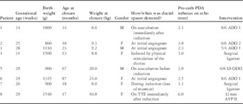
A total of eight infants born at gestational ages ranging from 25 to 34 weeks presented for transcatheter patent ductus arteriosus occlusion between 13 and 87 months of age (see Table 1). All had haemodynamically significant patent ducti with echocardiographic evidence of left ventricular and/or left atrial volume overload. Echocardiographically determined minimum patent ductus arteriosus diameters ranged from 2.3 to 6.0 mm.
Characteristics of the eight patients who experienced PDA spasm during attempted percutaneous closure of their PDAs, with their final interventions.

ADO=AmplatzerTM ductal occluder; AVP=AmplatzerTM vascular plug; F=female; LS=long shank; M=male; ODO=OcclutechTM ductal occluder; PDA=patent ductus arteriosus; TTE=transthoracic echocardiography
Before these eight cases, no formalised protocol to detect the possibility of ductal spasm was in place and detection methods of these cases were consequently varied. Ductal spasm of the eight cases was therefore detected at various times and in different ways, by either of the following:
-
∙ loss of ductal murmur before anaesthesia,
-
∙ loss of ductal murmur after full anaesthesia,
-
∙ on transthoracic echocardiographic screening after induction,
-
∙ on initial angiography, or
-
∙ after physical stimulation of the ductus.
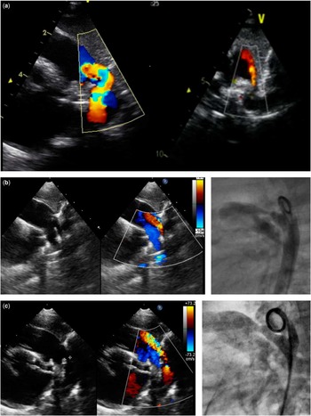
In each case, ductal spasm and the resultant changes in ductal diameter were confirmed and monitored by transthoracic echocardiography during cardiac catheterisation. As the ductal diameters were variable – from complete or partial spasm to full dilatation – they were not precisely measured after detection of spasm in each case: the precise size during partial spasm is immaterial, but its recognition is crucial. It was very clear – on two-dimensional echo and color Doppler – when ductal sizes were diminished during ductal spasm compared with pre-catheterisation parameters (Fig 2). Without such comparison, the ductal diameter could easily be underestimated, leading to erroneous conclusions. Note that, although the absence of a murmur indicates complete spasm, the presence of a murmur does not exclude partial spasm.
( a ) Echocardiogram of patent ductus arteriosus before catheterisation (left) and in partial spasm on transthoracic echocardiography screening immediately after induction of anaesthesia (right). ( b ) Partial spasm of patent ductus arteriosus on transthoracic echocardiography (left). Partial spasm of the same ductus confirmed on lateral angiography. ( c ) Full relaxation of the spasm (of the patent ductus arteriosus in Fig 2b) to pre-catheterisation dimensions observed on echocardiography (left), achieved after intravenous infusion of prostaglandin E1 for ~10 minutes; confirmed on lateral angiography (right).

On confirmation of spasm by echocardiography or angiography, the anaesthetist, in each case, changed the mode of anaesthesia from inhaled sevoflurane to total intravenous anaesthesia with propofol, reduced the inhaled oxygen fraction to 21%, and initiated a continuous intravenous effusion of prostaglandin E1 at 50–100 ng/kg/minute. Although these alterations in the anaesthetic procedure were similar in all cases, in the absence of an established protocol, management was by consensus between the interventional cardiologist and the anaesthetist.
In all eight infants, the first two steps (total intravenous anaesthesia and FiO2 0.21) resulted in only partial relaxation of the spasm. Usually, after ~5 minutes of the above changes in anaesthetic strategy, and always before infusion of intravenous prostaglandin E1, some relief of ductal spasm was observed by transthoracic echocardiography, but not to the echocardiographic diameters observed before catheterisation (Fig 2b). Only after IV prostaglandin E1 had been infused for ~10 minutes was full relaxation of the spasm to pre-catheterisation dimensions observed. This was subsequently confirmed on lateral angiography (Fig 2c).
Closures of six of the eight patent ducti were then performed by placement of AmplatzerTM ADO1, ADO2, AVP II, or OcclutechTM ODO occlusion devices in the routine manner. All were Krichenko Type C3 ducti, with some degree of central stenosis, typical of prematurely born (ex-prem) infants. Of the remaining two, one was judged to be anatomically unsuitable for device closure (Krichenko Type B3 patent ductus arteriosus) and the other was found to have severe pulmonary hypertension, with a pulmonary artery pressure at 70% of the systemic blood pressure; both were referred for ductal ligation.
While developing this protocol, one of us (R.D.D.) assisted a colleague at another paediatric cardiology unit with the percutaneous occlusion of a patent ductus arteriosus in a 9-year-old patient. On echocardiography, the ductal isthmus measured 6 mm, but at the initial aortic angiogram it was in spasm with an isthmus of only 1 mm, but with a large ampulla. We instituted the initial steps of the above protocol (FiO2 0.21 and total intravenous anaesthesia with propofol), but intravenous prostaglandin E1 was not available. We therefore elected to wait for 30 minutes, but on repeat angiography the isthmus had enlarged to only 3.3 mm. Consequently, being concerned that further dilatation may be possible, we elected to over-size the device and successfully occluded the patent ductus with an AmplatzerTM ADO 10/8 device (Fig 3). There was no evidence for coarctation of the aorta or stenoses of the branch pulmonary arteries on subsequent echocardiography. This case illustrates that intravenous prostaglandin may be essential for the confident resolution of the ductal spasm to allow for correct sizing of the occluding device. We were compelled to over-size the device significantly, which was only possible because of the presence of the large ampulla. The use of intravenous prostaglandin in this patient would have been valuable to shorten the procedure time and allow us to size the device accurately rather than require the use of a deliberately over-sized device. This latter strategy may not be possible in a patient without an adequate ampulla.
In the absence of an IV infusion of prostaglandin E1, the ductus may persist in partial spasm for >30 minutes. This ductus has a large ampulla, allowing over-sizing of an AmplatzerTM occluding device. Note the folding of the aortic disc (left), before release and after release (right), with minimal risk of embolisation.

Discussion
The phenomenon of ductal spasm during attempted device occlusions of patent ducti is well known (Batlivala et alReference Batlivala, Glatz, Gillespie, Dori and Jonathan Rome 5 ) and seems to occur only in prematurely born children (ex-prems), but in our opinion it is under-recognised. There are significant risks associated with under-recognition of ductal spasm, including occlusion failure – the false impression that the patent ductus has already permanently closed – and suboptimal occlusion with a residual shunt or device embolisation,Reference Guzeltas, Sengul, Tanidir and Odemis 6 , Reference Yates, Gautam and Syamasundar Rao 7 both due to under-sizing of the closure device.
As far as we are aware, this case series is the first to describe the utilisation of protocols to detect ductal spasms in ex-prems timeously, assess the ductal size correctly, and occlude the patent ducti safely, after fully relieving the ductal spasm rapidly and with confidence by altering anaesthetic management and infusing continuous intravenous prostaglandin E1. The first strategy is necessary as we postulate that inhaled anaesthesia contributes to the development of spasm and should be discontinued to initiate the onset of spasm relief; however, this strategy per se will not fully relieve ductal spasm. Within a few minutes of initiating a continuous infusion of alprostadil, the partially dilated ductus dilates to the maximum dimension seen at the pre-procedural echocardiography. Surprisingly, we have found clear evidence that the ducti of ex-prem children remain exquisitely responsive to infused prostaglandin E1 for as late as 7.3 years (87 months) of age!
A literature search on the responsiveness of older children – older than the generally accepted upper limit of 50 days – to alprostadil revealed only a single case: Kashani et alReference Kashani, Schmunk and Merritt 8 described an 11-month-old child with a patent ductus arteriosus responsive to prostaglandin E1.
Aetiology of ductal spasm
Our understanding of the precise aetiology of ductal spasm in prematurely born patients remains speculative. We have described observational evidence that the onset of severe spasm may occur before or after induction of anaesthesia, before venous or arterial access is obtained, or on physical stimulation of the ductus – for example, by a catheter or wire.
We postulate that several physiological, haemodynamic, and pharmacological triggers may lead to, or exacerbate, ductal spasm of ex-prem patients at the time of percutaneous occlusion of a patent ductus:
-
∙ An adrenergic state – inadequate pre-procedure sedation.
-
∙ Induction with supplemental face mask oxygen causing the following:
-
∙ stimulation of the ductal vasomotor tissue or,
-
∙ alteration of systemic and pulmonary vascular resistances with resultant response of the ductus to haemodynamic alterations.
-
-
∙ Direct contractile effect of gaseous – inhalational – anaesthesia.
-
∙ Direct physical stimulation of the ductus.
-
∙ Contractile response to intravenous paracetamol.
It is of note, however, that local anaesthesia, used to infiltrate the access site, does not influence ductal patencyReference Smith 9 but the pain of the injection, if given before anaesthesia, may do so.
Discussion of the relative influences of these various factors to the onset and maintenance of ductal spasm is speculative as no human studies have focussed on the long-term sensitivity of patent ducti to these modifying factors, especially for ex-prem patients. Animal studies of the agents influencing ductal patency have not focussed on the specific question of long-term sensitivity yet.
A crux observation/speculation is that of the increased sensitivityReference Heymann, Rudolph and Silverman 10 , Reference Popat, Kapoor and Travadi 11 of a ductus of a pre-term baby in the immediate postnatal period to endogenous prostaglandin E2, which may play a role in prolonged patency of that ductus and/or failure of medical treatment – for example, ibuprofen – to close a baby’s patent ductus in the postnatal period. This postulation may explain why the ducti of ex-prems remain exquisitely responsive to intravenous prostaglandin E1 at a much later age, when the patient may present for ductal closure. The question is whether a history of failure of cyclo-oxygenase inhibitor treatment, due to high prostaglandin sensitivity, may be useful to predict which children may be more at risk of ductal spasm. It begs the question whether the factors listed above that are known to be associated with spasm are antagonists of prostaglandin E2 action. Nevertheless, it is clearly evident that the persistent lability of the ex-prem ductus to varying conditions long after the postnatal period is not well understood. Therefore, any protocol to relieve ductal spasm must address the potential influences of all these factors.
Detection of ductal spasm
It is important to maintain a high index of suspicion when prematurely born children are considered for ductal occlusion, whatever their age at the time of a planned percutaneous occlusion; one patient in our case series was 7.3 years old and had ductal spasm before the onset of anaesthesia, detected simply by the absence of a murmur on auscultation in the induction room. It is important to auscultate each patient before anaesthesia, but the presence of a murmur does not exclude ductal spasm, as the patent ductus arteriosus may be in partial spasm. It is therefore preferable to perform a focussed echocardiogram on all ex-prem patients before draping, as illustrated by our case 7. Transthoracic echocardiography should confirm restitution of the ductal diameter and the presence of continuous wave Doppler gradients similar to those of the pre-catheterisation echocardiogram of that patient.
Immediate management of ductal spasm
Following our experience with the first two patients, we purposefully developed a protocol for the management of ductal spasm for the following eight patients that we encountered. The aim was to allow for confident and safe occlusion of the patent ductus arteriosus at one admission to the catheterisation laboratory and within minutes of detection of ductal spasm.
Protocol for the management on detection of ductal spasm
-
∙ Reduce inhaled oxygen and ventilate at FiO2 of 0.21.
-
∙ Change inhaled – gaseous – anaesthesia to total intravenous anaesthesia with propofol.
-
∙ Commence a continuous intravenous infusion of prostaglandin E1 – alprostadil – at 50–100 ng/kg/minute.
-
∙ While avoiding any physical stimulation of the ductus, follow the development of relief of ductal spasm by transthoracic echocardiography and confirm by lateral angiogram once echocardiographic findings conform to the pre-catheterisation appearance.
-
∙ Carefully consider the safest device to minimise risk of embolisation – for example, by over-sizing by one size or using an occluding device rather than a coil.
-
∙ Stop the continuous intravenous infusion of prostaglandin E1.
-
∙ Perform an angiogram.
-
∙ Confirm suitability for percutaneous closure in the routine manner.
-
∙ Perform a post-procedure echocardiogram before leaving the catheterisation laboratory, especially if a coil has been used, as well as later the same day as the device occlusion.
While using this protocol, six patent ducti were successfully occluded by AmplatzerTM (St Jude Medical, St Paul, Minnesota, United States of America) ADO1 (three cases), ADO2 (one case) devices, an OcclutechTM (Occlutech IS, Helsingborg, Sweden) device (one case), and an AmplatzerTM AVP2 device (one case). Only one patient was found to have a Krichenko Type B patent ductus arteriosus,Reference Krichenko, Benson and Burrows 3 without an adequate-sized ampulla, whereas another patient had severe pulmonary hypertension; both were referred for ductal ligation. On follow-up, all the patent ductus arteriosus had remained fully occluded.
Conclusion
Ductal spasm during transcatheter occlusion may be reliably resolved and the procedure safely completed by a careful approach, which includes the continuous intravenous infusion of prostaglandin E1 during the procedure. The influences of various anaesthetic and catheterisation factors remain speculative, but we suggest that our protocol is a safe approach to circumvent any and all of the postulated effects that may lead to ductal spasm, and thereby avoid significant complications. Further studies into the exact actions of the various factors influencing ductal spasm are required to prevent spasm ab initio or target the most important factor specifically and safely.
Acknowledgement
The authors would like to thank the enthusiastic assistance of the staff of the Cardiac Catheterisation Laboratory at the Red Cross Children’s Hospital in the performance of the cases and the development of the protocol. Dr Chris Hugo_Hamman is thanked for the use of his images in Figure 3.
Financial Support
This research received no specific grant from any funding agency, commercial, or not-for-profit sectors.
Conflicts of Interest
None.
Ethical Standards
The work reported in this paper was carried out in the cardiac catheterisation laboratory of the Red Cross Children’s Hospital on children who required the procedure as assessed by routine clinical criteria. Full informed consent was obtained from the parents or guardians of all the patients before the procedure. The data were captured into the routine databases of the Department of Cardiology. Ethics approval for the publication of these data was obtained from the Health Research Ethics Committee of the Faculty of Health Sciences of the University of Cape Town (HREC no: R017/2014, expiry date 30 May, 2017).