Hydatid disease is a zoonotic parasitic infection caused by Echinococcus granulosus, Echinococcus multilocularis, or Echinococcus vogeli, and is endemic in Turkey and other livestock-raising countries. Cardiac involvement is not a common presentation, and isolated cardiac disease is very rare.Reference Ipek, Omeroglu and Goksedef 1 Most patients with cardiac disease are asymptomatic.Reference Shojaei, Yassin and Rezahosseini 2 Chest pain, palpitations, cough, and dyspnoea are the most common symptoms, and in some cases the disease can mimic acute coronary syndrome.Reference Ipek, Omeroglu and Goksedef 1 , Reference Shojaei, Yassin and Rezahosseini 2 The diagnosis is made upon a combination of clinical suspicion, cardiac imaging modalities, and serologic tests. Here a case of isolated cardiac hydatid cyst in a female patient with chest pain and electrocardiographic changes mimicking myocardial ischaemia is presented.
Case report
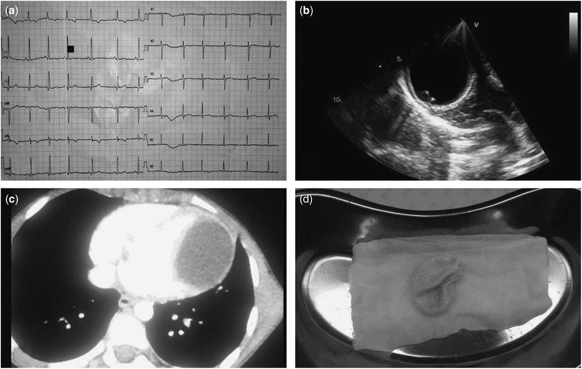
An 8-year-old girl presented to our clinic with complaints of chest pain that occurs at rest and with exertion and changes in electrocardiogram. She had no history of familial cardiac disease or sudden death. On physical examination, she had no abnormal findings. Electrocardiogram showed normal sinus rhythm with normal axis and negative T waves in leads I, AVL, and in leads V5-V6 (Fig 1a). Echocardiography revealed a large cyst with well-defined borders extending from the posterior wall of the left ventricle to the apex of the left ventricle with a diameter of 47×40 mm (Fig 1b). There was no mitral regurgitation or left ventricular outflow obstruction. The ejection fraction and shortening fraction were 68 and 36%, respectively. Abdominal ultrasound revealed normal findings. Thoracic CT showed a cystic lesion in the left ventricle with a diameter of 53×43 mm (Fig 1c). The patient was diagnosed with isolated cardiac hydatid cyst, and an enzyme-linked immunosorbent assay was positive for echinococcus antibodies. The patient underwent a successful surgery for excision of the cyst (Fig 1d) and is being treated with albendazole for 6 months. At the routine follow-up examination 6 months postoperatively, the patient has no trace of cysts on echocardiography.
(a) Electrocardiogram of the patient with negative T waves in leads I, AVL, and in leads V5-V6. (b) Echocardiographic image of the cyst with well-defined borders extending from the posterior wall of the left ventricle to the apex of the left ventricle. (c) Thoracic CT showing a cystic lesion in the left ventricle. (d) Surgically excised hydatid cyst.

Discussion
Hydatid disease is a zoonotic parasitic infection caused by E. granulosus, E. multilocularis, or E. vogeli, and is endemic in Turkey and other livestock-raising countries. The most common sites of hydatid cysts are the liver in 50–70%, lungs in 5–30%, muscles in 5%, bones in 3%, kidneys in 2%, spleen in 1%, and brain in 1% of the cases.Reference Ipek, Omeroglu and Goksedef 1 Cardiac hydatid cyst is a rare disease with varying degrees of symptoms in relation to the size and site of infection, and unless it is located in a critical site the diagnosis is late. The growth of hydatid cyst is usually slow and asymptomatic, but later it may cause symptoms owing to invasion of the surrounding structures, conductive system of the heart, and obstruction of the blood flow.Reference Shojaei, Yassin and Rezahosseini 2 Approximately 10% of patients with cardiac hydatid cyst are symptomatic. Chest pain, palpitations, cough, and dyspnoea are the most common presenting symptoms, and in some cases the disease can mimic acute coronary syndrome.Reference Ipek, Omeroglu and Goksedef 1 , Reference Shojaei, Yassin and Rezahosseini 2
Cardiac involvement is seen in only 0.5–2% of patients with hydatid disease,Reference Pakis, Akyildiz and Karayel 3 and, as in our patient, the left ventricle (55–60%) is the most commonly involved site because of the rich blood supply.Reference Ohri, Sachdeva, Bhatia and Shrivastava 4 Other parts of the heart such as interventricular septum is involved in 5–9%, right ventricle in 15%, and the right atrium may be involved in 3–4% of cases.Reference Shojaei, Yassin and Rezahosseini 2 Cardiac hydatid cysts can rupture and cause cardiac tamponade, fatal arrhythmias, or systemic infection and can mimic left ventricular aneurysm, and it should be one of the differential diagnoses of cystic cardiac lesions in endemic areas.Reference Ipek, Omeroglu and Goksedef 1 , Reference Shojaei, Yassin and Rezahosseini 2
Clinical suspicion, cardiac imaging, and serologic tests play a major role in the diagnosis of cardiac hydatid disease. In highly endemic areas, cardiac disease should be kept in mind even in the absence of past medical history of hydatid disease.Reference Canpolat, Yorgun, Sunman and Aytemir 5 Chest radiographs usually show a normal cardiothoracic ratio, but the presence of calcification may be a sign for hydatid disease.Reference Khosravi, Taghipour, Fanaei, Assar, Ghyasy and Mirlohi 6 Electrocardiographic findings vary according to the sites of the cysts, and non-specific changes such as T-wave abnormalities may be observed. Echocardiography is highly sensitive for the diagnosis of cardiac hydatid cysts.Reference Canpolat, Yorgun, Sunman and Aytemir 5 Localisation of the cyst and its effect on cardiac functions may be determined by echocardiography. However, in some cases, CT scan or MRI is necessary for the accurate location of lesion and relation of it with other structures. The presence of calcification in a cystic lesion, the presence of daughter cysts, and membrane detachment are other findings distinguishing the disease from other cardiac cystic lesions.Reference Shojaei, Yassin and Rezahosseini 2 Enzyme-linked immunosorbent assay is one of the most specific serologic tests, but serologic tests can be false negative in 50% of patients with cardiac cysts. Therefore, negative results do not exclude the diagnosis. Although anthelmintic drugs have been used in the preoperative and postoperative periods, surgical excision is the choice of treatment, and after a successful surgical treatment the duration of anthelmintic treatment is variable. Our patient had chest pain and electrocardiographic changes of ischaemia, but there was no calcification in the cystic lesion. Fortunately, enzyme-linked immunosorbent assay was positive for echinococcus antibodies in our patient, and the patient underwent a successful surgery for excision of the cyst and is being treated with albendazole for 6 months.
Conclusion
The clinical signs and symptoms of cardiac hydatid cyst are non-specific and may be difficult to diagnose. In particular, in livestock-raising countries such as Turkey where the disease is endemic, cardiac hydatid cyst can present with chest pain and electrocardiographic changes mimicking acute coronary syndrome. In patients living in endemic areas, even if the serologic tests are negative, the diagnosis should be made and confirmed by a combination of echocardiography, CT, and MRI.
Acknowledgements
None.
Financial Support
This research received no specific grant from any funding agency or from commercial or not-for-profit sectors.
Conflicts of Interest
None.
Ethical Standards
The authors assert that all procedures contributing to this work comply with the ethical standards of the Van Education and Research Hospital and with the Helsinki Declaration of 1975, as revised in 2008, and has been approved by the institutional committees of Van Education and Research Hospital, Van, Turkey.