

Introduction
Although antibiotic growth promoters (AGPs) improve the performance and health of farm animals (Feighner and Dashkevicz, Reference Feighner and Dashkevicz1987; Lin, Reference Lin2014), concerns about possible human health effects (Witte et al., Reference Witte, Klare and Werner1999; Wegener, Reference Wegener2003) led to the prohibition of their use within the European Union. In some cases the consequence has been increased digestive disorders and diseases as well as reduced bird performance (Van Immerseel et al., Reference Van Immerseel, Rood, Moore and Titball2009). Necrotic enteritis (NE) is caused by the ubiquitous soil bacterium Clostridium perfringens which can inhabit the gastrointestinal tract of broiler chickens without causing any disease (Timbermont et al., Reference Timbermont, Haesebrouck, Ducatelle and Van Immerseel2011). Predisposing factors, such as coccidiosis, high dietary concentrations of certain protein or non-digestible viscous fibre, contribute to the onset of NE (Van Immerseel et al., Reference Van Immerseel, De Buck, Pasmans, Huyghebaert, Haesebrouck and Ducatelle2004 and Reference Van Immerseel, Rood, Moore and Titball2009; Timbermont et al., Reference Timbermont, Haesebrouck, Ducatelle and Van Immerseel2011). In recent years research efforts have been directed to finding alternatives to AGPs (Lee et al., Reference Lee, Lillehoj, Jeong, Jeoung and An2011; Shojadoost et al., Reference Shojadoost, Vince and Prescott2012). The ideal candidate substance should present the same beneficial effects as AGPs, namely an improved feed conversion efficiency and inhibition of growth of pathogenic bacteria (Huyghebaert et al., Reference Huyghebaert, Ducatelle and Van Immerseel2011). Products such as exogenous enzymes, organic acids, yeast-derived beta-glucans, probiotic bacterial cultures, herbs and essential oils have been evaluated for this purpose (Huyghebaert et al., Reference Huyghebaert, Ducatelle and Van Immerseel2011).
Resin is a substance that coniferous trees secrete at the sites of mechanical injury to prevent the invasion of pathogenic bacteria and fungi and to deter herbivorous animals. Although resin acids are not a typical ingredient found in animal diets, they are commonly consumed by many wild herbivores of the northern hemisphere, including moose (Alces alces) and the popular game bird capercaillie (Tetrao urogallus; Wienemann et al., Reference Wienemann, Schmitt-Wagner, Meuser, Segelbacher, Schink, Brune and Berthold2011; Milligan and Koricheva, Reference Milligan and Koricheva2013). Resin contains compounds such as abietic, dehydroabietic, neoabietic, isopimaric, levopimaric and palustric acids, which are a diverse group of hydrophobic diterpene carboxylic acids with antimicrobial, antifungal and antiparasitic activity (San Feliciano et al., Reference San Feliciano, Gordaliza, Salinero and Miguel del Corral1993; Rubio et al., Reference Rubio, Calderón, Flores, Castroa and Céspedes2005; Savluchinske-Feio et al., Reference Savluchinske-Feio, Curto, Gigante and Roseiro2006). Gram-positive bacteria are especially sensitive to resin acids (San Feliciano et al., Reference San Feliciano, Gordaliza, Salinero and Miguel del Corral1993). For example, Streptococcus mutans (Ali et al., Reference Ali, Sangwan, Koul, Pandey, Bani, Abdullah, Sharma, Kitchlu and Khan2012) and multidrug-resistant and methicillin-resistant Staphylococcus aureus (Smith et al., Reference Smith, Williamson, Zloh and Gibbons2005), which are both Gram-positive species, are sensitive to resin acids. Traditional medicine in Scandinavia has used the antimicrobial properties of Norway spruce (Picea abies) and Scots pine (Pinus sylvestris) resin for centuries, and novel applications, like resin-salves for the treatment of severe wounds, have been developed in recent years (Sipponen and Laitinen, Reference Sipponen and Laitinen2011; Sipponen et al., Reference Sipponen, Kuokkanen, Tiihonen, Kauppinen and Jokinen2012).
The fact that C. perfringens is Gram-positive makes resin acids an interesting dietary supplement candidate for reducing the risk of NE in the broiler chickens fed antibiotic-free diets. The hypothesis for this study was that resin acids may decrease the growth of C. perfringens and favourably modulate the gastrointestinal microbiota, leading to improved performance in broiler chickens. The effects of a resin acid-enriched composition (RAC), a combination of fatty acids and resin acids from coniferous trees, using three model systems were evaluated. First, the product was tested in vitro to evaluate the effect of RAC against the multiplication of C. perfringens in pure cultures. Next, the effect of RAC on the structure and function of the entire ileal microbiota of broiler chicken was examined, using a unique ex vivo fermentation system. Finally, the effects of RAC in NE challenged broiler chickens was studies. Performance of the birds was monitored, and samples of ileal contents were analysed for the structure of intestinal microbiota and residual concentration of short chain fatty acids (SCFAs).
Material and Methods
Inhibition of C. perfringens in vitro
A resin acid-enriched composition (RAC; Suomen Rehu, Hankkija Ltd, Finland), containing natural resin acids of Scots pine and Norway spruce was produced by Forchem Ltd (Rauma, Finland). Depending on the experiment, RAC solution contained 5.0–8.5% resin acids, 88.5–93% free fatty acids, and 2–3% neutral components. The fatty acid profile of the product was 4-6% saturated acids, 27–31% monounsaturated fatty acids, and 63–69% of polyunsaturated fatty acids with an n-3 to n-6 -ratio of approximately 1:4.
Anaerobic trypticase soy-yeast extract medium (Lab M Ltd; Lancashire, UK) with 27.7 mM D-glucose, pH 7.0–7.2, was used as the basal growth medium for C. perfringens (type strain DSM 756) throughout the experiment. C. perfringens was grown at 37°C in 50 ml glass vessels with air-tight butyl rubber stoppers to ensure anaerobic status.
The inhibition study was conducted using 25 ml glass vials with butyl rubber stoppers with a reaction volume of 15 ml. The RAC preparation, used at 0, 0.1, 1, 5, and 10 mg/l, contained 5% resin acids. The study was initiated by injecting a 150 µl inoculum of the C. perfringens culture through the rubber stopper into the vessel with the growth medium and the chosen concentration of RAC. The bottles were kept at 37°C and continuously mixed by a gyratory shaker at 100 rotations per minute and growth was monitored for eight hours. There were three replicate vessels for each treatment, and the inoculation was done in a random order to avoid any potential systematic shifts.
Optical density (OD) was measured at 0, 3 and 6 h time points using a turbidometer with a wavelength of 690 nm. Total gas production in each vessel was measured at the 8 h time point by puncturing the rubber stopper with a needle connected to an accurate 20 ml glass syringe with a sensitive ground plunger. The volume of gas released from the vessels was recorded. C. perfringens produces gas while growing, thus the volume of gas was used as an indication of bacterial growth and metabolism.
Ileal fermentation in vitro
The ileal ex vivo fermentation study procedure was designed to model the luminal conditions of the ileum in broiler chickens. The study was carried out as described by Apajalahti et al. (Reference Apajalahti, Rademacher, Htoo, Redshaw and Kettunen2009) for piglet ileal fermentation. Three concentrations of RAC containing 7% resin acids were tested at 0, 0.1 and 1 g/kg total fermentation broth. For preparation of the fermentation medium, ileal contents (defined as the region of small intestine between the Meckel's diverticulum and the caecal junction) of 30 four-week old Ross 508 broiler chickens from a commercial farm were collected, pooled and mixed carefully. The digesta was combined with an equal volume of pH 6.5 buffer solution (0.02 M K2HPO4, 0.02 M NH4H2PO4, 0.0006 M MgSO4) and centrifuged at 18,000 x g for 20 min to pellet the solids. The pellet was discarded and the supernatant used as a substrate for the fermentation medium, ensuring it contained only authentic substrates of the intestinal origin. On the day the study was conducted, fresh ileal digesta was recovered from birds and kept on ice until used as a 1% inoculum for the fermentation study.
The study was conducted under N2 in an anaerobic glove box, at 42°C, using four replicate 2 ml microcentrifuge vials as fermentation vessels. The reaction volume was 1.5 ml. The vessels were inoculated in a random order to avoid any systematic shifts.
At the 10 hour time point, the fermentation was stopped by centrifuging the vials for 10 min at 18,000 × g. The supernatants were recovered for SCFA analysis and the pellets for bacterial analysis. The samples were stored in freezer at −20°C prior to the analysis of SCFAs and the density of total eubacteria and the densities of the Lactobacillus group and Enterococcus spp., as described below.
Challenge trial with broiler chickens
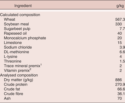
The experiment was conducted at the poultry facility of Alimetrics Ltd in Southern Finland (according to the ethical procedures of The National Animal Experiment Board of Finland). In total, 192 one-day-old male Ross 508 broiler chickens from a commercial hatchery (Suomen Broiler Oy, Masku, Finland) were randomly allocated to four dietary treatment groups, with eight replicate pens per diet and six chickens per pen. A commercial, wheat and soybean meal based diet for broiler chickens without coccidiostats, (Table 1) was used as a basis for the dietary treatments. RAC containing 5% of resin acids was hand-mixed with sugar beet pulp and then with the other feed components at the experimental feed mill of Agrifood Research Finland (Jokioinen, Finland). The concentration of RAC in the dietary treatments was 0, 0.5, 1 and 3 g/kg. Feed for the first four days was manufactured as 3 mm pellets and the rest of the feed was offered as 4 mm pellets. The chickens had a free access to feed and water throughout the experiment.
Calculated and analysed composition of the basal diet (g/kg dry matter, unless otherwise stated)

1 Providing the following per kg of feed: Ca, 0.6 g; Fe, 29 mg; Cu, 8 mg; Mn, 10 mg; Zn, 65 mg; I, 0.5 mg; Se, 0.2 mg. 2 Providing the following per kg of feed: Ca, 0.66 g; vitamin A, 12,000 IU (retinol);cholecalciferol, 112.5 µg; vitamin E, 54 IU (tocopheryl acetate 3a700); menadione, 3.1 mg; thiamine, 2.5 mg; riboflavin, 6 mg; pyridoxine, 4 mg; cyanocobalamin, 0.025 mg; biotin, 0.15 mg; folic acid, 1.0 mg; niacin, 40 mg; pantothenic acid, 15.0 mg.
The 1.125 m2 pens used were equipped with one feeder and two nipples of a watering line per pen. Wood shavings were used as a bedding material. During the first week, the ambient temperature was 29°C and brooder lamps provided additional heat to the pens. Thereafter, the brooder lamps were switched off and the temperature was gradually lowered to 25°C on d 17. The temperature and humidity were monitored and recorded on a daily basis. The lighting schedule was 23L:1D on day one. The dark period increased one hour per day until reaching 18L:6D which was maintained for the rest of the trial. The chickens were monitored twice per day, and, according to the Finnish practices, the chickens did not receive any vaccinations.
Feed intake was measured daily by weigh back from feeders. The chickens were individually weighed on days 1, 11, and 17, and the remaining feeds in the feeders were weighed on the same days. Birds found dead in the pens and birds culled because of health problems were weighed and recorded. Pen averages for body weight (BW), feed intake, feed conversion ratio (FCR), and daily mortality (as percentage) were calculated for the pre-challenge (d 1 – 11) and challenge (d 11 – 17) periods separately, and for the entire 17-d study period. FCR was calculated by dividing total feed intake by the total increase of body weight gain per pen for each time period without correcting for mortality. Birds in two adjoining pens, representing treatments RAC 0.5 g/kg and RAC 1 g/kg suffered from a technical problem in a nipple watering line on d 7. These pens were removed from the trial, which reduced the number of replicate pens to seven in these treatments.
On d 11, each chick received a coccidial challenge by an oral gavage of 5,000 sporulated oocysts of Eimeria maxima of the Weybridge strain (Biopharm Ltd, Czech Republic), suspended in 200 µl of tap water. On d 14, each chick was orally inoculated with 6 × 10⁸ cells of C. perfringens in 2 ml of growth medium, directly into the crop using a silicon hose and syringe. The C. perfringens strain used in the present trial was isolated from the ileal contents of a broiler chicken suffering from an acute NE case in Finland. To prepare the C. perfringens inoculum, the bacterium was grown overnight anaerobically at 37°C in trypticase soy-yeast extract medium with 27.7 mM D-glucose, pH 7.0–7.2. The culture was divided into 50 ml aliquots (one bottle per 25 birds) and sealed into anaerobic serum bottles under protective N2 gas flow to ensure that the bacterial culture was active at the time of inoculation.
On d 17, all remaining birds were weighed and killed by cervical dislocation. The abdominal cavity was cut open, and the ileum was excised and placed in plastic bags and frozen immediately. The samples were stored at −20°C until analysis.
Extraction of microbial DNA from intestinal samples
Four replicate samples from the RAC 0 g/kg and 1 g/kg treatments of the fermentation experiment and the ileal digesta samples of three birds per pen from seven pens per treatment of the NE challenge trial were chosen for the analysis of microbial community structure. They were analysed for the density of total eubacteria, Lactobacillus group and Enterococcus spp. by quantitative real-time PCR (qPCR). In addition, the samples of the NE challenge trial were analysed for the total density of alpha-toxin encoding C. perfringens.
For qPCR analysis, the bacterial DNA was extracted from the samples with the following protocol. The frozen ileal segments were thawed in room temperature and opened along the mesentery. The digesta was gently collected using a sterile wooden spatula and carefully mixed in a plastic test tube. One gram of digesta was homogenised and suspended in 5 ml of phosphate buffered saline with EDTA, vortexed vigorously for 5 minutes, and centrifuged at low speed (100 × g) for 10 minutes to remove the solid particles. Two ml of supernatant was transferred to a clean microcentrifuge tube and subjected to centrifugation at 17 000 × g for 10 minutes to pellet the bacterial cells. Subsequently, the pellet was re-suspended in 600 µl of lysis buffer containing EDTA and Tris HCl with 20 µl of proteinase K and the suspension was transferred to a screw-cap microcentrifuge tube containing 0.4 g of sterile glass beads and the suspensions were incubated at 65°C for 60 minutes with vortexing for 30 s (1400 rpm) at 10 minute intervals. The bacterial cells were disrupted by two 1 minute rounds of bead beating (MP Biomedicals, USA) at 6.5 m/s, after which genomic DNA was purified from the homogenates by adding 750 µl phenol-chloroform-isoamyl alcohol (25:24:1) and centrifuging at 10,000 × g for 10 minutes. 550 µl of the aqueous phase was collected, 600 µl of chloroform-isoamyl alcohol (24:1) was added and the mixture was centrifuged at 10,000 × g for 10 minutes. 350 µl of the aqueous phase was collected and DNA was precipitated by addition of 0.6 volumes of 100% isopropanol and 35 µl of Na-acetate, and pelleted by centrifugation at 18,000 × g for 10 minutes. Finally, the DNA pellet was washed twice with 1 ml of ice cold 70% ethanol, dried and re-suspended in 100 µl of Tris-EDTA buffer.
Real-time PCR analysis
Quantitative real-time PCR was performed with an ABI Prism Sequence Detection System 7500 instrument (Life Technologies, USA). The primers used for the analysis are presented in Table 2. The amplifications were performed in a volume of 15 µl with SYBR Select Master Mix (Life Technologies, USA), 0.25 µM of both primers, and 5 µl of 1:1000 diluted template DNA or deionised sterile water as a no-template control (NTC). The thermal cycling conditions used involved one cycle of preheating at 50°C for 2 minutes and initial denaturation at 95°C for 10 minutes followed by 40 cycles of denaturation at 95°C for 15 seconds and primer annealing and extension at primer-specific annealing temperature (Table 2) for 60 seconds. To determine the specificity of the PCR reactions, a melting curve analysis was carried out in conjunction with each amplification run by slow cooling from 95°C to 60°C, with fluorescence collection at 0.3°C intervals.
Target microorganisms and genes, annealing temperatures and primer sequences used in the qPCR analysis of samples from the ex vivo fermentation and the NE challenge trial with broiler chickens.
1phospholipase C (plc) gene of Clostridium perfringens
Tenfold serial dilutions ranging from 1 × 10⁸ to 1 × 10² 16S rRNA gene copies of the target microorganism were included on each 96-well plate. The exception was the alpha-toxin gene of C. perfringens, the phospholipase C (plc)-gene targeted assay in which genomic pure culture DNA was applied as a standard (range of standard curve was from 10 ng to 0.1 pg of C. perfringens genomic DNA, which corresponded to 2.8 × 10⁶ to 28 genomic equivalents). The fractional cycle number at which the fluorescence passes the threshold (set at 0.3 fluorescent units) was determined for the unknowns and compared with the standard curves. Taking into account the original mass of starting material, elution volume and PCR template dilution, the number of 16S rRNA gene targets or C. perfringens genomes was determined per gram (wet weight) of intestinal material.
Analysis of short chain fatty acids
Samples from the fermentation experiment and the NE challenge trial were analysed for the SCFAs as follows. A total of 0.4 g of digesta sample was weighed and 2.4 ml of pivalic acid was added as an internal standard. Volatile fatty acids were extracted by shaking the mixture for 5 min. The extract was subjected to centrifugation at 3,000 × g for 10 min. Following the centrifugation, 800 µl of the supernatant was precipitated by adding 400 µl of saturated oxalic acid solution and the mixture was incubated at 4°C for 60 min, then centrifuged at 18,000 × g for 10 min. Samples were analysed by gas chromatography using a glass column packed with 80/120 carbopack B-DA/4% Carbowax stationary phase, using helium as a carrier gas and a flame ionisation detector. The acids determined were acetic, propionic, butyric, iso-butyric, 2-methyl-butyric, valeric, iso-valeric and lactic acid.
Statistical Analysis
Data was subjected to one way ANOVA using the SPSS software (IBM, version 22). ANOVA P-values of less than 0.05 were considered significant, and those between 0.05 and 0.10 were considered a strong trend and shown in the tables. Means were separated by Tukey's HSD test. For the NE challenge data, pen means were used for statistical analysis of every parameter.
Results and Discussion
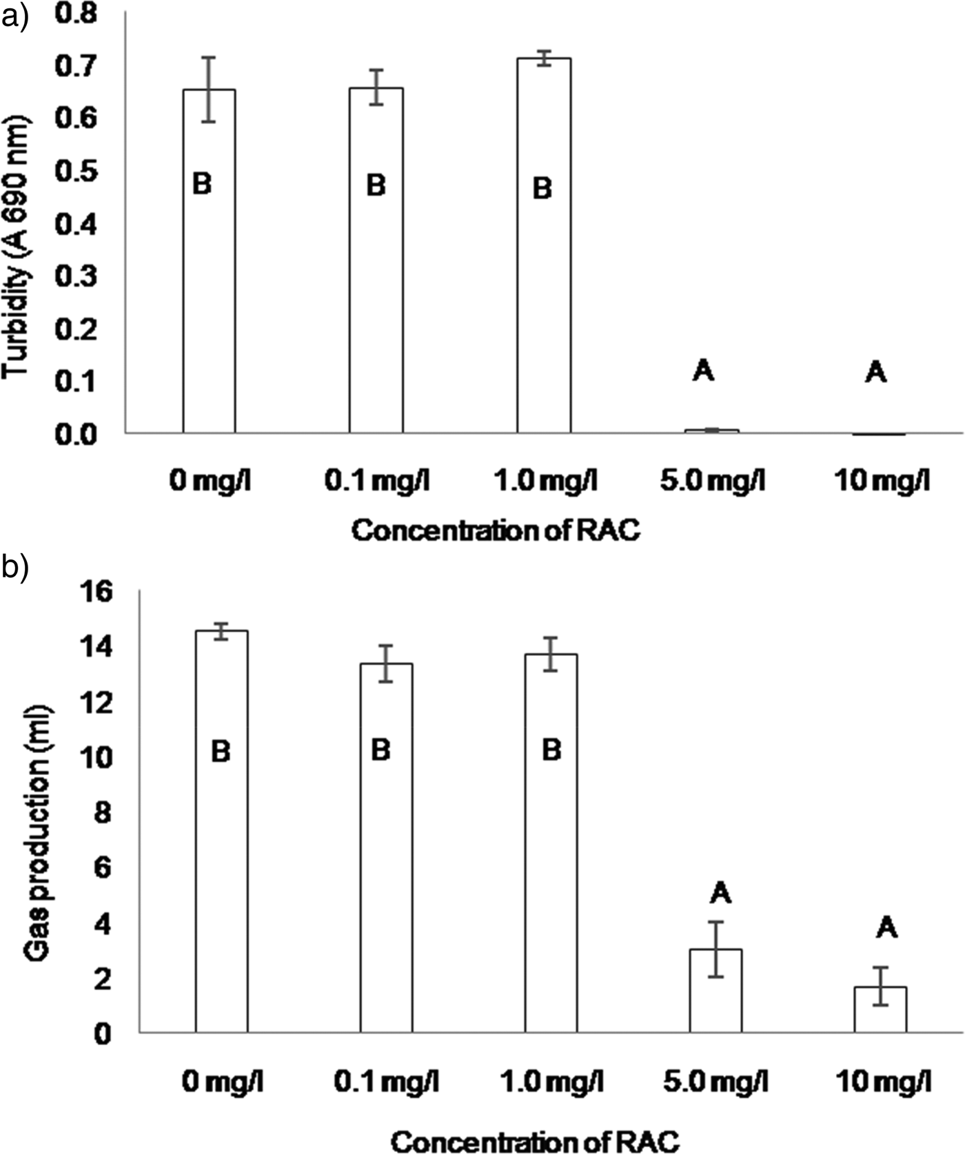
RAC significantly inhibited the growth of C. perfringens (P < 0.001), as indicated by the lack of increase of turbidity of the growth medium in the 5 and 10 mg/l RAC treatments (Figure 1a). The production of gas in the fermentation vessels was significantly inhibited by RAC (P < 0.001), this effect being visible at or above 5 mg/l (Figure 1b). However, RAC at 0.1 or 1 mg/l had little effect to the growth of C. perfringens. The resin acid concentration of the RAC batch used in the in vitro fermentation was 5%, so the lowest concentration of resin acids causing inhibition for the growth of C. perfringens was 0.25 mg/l. This suggested that the type strain of C. perfringens is very sensitive to RAC. The ability of resin acids to interact with phospholipid membranes of Gram-positive bacteria is the likely mode of action of their antimicrobial activity, as suggested by San Feliciano et al. (Reference San Feliciano, Gordaliza, Salinero and Miguel del Corral1993).
Turbidity at 690 nm (a) and cumulative volume of produced gas (b) as indications of the in vitro growth of Clostridium perfringens (mean ± SE, N = 3). Columns not sharing a letter differ significantly (P < 0.05)
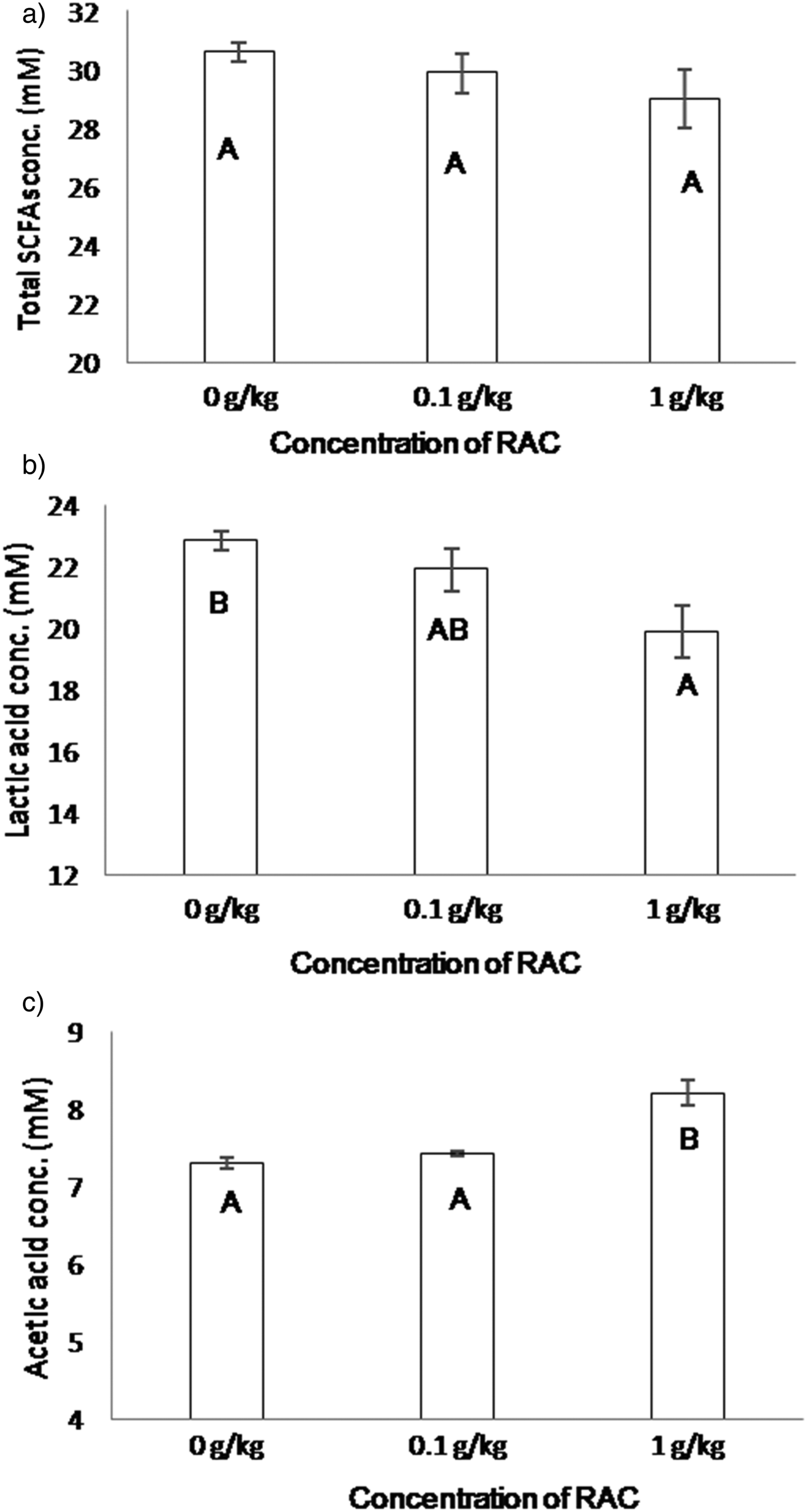
For the ex vivo laboratory fermentation experiment, the setting mimicked the physicochemical conditions of the distal small intestine but lacked the animal-to-animal variation typical of in vivo trials. RAC did not affect the total concentration of SCFAs produced by the microbes (Figure 2a). However, at the 1 g/kg dose it modulated the microbial fermentation by significantly decreasing the relative proportion of lactic acid (P = 0.006; Figure 2b) and the concentration of acetic acid was significantly increased by RAC addition (P < 0.001; Figure 2c).
Concentration of total short chain fatty acids (a), lactic acid (b) and acetic acid (c) from ex vivo ileal fermentation (mean ± SE, N = 4). Columns not sharing a letter differ significantly (P < 0.05)
The effect of RAC on the microbial community composition of the fermentation experiment was analysed from the 0 g/kg and 1 g/kg supplemented groups. The analysis was conducted by qPCR, so the result did not give the absolute number of bacterial cells, but rather the 16S rRNA copies per gram of sample fresh weight. The densities of the total eubacteria and Enterococcus spp. in the fermentation medium were not affected by RAC but the density of lactobacilli was significantly decreased by 1 g/kg RAC, compared with the 0 kg/t treatment (Table 3). This ~45% decrease in the density of lactobacilli by RAC was in line with the finding that RAC significantly decreased the concentration of lactic acid in the fermentation medium. The results of the ex vivo experiment are further discussed below with the results of the in vivo trial.
Microbial analysis of ex vivo fermentation including densities of the total eubacteria, Lactobacillus group and Enterococcus spp.
1Standard error of the mean of four replicate samples
216S rRNA gene copies per g sample fresh weight
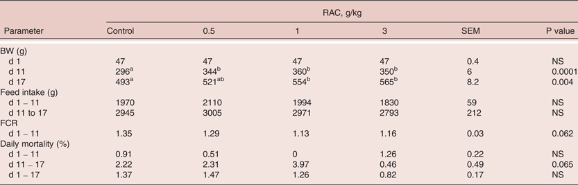
For the investigation into performance and ileal microbiota of NE challenged broiler chickens, BW, feed intake, FCR and mortality data are presented in Table 4. RAC at the of 0.5, 1 and 3 g/kg of inclusion in a wheat and soy -based starter formula did not affect feed intake, which suggested that RAC did not affect the palatability of the feed. Between days 1 – 11 (the pre-challenge period) the birds were apparently healthy, although the somewhat high FCR may suggest a slightly compromised digestion. RAC significantly increased BW at day 11 in a dose-dependent manner. The maximum increase in BW on day 11 was 17%, stimulated by RAC at 1 g/kg. FCR tended to be improved in the pre-challenge period (P = 0.062), the numerical effect being visible at all doses of RAC. For example, in the 1 g/kg group, FCR was 1.13, while it was 1.35 in the control treatment. Overall mortality was low and similar in all diet groups during the pre-challenge period.
Effects of RAC on body weight, feed intake, FCR (uncorrected for mortality) and daily mortality in a challenge trial with broiler chickens
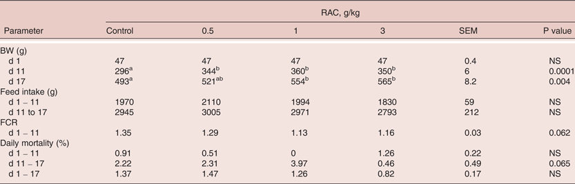
a, b Means in a column not sharing a common superscript are significantly different (P < 0.05)
As the intestinal microbiota and digestion of the birds was not studied during the pre-challenge period, the mode of action behind the performance-enhancing effect of RAC can only be speculated. Possible modes of action include suppression of microbial competition for nutrients, decreased pathogen pressure, activation of digestive functions or shifts in the microbiota towards composition capable of extracting more energy from the poorly digestible diet residue.
The challenge study induced the development of acute NE with high mortality during the last 24 hours of the trial. During the sampling of ileal segments, the jejunal epithelium of several chickens was visually examined, and lesions typical to necrotic enteritis were observed. The pen-to-pen variation in mortality was high, since the infection suddenly killed most of the chickens in some pens while birds in other pens were less severely affected. Thus, despite large differences in the mean daily mortality percentage between the treatments (for example 2.22% in the control group versus 0.46% in the RAC 3 g/kg group), the effect of RAC on mortality remained a trend (P = 0.065; Table 3). In contrast, RAC significantly increased BW on day 17 (P = 0.004), the effect being significant for the 1 and 3 g/kg groups, compared with the control treatment (Table 4).
Because of the severity of the NE infection and high mortality on d 17 in many pens, the FCR values for days 11 - 17 and days 1 – 17 showed an unusual amount of variation, and mean values without practical relevance. Therefore, FCR results for the challenge period and total study period are not shown. Considering that RAC at 3 g/kg had the highest day 17 BW and lowest days 1 - 17 mortality, it is possible that this high dosage of RAC gave some protection against the severe NE challenge. However, further studies are needed to support this finding.
One possible disease-alleviating mechanism of RAC might be linked to its fatty acid profile. Dietary oils high in n-3 fatty acids, like fish and flaxseed oil, may alleviate coccidiosis of broiler chickens (Korver et al., Reference Korver, Wakenell and Klasing1997; Allen et al., Reference Allen, Danforth and Augustine1998). Moreover, Gram-positive microbes in the ileal contents of broiler chickens may be reduced by dietary medium chain fatty acids (Van der Hoeven Hangoor et al., Reference Van der Hoeven Hangoor, van der Vossen, Schuren, Verstegen, de Oliveira, Montijn and Hendriks2013). Indeed, it is possible that part of the effect of RAC in the current experiments was due to the fatty acid content of RAC. As the effects of the fatty acids and resin acids of RAC were not separately studied, it is not possible to state whether one or the other fraction was more important, or whether the combination was critical.
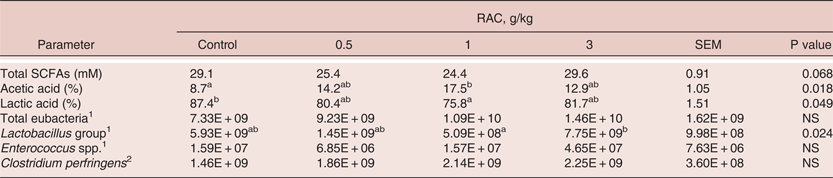
The ileal digesta samples of the NE challenge trial were collected from birds that were affected with acute NE, and their SCFA and microbial profiles (Table 5) should be interpreted with this in mind. The total concentration of SCFAs was similar in all diet groups, but RAC had significant dose-dependent effects on the relative proportions of the main individual SCFAs in the ileum, i.e. lactic and acetic acids. Supporting the findings of the present ex vivo fermentation study, the relative proportion of lactic acid decreased and acetic acid increased as a response to RAC (P = 0.049 and P = 0.018, respectively) in the NE challenge trial. The effect was statistically different in the control and RAC 1 g/kg groups.
Microbial analysis of the ileal digesta samples from disease challenged broiler chickens, including total short chain fatty acids, relative abundance of lactic and acetic acids and the density of total eubacteria and selected microbial groups are presented.
116S rRNA gene copies per g digesta fresh weight; 2Phospholipase C gene copies per g digesta fresh weight
a,bMeans in a column not sharing a common superscript are significantly different (P < 0.05)
The caecum is the main site of colonisation of C. perfringens in healthy broiler chickens (Stanley et al., Reference Stanley, Keyburn, Denman and Moore2012). There were no healthy control birds in this study, but the high abundance of C. perfringens in the ileum (1.46–2.25 x 10⁹ cells of C. perfringens per gram of fresh digesta) supported the diagnosis of acute NE. RAC did not affect the density of C. perfringens in the ileum during the acute NE phase, even in the 3.0 g/kg supplemented group. The concentration of RAC in the NE-infected ileal lumen may have remained too low to inhibit the growth of C. perfringens, but still may have altered the metabolism or virulence of the pathogen, since some indications for improved performance in this treatment group were observed.
RAC did not affect the total microbial density or the levels of Enterococcus spp. in the ileum, but had significant effects on the Lactobacillus group (P = 0.049). Although none of the RAC-treated groups differed from the control, the density of lactobacilli was significantly higher in the 3 g/kg group than in the 1 g/kg group.
The genus Lactobacillus dominates in the small intestine of broiler chickens (Gong et al., Reference Gong, Si, Forster, Huang, Yu, Yin, Yang and Han2007). Lunedo et al. (Reference Lunedo, Fernandez-Alarcon, Carvalho, Furlan and Macari2014) studied the ileal populations of Lactobacillus and Enterococcus spp. groups in sorghum and wheat-fed broiler chickens. According to them, the higher density of these microbes in the ileum of sorghum-fed birds was associated with the poorer nutritional value of sorghum compared with wheat. However, species identity and the metabolic characteristics of the lactobacilli were not determined in the present study, but it is possible that RAC shifted the lactic acid fermentation from homolactic (producing only lactic acid) towards heterolactic fermentation (producing lactic and acetic acid), both in the in vivo and ex vivo trials.
It has been shown that both therapeutic and prophylactic antibiotics suppress the numbers of lactobacilli in the small intestine of broiler chickens (Apajalahti and Kettunen, Reference Apajalahti, Kettunen, Barug, de Jong, Kies and Verstegen2006). Indeed, Lin (Reference Lin2014) suggested that the increasing growth performance effect of AGPs is associated with a reduction of the populations of gastrointestinal Lactobacillus group. One of the reasons could be that lactobacilli are the major producers of bile salt hydrolase (BSH) in the gastrointestinal tract. BSH has a negative effect on the utilisation of dietary fat, and therefore impairs FCR (Lin, Reference Lin2014). Van der Hoeven Hangoor et al. (Reference Van der Hoeven Hangoor, van der Vossen, Schuren, Verstegen, de Oliveira, Montijn and Hendriks2013) observed that supplementation of the diet with medium-chain fatty acids improved feed efficiency and suppressed Gram-positive bacteria of the phylum Firmicutes, including the Lactobacillus group, and at the same time promoted the numbers of Gram-negative bacteria. Therefore, the present findings that RAC decreases the density of lactobacilli both in vivo and ex vivo may suggest a mode of action behind the performance enhancing effect of RAC.
Conclusions
This paper described the results of three independent studies which suggested that the novel dietary ingredient RAC has the capability to modulate the small intestinal microbiota of broiler chickens and to improve the performance of the chickens even in the presence of a severe enteric infection. The potential of RAC to alter intestinal lactic acid fermentation may have a role in the performance-enhancing effect of products containing resin acids and fatty acids. The spread of antibiotic resistance genes is still growing (Koluman and Dikici, Reference Koluman and Dikici2013), and the diminishing portfolio of effective antibiotics against human pathogens is an increasingly serious threat. RAC and products with a similar impact on health and performance of poultry may help in fighting the increased need for antibiotic usage on farms and the consequent spread of resistance genes.
Acknowledgments
None
Declaration of Interest
The authors J. Vuorenmaa and E. Valkonen are employees of Hankkija Ltd. Hankkija Ltd fully funded this research and own the rights to the resin acid -based product ‘Progres’ used in this study.