Introduction
Sclerosing bone dysplasias are rare genetic disorders, characterised by generalised craniotubular bone modelling that results in an array of associated disorders, including auditory dysfunction.Reference Hamersma and Hofmeyr1, Reference Stein, Witkop, Hill, Fallon, Viernstein and Gucer2 Sclerosteosis is a benign form of excessive bone formation. Among the Dutch, French and German Afrikaner descendants of South Africa, 74 cases have been identified.Reference Hamersma and Hofmeyr1 The inheritance pathway for sclerosteosis is autosomal recessive, with a 25 per cent probability that each sibling of a proband will be affected.Reference Beighton, Hamersma, Brunkow, Pagon, Adam, Bird, Dolan, Fong and Stephens3 There is also a 50 per cent probability of being an asymptomatic carrier and a 25 per cent probability of being unaffected.Reference Beighton, Hamersma, Brunkow, Pagon, Adam, Bird, Dolan, Fong and Stephens3 The prevalence of sclerosteosis in the Afrikaner community in South Africa has been estimated to be 1 in 60 000.Reference Beighton and Hamersma4
Balemans and Van HullReference Balemans and Van Hul5 explained loss-of-function mutations in the SOST (two-exon) gene on chromosome 17, which codes for sclerostin, as the cause of sclerosteosis.Reference Balemans and Van Hul5 The localisation of the SOST gene is mostly restricted to the area where osteogenesis takes place.Reference Balemans and Van Hul5, Reference Balemans, Cleiren, Siebers, Horst and Van Hul6 Sclerostin is abundantly present in osteocytes and osteocytic canaliculi within bone.Reference Balemans, Cleiren, Siebers, Horst and Van Hul6 Osteocytes secrete sclerostin, which controls the proliferation and differentiation of osteoblastic cells and the activity of mature osteoblasts.Reference Balemans, Cleiren, Siebers, Horst and Van Hul6, Reference Leupin, Kramer, Collette, Loots, Natt and Kneissel7 Recent studies have discovered that excessive bone formation is due to the unusual activity of the sclerostin protein.Reference Hamersma and Hofmeyr1
A common clinical feature is gigantism, where individuals present with a tall stature during mid-childhood, with increased head circumference and excessive weight due to bony overgrowth.Reference Beighton and Hamersma4, Reference Beighton and Hamersma8, Reference Hamersma, Gardner and Beighton9 Syndactyly is also a characteristic feature of sclerosteosis, and its presence aids the early identification of sclerosteosis during the neonatal period.Reference Beighton, Hamersma, Brunkow, Pagon, Adam, Bird, Dolan, Fong and Stephens3, Reference Beighton and Hamersma4, Reference Beighton and Hamersma8, Reference Hamersma, Gardner and Beighton9 The severity of syndactyly is variable, ranging from minor skin webbing to complete bony union.Reference Beighton and Hamersma4, Reference Hamersma, Gardner and Beighton9 Progressive facial distortion during mid-childhood is mostly due to asymmetrical mandibular overgrowth.Reference Beighton, Hamersma, Brunkow, Pagon, Adam, Bird, Dolan, Fong and Stephens3, Reference Beighton and Hamersma4, Reference Beighton and Hamersma8, Reference Hamersma, Gardner and Beighton9
During the individual's progression towards adulthood, gross asymmetrical mandibular enlargement, as well as proptosis and mid-facial hypoplasia, may become evident.Reference Beighton and Hamersma4, Reference Hamersma, Gardner and Beighton9, Reference Hofmeyr and Hamersma10 The progressive bone formation may result in compression of the VIIth cranial nerve in the bony foramina, causing entrapment of the nerve. This may lead to acute recurrent attacks of facial palsy or facial weakness, mimicking Bell's palsy.Reference Beighton, Hamersma, Brunkow, Pagon, Adam, Bird, Dolan, Fong and Stephens3, Reference Hamersma, Gardner and Beighton9, Reference Hofmeyr and Hamersma10 A combination of these complications results in speech impairment due to insufficient lip closure and facial weakness.Reference Hamersma, Gardner and Beighton9, Reference Hofmeyr and Hamersma10
Elevation of intracranial pressure usually causes bothersome headaches during adolescence.Reference Beighton, Hamersma, Brunkow, Pagon, Adam, Bird, Dolan, Fong and Stephens3, Reference Beighton and Hamersma4, Reference Beighton and Hamersma8, Reference Hamersma, Gardner and Beighton9 The elevation of intracranial pressure is the consequence of progressive reduction in the size of the cranial cavity.Reference Beighton and Hamersma4, Reference Hamersma, Gardner and Beighton9 Sudden death is a serious risk for patients presenting with elevated intracranial pressure as a result of impaction of the medulla oblongata in the foramen magnum.Reference Beighton and Hamersma4, Reference Hamersma, Gardner and Beighton9
Some clinical features of sclerosteosis may have a direct influence on the auditory system. Bony overgrowth in the middle ear may cause a conductive hearing loss during the early stages of sclerosteosis.Reference Beighton and Hamersma4, Reference Hofmeyr and Hamersma10 The conductive hearing loss is mainly due to impaired movement of the middle-ear ossicles.Reference Hofmeyr and Hamersma10 Later, a sensorineural component may develop, caused by the closure of both the round and oval windows, and by compression of the VIIIth cranial nerve in the internal auditory canal.Reference Beighton and Hamersma4, Reference Hofmeyr and Hamersma10
Sclerosteosis leads to excessive bony overgrowth on the outer surface of the otic capsule, causing sclerosis of the periosteal layer of the cochlea.Reference Hofmeyr and Hamersma10, Reference Schuknecht11 The metabolic rate of cartilage and lamellar bone in the enchondral bony layer is very slow, thus sclerosteosis does not affect this layer.Reference Schuknecht11, Reference Jahn, Jahn and Santos-Sacchi12 The endosteal bone is also not involved in sclerosteosis, thus the lumen of the cochlea remains unaffected.Reference Schuknecht11, Reference Jahn, Jahn and Santos-Sacchi12
The symptoms and complications of excessive bony overgrowth are currently managed by surgical treatment.Reference Hofmeyr and Hamersma10 One of the major challenges, however, is the correction of hearing loss, which has limited success.Reference Beighton and Hamersma4, Reference Hofmeyr and Hamersma10 The dense bone that encroaches on the round and oval windows needs to be drilled away, which may damage hearing in the high frequencies.Reference Hofmeyr and Hamersma10 Furthermore, recurrent release of the ossicles is a temporary treatment as the progressive nature of the condition means that the ossicles will fixate again.Reference Hofmeyr and Hamersma10
Although there have been reports of otolaryngological, radiographical and genetic investigations related to sclerosteosis, there have been no studies to date that have systematically documented the auditory effects related to the condition. In addition, there is a shortage of research regarding the non-surgical management of sclerosteosis, despite the fact that most patients usually end up wearing a hearing aid.Reference Beighton and Hamersma4, Reference Hofmeyr and Hamersma10 Consequently, it is important to describe the auditory presentations of individuals with sclerosteosis in order to assist future diagnostic investigations and to direct management decisions. The current study therefore provides a cross-sectional description of the audiological profile of individuals with sclerosteosis.
Materials and methods
The institutional review board of the University of Pretoria approved the study before data collection commenced.
Participants
A cross-sectional, descriptive research design was followed. Participants were selected from a database of individuals with a confirmed diagnosis of sclerosteosis. The database was The South African Sclerosteosis database, which was started by Professor Herman Hamersma in 1964. The database is restricted to South Africans diagnosed with sclerosteosis. The study commenced in 2010 and ended in 2012.
There were 36 individuals with diagnosed sclerosteosis alive in South Africa; the 18 individuals that lived in the Gauteng region were contacted and invited to participate in the research study. Ten individuals responded and were included in the study; all provided written informed consent.
The participants were assessed using a comprehensive audiological test battery within a single test session, which lasted approximately 2 hours. The analysis of results was ear specific. Data for 20 ears were included in the analyses.
Measures
Test procedures included otoscopy, tympanometry, acoustic reflexes, diagnostic pure tone air and bone conduction audiometry, speech audiometry, distortion product otoacoustic emissions (DPOAEs), auditory brainstem responses (ABRs), and computed tomography (CT) scans. The otoscopic evaluation (carried out using a ReddyLite LED otoscope; GVR Products, Stoke-on-Trent, UK) was conducted to visualise any obstructive debris in the ear canal that may contraindicate other tests. Tympanometry (GSI Tympstar; Grason-Stadler, Madison, Wisconsin, USA) was conducted bilaterally to measure static compliance, middle-ear pressure and ear canal volume. The modified Jerger classification system for tympanometryReference Jerger13 was used to interpret the tympanogram type. Ipsilateral and contralateral acoustic reflexes were measured using a 226 Hz probe tone frequency, with pure tone octave stimuli from 500 to 2000 Hz for ipsilateral reflex recordings and from 500 to 4000 Hz for contralateral reflex recordings. The stimulus was initially presented at 75 dB HL. The intensity was increased in 10 dB increments and decreased in 5 dB increments until a reflex was elicited. A reflex threshold was the last visible deflection of 0.02 or more, and was only accepted if it was repeatable.
A clinical audiometer (GSI 61, Grason-Stadler, Milford, New Hampshire, USA) was used to conduct pure tone air conduction, bone conduction and speech audiometry. All testing was carried out in a soundproof booth using supra-aural earphones (TDH-49P; Telephonics, Farmingdale, New York, USA) for air conduction audiometry, and a bone oscillator (Radioear B-71; Radioear, New Eagle, Pennsylvania, USA) for bone conduction audiometry. The octave frequencies between 250 and 8000 Hz were used for pure tone air conduction audiometry, and frequencies between 250 and 4000 Hz were utilised for bone conduction audiometry. The pure tone and bone conduction threshold assessments were carried out according to the modified Hughson–Westlake method.Reference Carhart and Jerger14, Reference Hughson and Westlake15 The pure tone average (PTA) (500–2000 Hz) was used to categorise severity and audiometric configuration of hearing impairment, according to the criteria provided by Jerger and Jerger.Reference Jerger, Jerger, Paparella and Shumrick16
Speech audiometry stimuli were presented with a live voice speaking in Afrikaans, as all study participants were Afrikaans speaking. The Afrikaanse Spondee Woordelys Reference Laubscher and Tesner17 (a spondee word list) was used to obtain speech reception thresholds. A list of six spondee words was presented until a participant detected 50 per cent of the words correctly. The Afrikaanse Fonetiese Gebalanseerde Woordelys Reference Laubscher and Tesner18 (a phonetically balanced word list) was used to obtain speech recognition monaural performance scores across different intensities. A list of 25 phonetically balanced words was presented at three different intensities in order to obtain a percentage of words detected correctly. Agreement between the PTA and speech thresholds is considered reliable when within ±7 dB of each other.Reference Hall and Mueller19
Distortion product otoacoustic emissions were measured using the Bio-Logic Navigator Pro System (Natus Medical, Mundelein, Illinois, USA) with a 1.22 frequency ratio (f2/f1) at differential sound pressure levels (L1 = 65 dB SPL, L2 = 55 dB SPL). The DPOAE amplitude was measured at the frequencies 2f1–f2, with f2 varying from 1008 to 8016 Hz (1008, 1266, 1570, 1992, 2508, 3164, 4008, 5063, 6352 and 8016 Hz). The average noise floor was approximated around the 2f1–f2 frequency. The upper limits were 10 kHz frequency and 100 dB. The distortion product gram analysis ranged between –30 dB and 70 dB. The reference data used were according to the Vanderbilt 65/55 dB (L1/L2), 95–5th percentile norms. Interpretation of data followed the normative data system according to Hall and Mueller.Reference Hall and Mueller19
The same Navigator Pro system was used to measure the ABR in each participant. The ABR was elicited using air conduction click stimuli, with the participant lying supine on a bed in a soundproof booth. Following skin preparation, three electrodes were placed with the non-inverting electrode on the central midline (Cz), the inverting electrode on the test earlobe and the ground electrode on the contralateral earlobe. Impedance was kept below 5 kΩ. Participants were instructed to lie comfortably on the bed with their eyes closed. Rarefaction clicks were delivered monaurally through insert earphones. The stimulus rate was 11.1 stimuli per second. Participants were tested at an intensity of 90 dB nHL, with a recording time window of 21 ms. The maximum number of averaged recordings was 2000 sweeps. Artefact rejection was set at 23.3 µV, and the recordings were filtered between 100 and 3000 Hz. Latencies of waves I, III and V were measured and interpreted by three experienced clinicians.Reference Hall and Mueller19, Reference Hall and Swanepoel20
Nine participants underwent CT (one participant was unable to attend the appointment). The CT scans were conducted and interpreted by an experienced radiologist (AASBurger) at the Muelmed Mediclinic in Pretoria.
Data analysis
Test results were examined and descriptive analyses were used to illustrate the average distributions (mean, standard deviation, minimum and maximum values). The independent samples Mann–Whitney U test was used to assess whether older participants had larger air–bone gaps (ABGs) than younger participants (5 per cent significance level was used).
Results
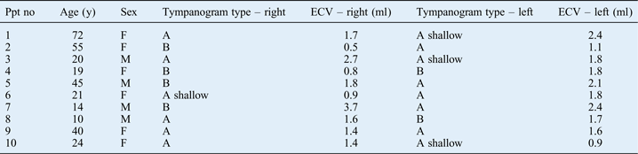
The mean age of the 10 sclerosteosis participants (4 male, 6 female) was 32 years (±20.2 standard deviation (SD)), with a range of 10–72 years (Table I). None of the participants reported otalgia; however, tinnitus was reported by seven participants (70 per cent; two unilateral and five bilateral), and all participants reported dizziness. The dizziness was attributed to low blood pressure in six participants (60 per cent). Subjective dizziness associated with hearing loss was reported by four participants (40 per cent). Three participants (30 per cent) reported problems with balance.
Examples of three hearing loss configurations recorded in the sample of sclerosteosis participants (air conduction thresholds and masked bone conduction thresholds).

Characteristics of sclerosteosis participants
Ppt no = participant number; y = years; ECV = ear canal volume; F = female; M = male
Otoscopic examination indicated a clear external auditory meatus, free from any debris, and a healthy tympanic membrane in all participants. Tympanometry (Table I) results included type A tympanograms for 50 per cent (n = 10) of ears, with large ear canal volumes in seven ears and normal ear canal volume (1.0–1.5 ml) in three ears. Half of the ears had type A shallow (n = 4) or type B (n = 6) tympanograms. Two type B tympanograms had an ear canal volume of less than 1.0 ml and four had an ear canal volume of more than 1.5 ml. Two type A shallow tympanograms had ear canal volumes of more than 1.5 ml and two ears had ear canal volumes of less than 1.0 ml. Ipsilateral and contralateral acoustic reflexes were absent in all participants.
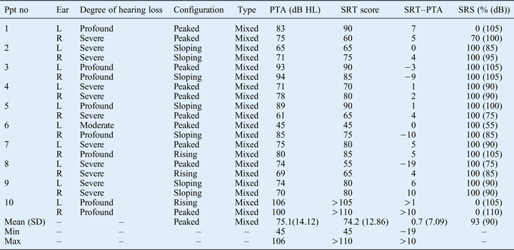
The degree of hearing loss varied from moderate (5 per cent; n = 1), to severe (55 per cent; n = 11) to profound (40 per cent; n = 8) across the 20 ears. Hearing losses were symmetrical in six participants and asymmetrical (comparing PTAs) in four. Hearing loss configurations were: rising in 3 ears (15 per cent) and sloping in 7 (35 per cent), and air conduction thresholds peaked at 2000 Hz in 10 ears (50 per cent) (Figure 1; Table II). The mean 2000 Hz air conduction peaks differed from the thresholds at 1000 Hz by 15 dB (±7.89 SD, range 5–25 dB), while the mean difference between the thresholds at 4000 Hz was 20 dB (±11.28 SD, range 5–45 dB). Speech audiometry results (speech reception threshold and speech recognition score) are compared with the PTA in Table II.
Pure tone and speech audiometry findings*
* For participants with sclerosteosis (n = 20 ears). Ppt no = participant number; PTA = pure tone average; SRT = speech reception threshold; SRS = speech recognition score; L = left; R = right; SD = standard deviation
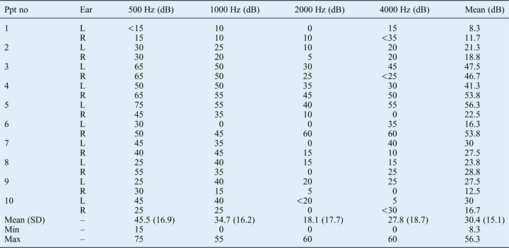
Mixed hearing losses were found in all ears across the frequencies of 500 to 4000 Hz (Table II and III). Air–bone gaps in six ears (30 per cent) demonstrated a decline towards the higher frequencies. Participants were divided into two age groups to determine whether there was a difference in ABGs with age. The first group ranged from 10 to 39 years (12 ears) and the second group from 40 to 72 years (8 ears). Mean ABG results for group 1 were 47 dB (±14.7 SD) at 500 Hz, 39 dB (±15 SD) at 1000 Hz, 21 dB (±21 SD) at 2000 Hz and 32 dB (±18 SD) at 4000 Hz. The mean ABGs for group 2 were 36 dB (±20 SD) at 500 Hz, 26 dB (±16 SD) at 1000 Hz, 13 dB (±13 SD) at 2000 Hz and 19 dB (±19 SD) at 4000 Hz. Although there were larger ABGs in the younger age group, the descriptive difference was not statistically significant (p > 0.05; independent samples Mann–Whitney U test), but this may be partly because of the small sample size.
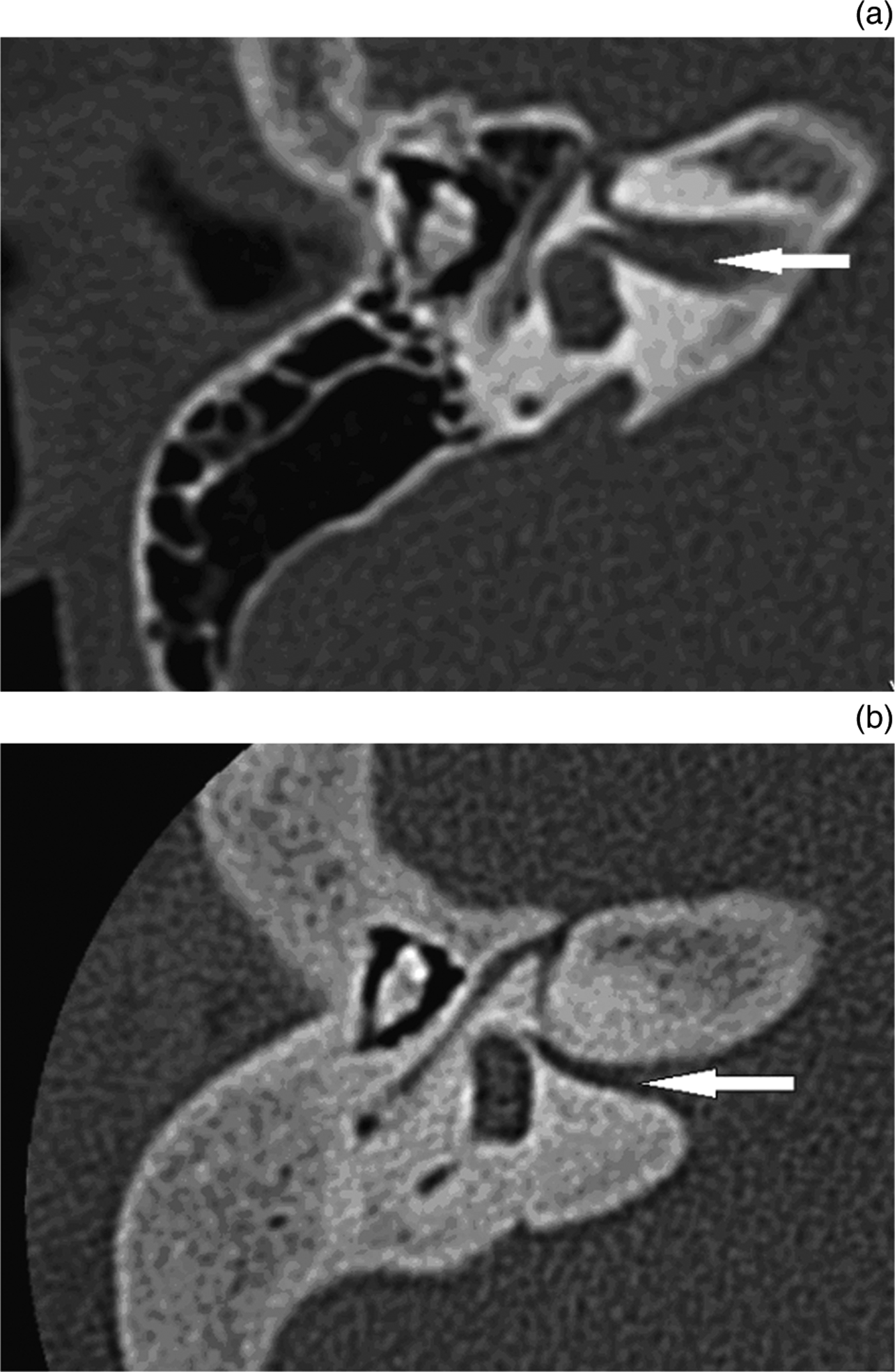
Axial computed tomography through the internal auditory canals, showing: (a) a normal internal auditory canal in a normal developed ear (white arrow), and (b) a narrowed internal auditory canal (white arrow) in sclerosteosis (the auditory nerve is compressed in the internal auditory canal).
Air–bone gap data*
* For participants with sclerosteosis (n = 20 ears). Ppt no = participant number; L = left; R = right; SD = Standard deviation
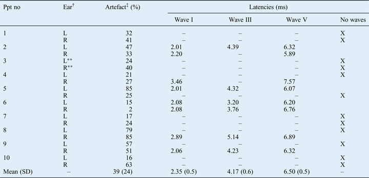
The distortion product otoacoustic emission magnitudes for all ears across all frequencies could not be repeated, indicating a lack of stability for the findings. Despite taking precautions to minimise internal and external noise during the ABR recordings, a high number of ABR epochs were rejected because of excessive electroencephalogram (EEG) noise (artefacts) across the 20 ears (Table IV). Only 4 recordings of 2000 sweeps could be made with less than 20 per cent of the sweeps being rejected because of excessive EEG activity. No waves were recorded in 12 ears (60 per cent). Waves I, III and V were recorded in six ears (30 per cent). Waves I and V were recorded in two ears (10 per cent), with wave III being absent.
Auditory brainstem response data*
* For participants with sclerosteosis (n = 20 ears). †Auditory brainstem response (ABR) recording conducted at 95 dB nHL. ‡Mean number of artefact and rejection sweeps for each ABR recording. **A single ABR recording conducted at 95 dB nHL. Ppt no = participant number; L = left; R = right; SD = standard deviation; X = no waves present
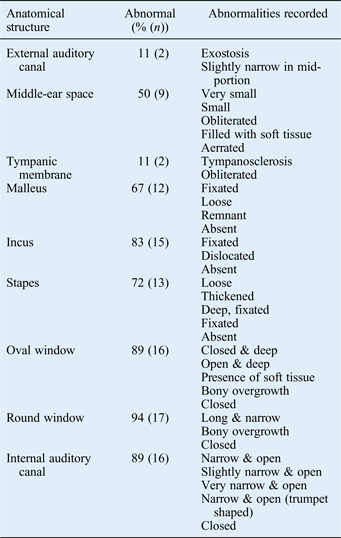
Nine participants underwent CT. The anatomical structures analysed (reported in Table V) indicated abnormalities of the external auditory canal, tympanic membrane, middle-ear space, ossicles, oval window, round window and the internal auditory canal (Figure 2).
Auditory system anatomical abnormalities: ct findings*
* In participants with sclerosteosis (n = 18 ears). CT = computed tomography
Discussion
This is the first study to provide a comprehensive auditory profile of individuals with sclerosteosis, including functional measures of the auditory system and imaging studies. Results indicate that mixed hearing loss is typical, with structural abnormalities in the middle ear, oval and round windows of the cochlea, and internal auditory canal.
Beighton and HamersmaReference Beighton and Hamersma8 were the first investigators to document the clinical, radiological and genetic features of sclerosteosis. In their study, they combined the documentation of individuals affected by sclerosteosis in order to describe the clinical features of the condition. Temporal bone and other radiology scans of affected patients showed that sensorineural hearing loss was due to the closure of the oval and round windows, as well as the narrowing of the internal auditory canal, with resultant compression of the auditory nerve.Reference Hamersma and Hofmeyr1, Reference Beighton and Hamersma8, Reference Hofmeyr and Hamersma10 The current study findings concur with these earlier reports, showing that internal auditory canals narrowed to some degree in the majority of ears (16 out of 18 ears). Several anatomical deformities, including closed, deep, narrow and long oval and round windows, were noted in the current study; these were attributed to both soft tissue and bony overgrowth.
Normal middle-ear functioning, as measured by tympanometry, was indicated for 50 per cent of ears, whereas 50 per cent showed abnormal functioning with type A shallow and type B tympanograms. However, CT (performed on 9 of the 10 participants, i.e. 18 ears) indicated abnormalities of the malleus (67 per cent, n = 12), incus (83 per cent, n = 15) and stapes (72 per cent, n = 13), the majority of which were fixated. The ossicular abnormalities were attributed to obliteration of the middle-ear space, due to overgrowth of bone and soft tissue. Interestingly, normal tympanograms could still be recorded, suggesting that the major conductive hearing loss component may be related to fixation of the stapes. Hamersma and HofmeyrReference Hamersma and Hofmeyr1, Reference Hofmeyr and Hamersma10 reported middle-ear abnormalities through CT scans, and visualised abnormalities during surgery. The authors stated that the middle-ear condition was surgically treatable, but that improvements in hearing were expected to be temporary, as bone encroaches on the middle ear and fuses the ossicles again.Reference Hamersma and Hofmeyr1, Reference Hofmeyr and Hamersma10
The abnormal bony growth in the middle ear and round and oval windows of the cochlea associated with sclerosteosis, and the resulting audiological findings, show similarities to otosclerosis. Hearing loss configurations in sclerosteosis were: rising or sloping, or air conduction thresholds which peaked at 2000 Hz. Yasan'sReference Yasan21 research findings on Carhart's notch (2000 Hz) have been documented in relation to various disorders, such as otitis media, tympanosclerosis and congenital ossicular abnormalities. Carhart's notch also relates to the air conduction peak at 2000 Hz in sclerosteosis. Air conduction configurations in sclerosteosis can indicate middle-ear obliterations, fixation or absence of various middle-ear ossicles, cochlear abnormalities at the round and oval windows, and compression of the internal auditory canal. Slight asymmetrical hearing loss was reported in a minority of participants (n = 4) in the current study, but there was no obvious difference between the structures of the middle ear, oval and round windows of the cochlea, and internal auditory canal (as observed on the CT scans) between these sets of ears. The asymmetrical hearing loss is therefore most likely due to functional impairment in the auditory system or structural abnormalities not visualised on CT scans.
Previous reports have demonstrated ABGs related to otosclerosis that are as large as 40 dB in the low frequencies, decreasing towards 2000 Hz where Carhart's notch is typically found.Reference Azlan, Asma and Saim22 Mixed hearing loss has been recorded in isolated cases of otosclerosis involving the middle ear and cochlea.Reference Velegrakis23 Middle ear, and oval and round window involvement are common in otosclerosis as well as in sclerosteosis. Fixation of the middle-ear ossicles, and closure of the round and oval windows have been reported in both conditions, but the degree to which these structures are affected leads to a more severe sensorineural hearing loss in sclerosteosis.Reference Hofmeyr and Hamersma10, Reference Velegrakis23–Reference Perez, de Almeida, Nedzelski and Chen25 In addition, sclerosteosis includes involvement of the internal auditory canal, which is not typical in otosclerosis. Audiometric findings related to age and otosclerosis have indicated that air and bone conduction thresholds deteriorate at all frequencies with increasing age.Reference Topsakal, Fransen, Schmerber, Declau, Yung and Gordts26 In the current study, the ABGs were generally smaller in the older age group, but the results were not statistically significant.
Van Buchem's disease and sclerosteosis both belong to the family of osteopetroses, and are characterised by skeletal density and bone modelling.Reference Beighton, Horan and Hamersma27 Clinical features of Van Buchem's disease closely resemble those of sclerosteosis, but are present in a milder form. The auditory profile on audiometric testing for Van Buchem's disease has not been systematically documented, but hearing loss has been reported in 40 per cent of individuals presenting with the disease, compared with 92 per cent in individuals with sclerosteosis.Reference Beighton, Horan and Hamersma27, Reference Beighton, Barnard, Hamersma and Van der Wouden28 In the current study, all participants with sclerosteosis presented with mixed hearing loss, compared with 92 per cent of the 25 individuals investigated by Beighton et al. Reference Beighton, Barnard, Hamersma and Van der Wouden28
Osteogenesis imperfecta is another disorder with similarities to sclerosteosis in terms of auditory involvement related to bone abnormalities. This bone mineral density disorder affects the connective tissue of the musculoskeletal system, causing otosclerotic-like lesions that affect the ears.Reference Swinnen, De Leenheer, Goemaere, Cremers, Coucke and Dhooge29 Sclerosteosis and osteogenesis imperfecta are both associated with middle-ear abnormalities and involvement of the oval window of the cochlea. Ossicular discontinuity and fixation of the stapes has been reported in osteogenesis imperfecta as in sclerosteosis, with the middle ears affected by abnormal bony growth in both disorders.Reference Hofmeyr and Hamersma10, Reference Swinnen, De Leenheer, Goemaere, Cremers, Coucke and Dhooge29 The oval window is affected by encroachment of bony overgrowth in both disorders, but in sclerosteosis the round window is also affected.Reference Hamersma and Hofmeyr1, Reference Hofmeyr and Hamersma10, Reference Swinnen, De Leenheer, Goemaere, Cremers, Coucke and Dhooge29 The sensorineural hearing loss in osteogenesis imperfecta is a result of abnormal bone encroachment, which causes haemorrhage into the labyrinth, resulting in cochlear hair cell damage.Reference Swinnen, De Leenheer, Goemaere, Cremers, Coucke and Dhooge29 In comparison, sensorineural hearing loss in sclerosteosis has been attributed to closure of the round and oval windows, and narrowing of the internal auditory canal, which compromises auditory nerve functioning.Reference Hamersma and Hofmeyr1, Reference Hofmeyr and Hamersma10 Swinnen et al.Reference Swinnen, De Leenheer, Goemaere, Cremers, Coucke and Dhooge29 documented a variety of mild to profound, mixed hearing losses (23–78 per cent) in osteogenesis imperfecta, whereas the current study recorded only moderate to profound, mixed hearing losses (100 per cent) in sclerosteosis. In contrast to sclerosteosis, osteogenesis imperfecta has no documented effect on the internal auditory canal.
In the current study, ABR recordings were challenging, as excessive EEG activity caused high numbers of artefacts and rejected sweeps, resulting in unreliable ABR recordings. Auditory brainstem response waves were absent at a maximum intensity of 90 dB nHL in most ears (n = 12), but when present they were significantly delayed. The CT scans confirmed conductive and sensorineural abnormalities due to a combination of middle-ear irregularities, round and oval window closure, and internal auditory canal involvement. The internal auditory canal was unaffected in only one of the nine participants who underwent CT. In contrast to the ABR results for most other participants, this particular participant presented with waves I, III and V. All ABR waves were present unilaterally in only four other participants. Three of these four participants underwent CT scans, which demonstrated that the internal auditory canals were only mildly affected, with slight narrowing. Auditory brainstem response waves in these cases were also delayed, most likely as a result of the conductive hearing loss and some sensorineural contribution.Reference Musiek, Shinn, Jirsa, Burkard, Eggermont and Don30 Two of the remaining 14 ears had an absent ABR wave III, and the other 12 ears had no ABR waves. The CT scans showed that 16 ears had internal auditory canals that were either narrow, very narrow (slightly open) or completely closed. Retrocochlear auditory involvement was indicated in two participants by the performance-intensity function for the phonetically balanced word list (the ‘rollover’ effect).Reference Miranda and Pichora-Fuller31–Reference Jerger and Hayes33 Computed tomography scans showed very narrow internal auditory canals and no ABR waves in one of those two participants and an absent wave III in the other. Auditory brainstem response and CT scan findings are in agreement with the speech audiometry results, indicating possible retrocochlear involvement.
• Bony overgrowth in sclerosteosis causes anatomical abnormalities of: the middle ear, oval and round windows, and internal auditory canal
• Sclerosteosis causes moderate to profound, mixed hearing loss, with similarities to other conditions affecting auditory system bony structures (e.g. otosclerosis)
• Abnormalities of the oval and round windows, and internal auditory canal are primary causes of sensorineural involvement
• Air–bone gaps seem to decrease with age as sensorineural component in sclerosteosis becomes more severe
• Auditory brainstem responses and speech audiometry may elucidate functional involvement of internal auditory nerve in a narrowed internal auditory canal
In young children (before the age of six years), surgical decompression of the internal auditory canal is recommended, as early as possible. This can prevent narrowing of the internal auditory canal and damage to the auditory nerve.Reference Hamersma and Hofmeyr1 If surgical decompression of the internal auditory canal proves to have sustainable success and the auditory nerve is not damaged, future studies should document the possible management of hearing loss with cochlear implants. It would be hazardous to decompress the internal auditory canal in older individuals because the auditory nerve may sustain damage during the surgical intervention. Auditory brainstem implants may be a possible option for the management of retrocochlear involvement, but future investigations need to confirm the benefit of implants as a last resort treatment of sclerosteosis-related auditory dysfunction. Conservative management of hearing loss includes a conventional hearing aid or a bone-anchored hearing aid, depending on the severity of the loss.Reference Hofmeyr and Hamersma10
Conclusion
Individuals with sclerosteosis present with anatomical abnormalities of the middle ear, the oval and round windows of the cochlea, and the internal auditory canal. These abnormalities result in moderate to profound, mixed hearing loss. The audiometric findings show similarities to those of otosclerosis and other conditions characterised by abnormal bony growth, although sclerosteosis-related auditory dysfunction is typically more severe. Abnormalities of the oval and round windows of the cochlea and internal auditory canal cause sensorineural involvement, as evidenced by absent distortion product otoacoustic emissions and absent or abnormal ABR findings. Auditory brainstem response testing and speech audiometry may assist in differentiating cases with significant involvement of the internal auditory canal. The progressive abnormal bony overgrowth, which is the hallmark of sclerosteosis, leads to functional impairment at various levels in the auditory system. The current findings provide a comprehensive auditory profile for sclerosteosis, and may be utilised alongside future research findings to direct criteria and audiological indications for surgical and audiological intervention.