Introduction
The snakehead, Channa argus, is native to the North Asian continent (Maruyama et al. Reference Maruyama, Fujii, Kijima and Maeda1987) and was introduced to the Japanese archipelago from the Korean peninsula, specifically to Nara Prefecture, Honshu, between 1923 and 1924 (Maruyama et al. Reference Maruyama, Fujii, Kijima and Maeda1987). Spread, likely facilitated by human activities (Maruyama et al. Reference Maruyama, Fujii, Kijima and Maeda1987), has resulted in its current status as a common fish species in Japanese freshwaters, including ponds and streams (Seno and Matsuzawa Reference Seno and Matsuzawa2008). Although used as food and in the aquarium trade, large-scale commercial fishing is not practiced (Seno and Matsuzawa Reference Seno and Matsuzawa2008). The effects of this introduced species on native Japanese fauna are not fully understood, though predation on small native freshwater animals, such as insect larvae and fish, has been documented (Maruyama et al. Reference Maruyama, Fujii, Kijima and Maeda1987; Seno and Matsuzawa Reference Seno and Matsuzawa2008; Yasuno et al. Reference Yasuno, Fujimoto, Shimada, Shikano and Kikuchi2019).
The trematode genus Azygia includes species whose adults parasitize piscivorous fish (e.g., Shimazu Reference Shimazu2014). Based on phylogenetic analyses, López-Hernández et al. (Reference López-Hernández, Reyda, Li, Pinto and Locke2025) proposed the synonymization of Leuceruthrus (which comprises five species; WoRMS 2024b) with Azygia. Azygia species generally utilize intermediate, paratenic, and definitive hosts in their life cycles. Adult worms reside in the digestive tracts of their definitive fish hosts, and eggs containing miracidia are shed in the host feces. These eggs subsequently infect freshwater snails (the intermediate hosts), where they develop into sporocysts and rediae within the snail’s hepatopancreas. Cercariae are released from the infected snails into the water. Upon ingestion by a definitive fish host, the cercarial body proper (the juvenile trematode located in the anterior region of the mature cercaria) develops into an adult worm (Shimazu Reference Shimazu1979, Reference Shimazu2014; Stunkard Reference Stunkard1956; Szidat Reference Szidat1932). When small fish (paratenic hosts) ingest the cercariae, the cercarial body proper infects the fish’s digestive tract, where it is termed a ‘juvenile’ and exhibits slow growth (Shimazu Reference Shimazu2014). The life cycle is completed when the piscivorous fish (definitive host) consumes the paratenic host containing the juvenile trematode (Shimazu Reference Shimazu1979; Stunkard Reference Stunkard1956).
Azygia gotoi, A. perryii, and A. rhinogobii are three species of the trematode genus Azygia known from Japan (Shimazu Reference Shimazu2014). We reported the first Japanese record of A. hwangtsiyui, recovered from the introduced snakehead C. argus. This trematode was previously reported only from the snakehead in mainland China (Tsin Reference Tsin1933). Based on our additional survey, we have also identified the intermediate and paratenic hosts in Japan. This study aims to report the introduction of A. hwangtsiyui into Japan and to detail its lifecycle in this country.
Material and methods
Sampling and observations
Between 2014 and 2024, 34 snakeheads (ranging from 40 to 810 mm in standard length [SL]) were collected from 15 sites in Japan (Table 1, Figures 1 and 2a). Sites were selected for collection based on public accessibility and the known presence of the host fish. A subset of these snakeheads had been previously reported by Saito et al. (Reference Saito, Iwata, Hayashi, Nitta, Ishikawa, Hagiwara, Ikezawa, Mano and Waki2025) in a study focused on bucephalid trematodes. Our sampling also included fish collected commercially from Gunma Prefecture (site 6, May 2024), Hiroshima Prefecture (site 17, October 2014), and Fukuoka Prefecture (site 18, November 2023). The fish samples were transported to the laboratory and dissected to examine them for trematodes with the naked eye and under the stereomicroscope. When adult trematodes were found, the affected organs were documented. All adult trematodes, except one specimen retained for histological examination, were fixed between coverslips and slides in 70% ethanol, AFA or 99% ethanol. For some ethanol-fixed adult trematodes, tissue from the posterior body region was used for polymerase chain reaction (PCR), as described subsequently. The remaining tissues from these adults, as well as other adults fixed between coverslips and slides, were stained with either acetocarmine or Heidenhein’s iron-hematoxylin, dehydrated using an ethanol series, cleared in xylene, and mounted on slides with Canada balsam for measurement of body parts. A light microscope (Olympus BX53) with a digital camera unit (Advan vision AdvanCam-U3II) was used for morphological observation, and body part measurements were taken from digital images for each developmental stage. Histological analysis of trematode genitalia was performed on a single adult specimen collected at site 4. This specimen was fixed in hot AFA, preserved in 70% ethanol, embedded in paraffin, sectioned, and stained with hematoxylin and eosin. The stained sections were examined under a light microscope. Line drawings were created using ArtCam and InkScape software packages.
Detection of adult and juvnile of Azygia hwangtsiyui from fishes in Japan. Site numbers correspond to those given in Figure 1. Only sites where fish were sampled are shown. Pref.: Prefecture

* Details of sampling locality was unclear.
** Sampling date was unclear.
Sampling sites (numbered 1–18) used in this study. Site numbers correspond to those in Table 1.

Azygia hwangtsiyui Tsin, Reference Tsin1933 and its hosts. (a) The snakehead Channa argus (Cantor, 1842), the definitive host. (b) Adults (arrowheads) detected in the stomach of the snakehead Channa argus. (c) Sinotaia quadrata histrica (W.H. Benson, 1842), the intermediate host snail. (d) Rediae in the hepatopancreas of the host snail. (e) Cercaria. (a, c, d, e) Non-scale figures.

During the period 2022–2024, snail sampling was conducted at five sites in the Kanto region (Table 2, Figure 1). Sampling site selection was based on public accessibility and the presence of freshwater snails. Collected snails were transported to the laboratory and morphologically identified according to Dillon et al. (Reference Dillon, Wethington, Rhett and Smith2002) and Masuda and Uchiyama (Reference Masuda and Uchiyama2004). The scientific names of the snails followed MolluscaBase (2025). Following shell crushing, snails were dissected in a 0.4% NaCl solution and examined under a stereomicroscope. Any sporocysts or rediae detected were fixed and preserved in either 70% or 99% ethanol for subsequent molecular analysis (see below) after initial observation under the stereomicroscope. Cercariae exhibiting morphological characteristics consistent with Azygia species (Shimazu Reference Shimazu1979) that were observed within rediae were fixed in heated 5% formalin. A subset of the fixed cercariae was stained with alum carmine and mounted on slides following the same protocols used for the adults, followed by stereomicroscopic examination. A further subset of fixed cercariae was mounted on slides with Hoyer’s medium and observed under a microscope. For sporocysts other than Azygia species, cercariae were mounted on slides with Hoyer’s medium and observed under the microscope when mature cercariae were obtained. Line drawings and measurements were generated as described previously.
Detection of rediae of Azygia hwangtsiyui and other trematode species from snails. Site numbers correspond to those given in Figure 1. Only sites where snails were sampled are shown. Pref.: Prefecture

Supplementing our newly collected samples, we also examined existing slide specimens of juvenile Azygia species prepared and preserved in our laboratory. These specimens originated from four short-spined Japanese trident gobies Tridentiger brevispinis (46.2–67.2 mm SL) collected by angling at site 11 (Kasumigaura Lake, Ami Town, Ibaraki Prefecture) on May 30, 2015. The fish were dissected in the laboratory, and juvenile trematodes were found in their stomachs and fixed in AFA between coverslips and slides. These juvenile trematodes were not used for molecular analyses. The slide specimens and associated line drawings were prepared using the methods described previously.
Molecular analysis
As previously described (Nakao et al. Reference Nakao, Waki, Sasaki, Anders, Koga and Asakawa2017), alkaline lysates of trematodes at each developmental stage were prepared and used as templates for PCR. The nuclear 28S ribosomal RNA (rDNA) gene and the mitochondrial cytochrome c oxidase subunit 1 (COI) gene were amplified using the following primer sets: 28S rRNA: dig12 and 1500R (Tkach et al. Reference Tkach, Kudlai and Kostadinova2016) or LSU5 (Littlewood et al. Reference Littlewood, Curini-Galletti and Herniou2000) and 1600R (Williams et al. Reference Williams, Reid and Littlewood2003); COI: JB3 and CO1-R trema (Miura et al. Reference Miura, Kuris, Torchin, Hechinger, Dunham and Chiba2005), JB3 and cox1rrnL (Králová-Hromadová et al. Reference Králová-Hromadová, Špakulová, Horáčková, Turčeková, Novobilský, Beck, Koudela, Marinculić, Rajský and Pybus2008), or JB3 and JB4.5 (Bowles et al. Reference Bowles, Blair and McManus1992). These primers were also used for sequencing. The following internal sequencing primers were used as needed for 28S rDNA: ECD2S (reverse, Williams and Ozawa Reference Williams and Ozawa2006), D3m (reverse, Takano and Kano Reference Takano and Kano2014), and 900F (forward, Lockyer et al. Reference Lockyer, Olson, Østergaard, Rollinson, Johnston, Attwood, Southgate, Horak, Snyder, Le, Agatsuma, McManus, Carmichael, Naem and Littlewood2003). Sanger sequencing of the PCR amplicons was outsourced to Eurofins Genomics Inc. (Tokyo).
Sequence alignment of 28S rDNA and COI gene sequences for adults and rediae were performed using BioEdit (Hall Reference Hall1999). DNA sequences of related taxa were obtained from the International Nucleotide Sequence Database Collaboration (INSDC: DDBJ/EMBL/GenBank). MEGA X (Kumar et al. Reference Kumar, Stecher, Li, Knyaz and Tamura2018) was used for computing genetic divergence, estimating the appropriate substitution model, and inferring the maximum-likelihood (ML) phylogenetic tree using sequences of related species retrieved from online databases. The 28S rDNA ML tree was constructed using the Hasegawa-Kishino-Yano (HKY) + G model (Nei & Kumar Reference Nei and Kumar2000). Pairwise comparisons of p-distance values were calculated in MEGA X for both the 28S rDNA and COI datasets.
For sporocysts other than Azygia species, 28S rDNA sequences were compared with published data in the DDBJ/EMBL/GenBank databases using the Basic Local Alignment Search Tool (Blast) to determine their systematic position at the species or higher taxonomic level. The family and lower taxonomic classification of the sporocyst detected in S. q. histrica at site 10 could not be determined by this initial analysis. Therefore, phylogenetic analyses were performed to determine its family using 28S rDNA sequences of the Microphallidea superfamily available in online databases. MEGA X was used for these phylogenetic analyses, employing the ML method with 1000 bootstrap replicates and the HKY + G model (Nei and Kumar Reference Nei and Kumar2000). COI sequences were not amplified for these sporocysts.
The sequences obtained in this study were deposited in the INSDC through DDBJ under accession numbers.
Results
Molecular analysis
Our morphological analyses revealed that our adults were identified as A. hwangtsiyui as described later. COI sequences (424–861 bp), representing three distinct haplotypes, were obtained from these adults. Pairwise p-distances of an overlapping 344 bp region of the COI sequences between our adults and A. hwangtsiyui in the online databases ranged from 0 to 0.0116. This range is consistent with the level of intraspecific variation (0–0.0145) calculated among other Azygia species in online databases, supporting our morphological identification. Two COI sequences (886 and 917 bp) were obtained from the rediae. The p-distance calculated for the overlapping region between the rediae and our adult COI sequences ranged from 0 to 0.0116, indicating that our rediae were identified as A. hwangtsiyui.
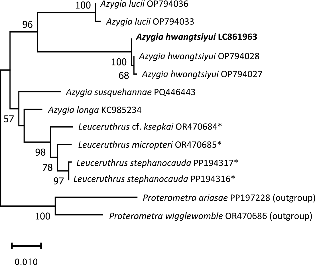
For Azygia species present in the INSDC, two 28S rDNA sequences for ‘A. sinipercae’ were available. One (MG976845) was identical to our A. hwangtsiyui sequences. The other unpublished ‘A. sinipercae’ sequence (MH777021) differed significantly from MG976845 and exhibits substantial variation in its 3’ region, resulting in 20 nucleotide gaps when aligned with other Azygia species sequences. In addition, the 28S rDNA sequence of ‘A. longa’ (KC985234) was found to be identical to that of A. susquehannae (PQ446443). Based on these, we excluded ‘A. sinipercae’ (MG976845 and MH777021) and ‘A. longa’ (KC985234) from our analyses of 28S rDNA, as discussed later. Identical 28S rDNA sequences (876–1648 bp) were obtained from both our rediae and adults. Based on the sequences of Azygia species in online databases, intraspecific p-distances for an 868-bp overlapping region of the 28S rDNA sequences ranged from 0 to 0.0012, which supports the identification of rediae as A. hwangtsiyui. The p-distance between specimens obtained in this study and A. hwangtsiyui sequences available in online databases ranged from 0.0012 to 0.0023, slightly higher than the maximum intraspecific variation (0.0012) previously observed within this genus. Given that our adults were morphologically identified as A. hwangtsiyui, and this identification was supported by the molecular analyses of the COI sequences, the observed variation in the 28S rDNA sequences is considered to represent intraspecific variation within A. hwangtsiyui. The phylogenetic tree generated from the 28S rDNA sequences (1216 sites) revealed that our sequence and A. hwangtsiyui sequences from online databases clustered together in a clade with strong bootstrap support (BP = 100) (Figure 3), supporting our species identifications.
Maximum-likelihood phylogenetic tree of Azygia trematodes inferred from partial 28S rDNA sequences (1216 sites) using the Hasegawa–Kishino–Yano (HKY) + G model. An isolate from this study is in bold. Bootstrap values >50% are shown. GenBank accession numbers follow species names. Proterometra spp. (PP197228 and OR194317) were used as outgroup taxa. Asterisks indicate sequences depositted as Leuceruthrus spp. (Leuceruthrus is a synonym of Azigya; López-Hernández et al. Reference López-Hernández, Reyda, Li, Pinto and Locke2025). Scale bar represents substitutions per site.

Morphological identification
Family Azygiidae Lühe, 1909
Genus Azygia Looss, 1899
Species Azygia hwangtsiyui Tsin, Reference Tsin1933
[New Japanese name: Raigyo-no-nedoko-mushi]
Materials examined. Adults (IMN-1-130008–130011) from the stomach of the snakehead Channa argus (Cantor, 1842) (Figure 2a, b), Toda City, Saitama Prefecture, Japan, May 27, 2023 (IMN-1-130008); Fukuoka Prefecture, Japan, Nov., 2023 (IMN-1-130009); Oyama City, Tochigi Prefecture, Japan, Oct. 19, 2015–June 25, 2017 (IMN-1-130010 and 130011). Juvenile (IMN-1-130015) from the short-spined Japanese trident goby Tridentiger brevispinis Katsuyama, Arai & Nakamura, 1972, Kasumigaura Lake, Ami Town, Ibaraki Prefecture, Japan, May 30, 2015. Cercariae (IMN-1-130016) from the hepatopancreas of the freshwater snail Sinotaia quadrata histrica (Figure 2c, d, e), Inashiki City, Ibaraki Prefecture, Japan, Nov. 6, 2022.
Depository. Ibaraki Nature Museum, Ibaraki Prefecture, Japan.
Description.
Adult (Figures 2b, 4a–c, measurements in Table 3). Body elongated, variable in size, with round ends. Oral sucker rounded, subterminal. Prepharynx absent. Pharynx muscular. Esophagus short, T or Y-shaped, bifurcating just anterior to prostatic sac. Drüsenmagen distinct. Caeca long, extending to posterior body terminal. Ventral sucker smaller than oral sucker, rounded, situating anterior to level of anterior one-fifth of body. Testes at posterior one-fourth of body, situated very close and tandem. Anterior testis smaller than or subequal to posterior testis. Seminal vesicle and pars prostatica surrounded by prostatic glandular cells in prostatic sac. Prostatic sac ovid, small, thin walled, just anterior to ventral sucker. Ejaculatory duct running and opening in sinus organ. Hermaphroditic duct short. Hermaphroditic pore on ventral margin of sinus organ. Genital atrium well developed. Genital pore large, anterior to ventral sucker. Vitellaria situated bilaterally, its posterior margin and anterior to posterior margin of caecum and not close to posterior terminal of body. Ovary elliptical shape, just anterior to anterior testis. Ootype pouch distinct. Laurer’s canal indistinct. Ootype vesicular, Mehlis’ gland well developed. Metraterm developed, opening in sinus organ, close to opening of ejaculatory duct. Most of uterus posterior to ventral sucker to anterior margin of ovary. Eggs numerous, elongate oval. Excretory vesicle I-shaped in posterior body. Excretory pore at posterior margin of body.
Drawings of Azygia hwangtsiyui (a–g) collected from the Tone River system. (a–c) Adult. (d–e) Juvenile. (f–g) Cercaria. (b, e) Terminal genitalia (b. lateral view; e. ventral view). (c) Ootype complex, ventral view. Abbreviations: ca, caecum; cvd, common vitelline duct; d, drüsenmagen; e, excretory pore; ed, ejaculatory duct; ga, genital atrium; gp, genital pore; hd, hermaphroditic duct; hp, hermaphroditic pore; Mg, Melhlis’ gland; mt, metraterm; o, ovary; op, ootype pouch; os, oral sucker; ot, ootype; ov, oviduct; p, pharynx; pc, prostatic gland cells; pp, pars prostatica; ps, prostatic sac; so, sinus organ; t, testis; u, uterus; v, vitellarium; vd, vitelline duct; vs, ventral sucker.

Comparison of morphological measurements of Azygia species in different references. All measurements are in μm unless indicated otherwise. Prov.: Province

* Diameter
** Sizes of two testes
Juvenile (range for three juveniles) (Figure 4d, e)
Body proper obovate, 1.3 mm (1.1–1.4 mm) in length and 371 (364–378) in width. Oral sucker subglobular, subterminal, 254 (218–279) in length and 253 (241–266) in width. Prepharynx absent. Pharynx muscular, 84 (78–91) in length and 93 (88–102) in width. Esophagus short. Caecum extending posterior terminal of body. Ventral sucker anterior to middle of body, subglobular, 271 (192–241) in length and 230 (223–238) in length. Ovary subglobular, situating just anterior to anterior testis. Testes subglobular, situating very close and tandem. Ejaculatory duct and metraterm open in sinus organ. Hermaphroditic pore opening ventral margin of sinus organ.
Cercaria (Figures 2e, 4f, g).
Furcocystocercous type. Body (including body proper and tail) with many small papillae. Tail large, distally forked. Tail furcae flattened. Cercarial body proper obovate. Oral sucker subglobular, subterminal, opening ventrally. Prepharynx absent. Pharynx muscular. Esophagus indistinct. Caecum undulating, extending posterior terminal of body. Ventral sucker at middle of body, subgloblar. Ovary subglobular, situating anterior to testes. Testes subgloblar.
Remarks. Morphological analysis of the adults sampled in this study identified them as members of the trematode genus Azygia. This identification was based on the following characteristics: oral sucker larger than ventral sucker; testes tandem, posterior to ovary and far from excretory pore; vitellaria situated bilaterally; majority of uterus posterior to ventral sucker (Gibson et al. Reference Gibson, Jones and Bray2002; López-Hernández et al. Reference López-Hernández, Reyda, Li, Pinto and Locke2025). Furthermore, these adults were identified as A. hwangtsiyui based on the following morphological features: body elongated; oral sucker larger than ventral sucker; ventral sucker positioned anterior to anterior one-fifth of body; testes at posterior one-fourth of body; testes situating very close to each other; testis width subequal to or smaller than 1/2 of body width; elliptical ovary just anterior to anterior testis; prostatic sac ovid; vitellaria not extending close to posterior margin of body; ejaculatory duct and metraterm opening within sinus organ; hermaphroditic pore on ventral margin of sinus organ.
Thirty-one species are currently classified within the genus Azygia (López-Hernández et al. Reference López-Hernández, Reyda, Li, Pinto and Locke2025; Shimazu Reference Shimazu1979; WoRMS 2024a, b). Here, A. asiatica, A. cyprinus, A. gotoi, A. perryii, and A. sinipercae had long body, oral sucker larger than ventral sucker, ventral sucker situating anterior to anterior one-fifth of body, testes situated very close to each other, elliptic ovary just anterior to anterior testis (Fujita Reference Fujita1918; Institute of Hydrobiology, Hubei 1973; Ozaki Reference Ozaki1924; Simha and Pershad Reference Simha and Pershad1964; Wu et al. Reference Wu, Sun and Song1991). Of these, A. asiatica and A. sinipercae are distinguished from A. hwangtsiyui by the posterior extent of their vitellaria, which reach close to the posterior extremity of the body (vs. not close in A. hwangtsiyui). A. sinipercae also differs in the location of its ventral sucker (at anterior one-third of body vs. anterior one-fifth in A. hwangtsiyui) and testes (at posterior one-fifth of body vs. posterior one-fourth in A. hwangtsiyui). The testes of A. cyprinus are significantly larger (width nearly equal to body width) than those of A. hwangtsiyui (width approximately half or less of body width). In A. gotoi, the ejaculatory duct and metraterm open on the ventral margin of the sinus organ, whereas they open within the sinus organ in A. hwangtsiyui. Azygia perryii has a rounded prostatic sac, while it is ovid in A. hwangtsiyui.
Despite the absence of molecular data for the juveniles collected from the short-spined Japanese trident goby, they were identified morphologically as A. hwangtsiyui based on the following features: ovary and testes situating close together and tandem; ejaculatory duct and metraterm opening within the sinus organ; hermaphroditic pore on ventral margin of sinus organ.
Three Azygia species are known to occur on Honshu Island, Japan (A. hwangtsiyui, A. gotoi, and A. rhinogobii; Shimazu Reference Shimazu2014). Among these, A. hwangtsiyui is unique in having the ejaculatory duct and metraterm opening within the sinus organ. Moreover, A. rhinogobii is characterized by diagonally arranged testes (vs. tandem in our specimen) and an ovary positioned lateral to the anterior testis (vs. anterior to the testis in our specimen). Although A. rhinogobii adults have been reported from gobies in Kasumigaura Lake (Shimazu Reference Shimazu2014), juveniles in the present study were determined to be immature A. hwangtsiyui based on the morphology of their reproductive organs.
As described above, our cercariae were molecularly identified as A. hwangtsiyui. Our cercariae were smaller than those of A. hwangtsiyui detected from freshwater snail Cipangopaludina ussuriensis and observed in a live condition (Table 4, Besprozvannykh Reference Besprozvannykh2005). The differences were based on differences in host species (Sinotaia quadrata histrica in the present study) and conditions of cercariae used for observations (fixed in the present study). Shimazu (Reference Shimazu1979) reported the furcocystocercous cercaria of A. gotoi from Cipangopaludina japonica. Our cercariae were morphologically distinct from those of A. gotoi, which possess few, small papillae on the tail furcae compared to the numerous papillae on the body proper and tail in A. hwangtsiyui. The cercarial morphology of the other two Azygia species found in Japan is currently unknown.
Comparison of morphological measurements of cercaria of Azygia hwangtsiyui in different references. All measurements are in μm unless indicated otherwise

* Not described
** Diameter
Etymology. The Japanese name ‘Raigyo-no-nedoko-mushi’ is derived from ‘unagi no nedoko’ (‘eel’s bed’), alluding to the worm’s long, narrow shape. ‘Raigyo’ means ‘snakehead’, and ‘mushi’ means ‘worm’ in Japanese.
DNA markers. DDBJ accession numbers: LC861966–861968 (COI, 861–917 bp). LC861961–861963 (28S rDNA, 1254–1648 bp)
Detection of other sporocysts from snails
Larval stages of two trematode species, in addition to A. hwangtsiyui, were detected in the freshwater snails S. q. histrica and P. acuta at site 12. COI sequences were not obtained for these sporocysts.
Telorchis sp.
Family: Telorchiidae Looss, 1899
Genus: Telorchis Lühe, 1899 nec Looss, 1899
Two identical 28S rDNA sequences (891 bp; LC861964) were obtained from two sporocysts found in P. acuta. The 28S rDNA sequences of these sporocysts were identical, within a 720-bp overlap, to sequences from unidentified sporocysts assigned to the genus Telorchis (LC818885 and LC 794207). As interspecific divergence in 28S rDNA among identified Telorchis species ranges from 0.0111 to 0.0389 (in the overlapped region), sporocysts are likely members of this genus. However, species-level identification was precluded by the absence of data on intraspecific 28S rDNA variation within Telorchis. Mature cercariae were not obtained for this species.
Microphalloidea fam. gen. sp.
A sporocyst collected from S. q. histrica was tentatively assigned to the superfamily Microphalloidea; however, 28S rDNA sequences from closely related species were absent from the databases. Our sequences (1270 bp, LC861965), for instance, shared only 94% similarity with Gyrabascus oppositus (Microphalloidea, ON036090). Phylogenetic tree of 28S rDNA sequences from members of the superfamily placed sporocysts in a clade that also contained several families, including Lecithodendriidae, with moderate bootstrap support (BP = 86) (Figure 5a). However, its position near the poorly supported basal node of this clade (BP <50%) did not provide conclusive evidence for its family placement. Consequently, we labelled our sporocyst as Microphalloidea fam. gen. sp.
(a) Maximum-likelihood phylogenetic tree of Microphallidea inferred from partial 28S rDNA sequences (1011 sites), using Mesocoelium spp. (LC770431 and LC770435) as outgroup taxa. Bootstrap values >50% are shown. Database accession numbers follow species names. Newly obtained sequences are in bold. Scale bar represents substitutions per site. (b) Cercaria of Microphalloidea fam. gen. sp. detected from Sinotaia quadrata histrica (W.H. Benson, 1842). Abbreviations: os, oral sucker; p, pharynx; s, stylet; vs, ventral sucker.

Description (Figure 5b, based on three cercariae). Body oval, 151 (137–163) in length and 70 (48–83) in width. Tegmental spines on body surface. Oral sucker subglobular, subterminal, 32 (30–34) in length and 31 (30–34) in width. Stylet anterior terminal of oral sucker, 14 (13–16) in length. Prepharynx not observed. Pharynx muscular, 11 (10–13) in length and 13 (11–15) in width. Esophagus and caecum not well observed. Ventral sucker posterior to middle of body, subglobular, 24 (22–26 in length and 31 (30–31) in length. Tail half as long as body, 85 (71–104) in length and 23 (21–27) in width.
Discussion
This study provides the first record of the trematode parasite A. hwangtsiyui in Japan. The snakehead, its definitive host, was introduced to Japan between 1923 and 1924 (Maruyama et al. Reference Maruyama, Fujii, Kijima and Maeda1987) and is now widely distributed in freshwater habitats throughout the country (Seno and Matsuzawa Reference Seno and Matsuzawa2008). Because A. hwangtsiyui adults have only been reported from snakeheads (Tsin Reference Tsin1933), this fish is likely the only definitive host species. Of the several snail species sampled, A. hwangtsiyui rediae were found only in the freshwater snail S. q. histrica. Recent surveys of Japanese freshwater snails, which largely focused on snails other than S. q. histrica, have not detected A. hwangtsiyui sporocysts or rediae (Ansai et al. Reference Ansai, Nitta, Saito, Kojima and Waki2024; Miura and Takisawa Reference Miura and Takisawa2024; Seo et al. Reference Seo, Ansai, Sase, Saito, Takano, Kojima and Waki2024). These results indicate that S. q. histrica is the primary host for the sporocyst and redia stage. While Azygia cercariae typically develop into adults when ingested by their definitive hosts, small fish can act as paratenic hosts (Shimazu Reference Shimazu1979; Stunkard Reference Stunkard1956). In this study, the short-spined Japanese trident goby is identified as a paratenic host for juvenile A. hwangtsiyui. However, juvenile trematodes of the genus Azygia are known to utilize a wide range of paratenic hosts (Shimazu Reference Shimazu2014; Szidat Reference Szidat1932). While the digestive tracts of other small fish were not examined in the present study, probably other small fish can also serve as paratenic hosts for juvenile A. hwangtsiyui when they ingest cercariae. Based on these results, the life cycle of this trematode in Japan is hypothesized to be as follows: A. hwangtsiyui cercariae are released from the snail host S. q. histrica. The released cercariae orally infect small snakehead larvae, which have been reported to feed on small freshwater insects (e.g., mosquito larvae) (Yasuno et al. Reference Yasuno, Fujimoto, Shimada, Shikano and Kikuchi2019), and subsequently develop into adults within the stomachs of the fish. Paratenic hosts are thought to be the main route of infection for larger snakeheads, which are known to prey on freshwater animals such as fish (Yasuno et al. Reference Yasuno, Fujimoto, Shimada, Shikano and Kikuchi2019).
The distribution of this trematode from Kyushu Island to the Kanto area indicates that A. hwangtsiyui is not a recent introduction. The presence of three the distinct COI haplotypes suggests multiple independent introductions. While previous studies have examined snakehead parasites (e.g., Moravec and Nagasawa, Reference Moravec and Nagasawa2018), these investigations have largely concentrated on nematodes, and we found no evidence that trematodes, including A. hwangtsiyui, have been surveyed in Japanese snakeheads. Therefore, the exact date of introduction of A. hwangtsiyui to Japan remains uncertain. However, as mentioned previously, the snakehead was introduced to Japan between 1923 and 1924 (Maruyama et al. Reference Maruyama, Fujii, Kijima and Maeda1987), and we found no record of later introductions. Moreover, both the intermediate and paratenic hosts of A. hwangtsiyui were distributed in Japan when the snakehead was first introduced (Matsuzawa and Matsuura Reference Matsuzawa and Matsuura2011; Ye et al. Reference Saito, Hirano, Dong, Do and Chiba2020). This suggests that the trematode may have been introduced along with the initial snakehead introduction. Given the frequent commercial and accidental transport of freshwater fishes and mollusks from the Asian continent to Japan (Ito Reference Ito2007; Takano et al. Reference Takano, Uchida and Yamakawa2023), A. hwangtsiyui may also have been introduced into Japan via this route. The wide distribution of A. hwangtsiyui is likely linked to the broad ranges of both its intermediate and definitive hosts in Japan (Masuda and Uchiyama Reference Masuda and Uchiyama2004; Seno and Matsuzawa Reference Seno and Matsuzawa2008). Furthermore, the trematode has likely spread within Japan through the commercial transport of snakeheads (National Institute of Environmental Studies 2025). Recently, freshwater fishes, including snakeheads and gobies, and the snail S. q. histrica have recently been sold in Japanese fish markets and online shops and auctions (e.g., Yahoo auctions, https://auctions.yahoo.co.jp/). Such commercial trade in these hosts may further contribute to the trematode’s dispersal.
The short-spined Japanese trident goby, serving as a paratenic host for the juvenile trematode, is thought to be native to Japan (Matsuzawa and Matsuura Reference Matsuzawa and Matsuura2011). The freshwater snail S. q. histrica in Japan, however, has a complex evolutionary origin, with Japanese populations resulting from natural migrations and/or artificial introductions from mainland China over a millennium ago (Matsuzawa and Matsuura Reference Matsuzawa and Matsuura2011; Ye et al. Reference Saito, Hirano, Dong, Do and Chiba2020). The establishment of these snail populations, combined with the artificial introduction of the snakehead, enabled A. hwangtsiyui to establish a population in Japan. Since the snakehead is crucial for the A. hwangtsiyui life cycle, the trematode’s introduction to Japan represents an example of an invasion meltdown, where the introduction of one invasive species accelerates the introduction of another in a cascading fashion (Simberloff and Von Holle Reference Simberloff and Von Holle1999). If the snail had only been introduced artificially, introductions of this snail by human activity would also have contributed to the invasion of A. hwangtsiyui in Japan. We believe that the introduction of invasive host species should be avoided, regardless of whether they are infected or not, to prevent the invasion of introduced parasites.
Two 28S rDNA sequences for ‘A. sinipercae’ were found in online databases, and one (MG976845) of the two was identical to our A. hwangtsiyui sequences. Our adult specimens were morphologically identified as A. hwangtsiyui, which is morphologically distinct from A. sinipercae. Vainutis et al. (Reference Vainutis, Voronova, Mironovsky, Zhigileva and Zhokhov2023) proposed that these two species are a single species; however, morphological data for the ‘A. sinipercae’ specimen (MG976845) have not been published. Therefore, we are unable to discuss and explain this sequence identity. The other unpublished ‘A. sinipercae’ sequence (MH777021) differed significantly from MG976845, suggesting that these two ‘A. sinipercae’ sequences represent distinct species. As discussed in López-Hernández et al. (Reference López-Hernández, Reyda, Li, Pinto and Locke2025), the sequences attributed to ‘A. sinipercae’ on the online databases may be problematic. In addition, the 28S rDNA sequence of ‘A. longa’ (KC985234) was found to be identical to that of A. susquehannae (PQ446443). The specimen for ‘A. longa’ (KC985234) were certainly identified as A. susquehannae as discussed in López-Hernández et al. (Reference López-Hernández, Reyda, Li, Pinto and Locke2025). Consequently, the sequences of ‘A. sinipercae’ (MG976845 and MH777021) and ‘A. longa’ (KC985234) were not used in our 28S rDNA p-distance calculations in this study.
Acknowledgements
The present study was kindly helped by Naoya Anzai, Kazuo Arai, Hiroshi Eguchi, Toshio Furota, Takeru Hasumi, Hiromi Ikezawa, Fumiko Kaburagi, Koichiro Kawai, Takashi Kimura, Kei Kobayashi, Tsubasa Miyoshi, Yasushi Morooka, Kazuya Nagasawa, Mizuki Ohtsubo, Yoshiki Saito, Minoru Sato, and Tomoji Somemiya.
Financial support
This work was partially supported by a JSPS KAKENHI Grant (No. 15J05777 to MN).
Competing interests
The authors declare none.
Ethical standard
Formal ethical approval was not required for this study because it involved unregulated fish and invertebrate species.