Implication
This study has demonstrated that regulation of fatty acid composition by the dietary n-3 and n-6 fatty acids in cattle is mediated, at least partially, through tissue-specific regulation of lipogenic enzymes expression. This finding is important for understanding the mechanisms underlying the accumulation and tissue distribution of n-3 and n-6 polyunsaturated fatty acid. Therefore, this research is an important step in the development of a complex (diet- and genetic-based) approach for improvement of eating quality of beef. Improving of beef fatty acid composition would be of direct benefit to human health and to the beef-producing industry.
Introduction
It is well known that the amount and type of fat in the diet is closely related to human health. A high intake (more than 15% of daily energy intake) of saturated fatty acids (SFAs) (mainly myristic, lauric and palmitic acid) is positively associated with coronary heart disease and mortality rate (Rioux and Legrand, Reference Rioux and Legrand2007). The negative effect of SFA is mediated through increase in blood cholesterol concentrations, especially plasma total and low-density lipoprotein cholesterol (Williams, Reference Williams2000). Moreover, the diets high in SFA reduce anti-inflammatory properties of high-density lipoprotein (Rioux and Legrand, Reference Rioux and Legrand2007). In contrast to SFA, unsaturated fatty acids are recognised as health beneficial. A number of studies demonstrated hypo-cholesterolemic effects of monounsaturated fatty acids (MUFAs) and some polyunsaturated fatty acids (PUFAs) (Demaison and Moreau, Reference Demaison and Moreau2002). MUFA have also been reported to possess anti-thrombogenic properties (Smith et al., Reference Smith, Kelly, Fielding, Hauton, Silva, Nydahl, Miller and Williams2003). In countries with the Mediterranean diet (low content of SFA and high content of MUFA) the incidence of coronary heart disease is much lower when compared with the countries with the diets which are high in SFA (Lada and Rudel, Reference Lada and Rudel2003). The particular health benefit has been linked to n-3 PUFA which are essential for normal growth and development (Simopoulos, Reference Simopoulos1991). Long chain n-3 PUFA (docosahexaenoic acid and eicosapentaenoic acid) demonstrated anti-atherogenic, anti-thrombotic, anti-inflammatory and immunosuppressive properties in experimental studies in man (Williams, Reference Williams2000; Garg et al., Reference Garg, Wood, Singh and Moughan2006). In addition to n-3 PUFA an increasing attention has been paid to another PUFA, namely conjugated linoleic acids (CLAs). CLA is a group of isomers, which have been demonstrated to enhance immune system, to reduce body weight and to decrease the risk of cancer (Tricon et al., Reference Tricon, Burdge, Williams, Calder and Yaqoob2005; Bhattacharya et al., Reference Bhattacharya, Banu, Rahman, Causey and Fernandes2006). The main natural source of cis-9, trans-11 CLA is meat and milk of ruminants (Song and Kennelly, Reference Song and Kennelly2003).
According to national and international dietary guidelines, SFA contribution to dietary energy intake should be not more than 10% of dietary energy (Wahrburg, Reference Wahrburg2004), and the inclusion of MUFA and PUFA into human diet should be up to 15% of total daily energy for MUFA (Wahrburg, Reference Wahrburg2004) and to a maximum of 10% of total daily energy for PUFA (Wahrburg, Reference Wahrburg2004). One way to achieve this is to decrease SFA and increase MUFA and PUFA content in food and in meat in particular. This is particularly related to ruminant meat which is higher in SFA when compared with pig or poultry (Valsta et al., Reference Valsta, Tapanainen and Mannisto2005).
The fatty acid composition of adipose and muscle tissues depend on a number of factors such as diet, fatness of animals, age/weight, gender, breed, season and others (Nuernberg et al., Reference Nuernberg, Wegner and Ender1998; Scollan et al., Reference Scollan, Hocquette, Nuernberg, Dannenberger, Richardson and Moloney2006; Wood et al., Reference Wood, Enser, Fisher, Nute, Sheard, Richardson, Hughes and Whittington2008). In monogastric animals the quality of meat can be relatively easy improved by dietary manipulations. However in ruminant’s dietary manipulations of fatty acid composition proved to be difficult because of rumen biohydrogenation. Therefore, there is an increasing interest in manipulation of fatty acid composition in ruminant tissues through the regulation of tissue fatty acid biosynthesis. The key enzymes involved in SFA, MUFA and PUFA biosynthesis are acetyl-CoA carboxylase (ACC) (EC 6.4.1.2), stearoyl-CoA desaturase (SCD) (EC 1.14.19.1) and delta 6 desaturase (Δ6d) (EC 1.14.19.3), respectively. These enzymes have been extensively studied in a number of species, including pigs. However, there is very limited information about tissue distribution and regulation of expression of these enzymes in cattle (Chang et al., Reference Chang, Lunt and Smith1992; Smith et al., Reference Smith, Hively, Cortese, Han, Chung, Casteñada, Gilbert, Adams and Mersmann2002; Archibeque et al., Reference Archibeque, Lunt, Gilbert, Tume and Smith2005).
The aim of this study was to investigate effects of the diet enriched in linolenic acid (as a source of n-3 PUFA) v. linoleic acid (source of n-6 PUFA) on fatty acid composition and protein expression of the key lipogenic enzymes in cattle tissues. The particular questions which we aim to answer in this study are: (i) whether the effects of PUFA-enriched diets on fatty acid composition of cattle muscle and adipose tissue is mediated through the regulation of lipogenic enzyme expression and (ii) whether the effects of the diet on enzyme expression are tissue-specific.
Material and methods
Animals and diets
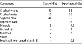
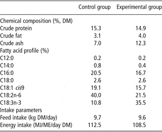
Fourteen German Holstein bulls were assigned to two dietary treatments: (i) maize silage with soybean-based concentrate (control group) result in a 1.4 times higher content of C18:2n-6 and (ii) grass silage with linseed oil and rapeseed cake supplemented concentrate (experimental group) with a four times higher concentration of C18:3n-3 compared with control group. There were eight animals in control group and six animals in experimental group (initial number of animals in experimental group was eight but two animals died at the beginning of the trial). Bulls were kept in-door in groups over the whole fattening period and were fed a total mixed ration (TMR). Table 1 presents the composition of the two concentrates used. The chemical and fatty acid composition of the TMRs, the feed and the energy intake is given in Table 2. The animals received feed two times a day adapted to fit the needs of bulls in every state of growth. The feed was weighed and prepared with a fodder-mixing trailer and the daily feed intake was recorded through the car computer in the fodder-mixing trailer for each group during the trial. The metabolisable energy of the TMRs was 11.6 and 11.3 MJ/kg DM for the control and experimental groups, respectively. Daily feed intake was 9.7 and 9.6 kg DM/day for the control and experimental groups, respectively. The 14 bulls were fed the control and experimental ration for 209 ± 7 and 216 ± 8 (P = 0.5073) days, respectively. The average live weight of bulls at the slaughter was 621.1 ± 10.4 kg in control group and 627.7 ± 12.0 kg (P = 0.6871) in experimental group, respectively. The bulls were slaughtered at the facilities of the Institute for the Biology of Farm Animals, Dummerstorf, Germany and were killed by captive bolt stunning followed by exsanguinations in accordance with EU regulations.
Composition of the concentrate compound of the diets (wt %)

Chemical and fatty acid composition (mg/100 g) of total mixed ration and intake parameters

DM = dry matter.
Samples of longissimus muscle and subcutaneous adipose tissue for protein expression analyses (approximately 50 g) were collected from the right side of carcass, between the thirteenth and fourteenth rib, within 30 min after slaughter. All the samples were snap-frozen in liquid nitrogen and stored at −70°C until analysed. It has been previously demonstrated that the storage conditions do not affect the lipogenic enzyme expression (Doran et al., Reference Doran, Moule, Teye, Whittington, Hallett and Wood2006).
Isolation of microsomal and cytosolic fractions
Microsomal and cytosolic fractions from muscle and subcutaneous adipose tissues were isolated by differential centrifugation. The tissue samples (10 g) were defrosted and homogenised (by using Potter homogenizer) in 20 ml of Tris-sucrose buffer composed of 10 mmol Tris-HCl and 250 mmol sucrose (pH 7.4) at room temperature. After centrifugation at 12 000×g for 10 min at 4°C, the supernatant (in the case of muscle) and the infranatant (in the case of subcutaneous adipose tissue) were collected. About 8 mmol of CaCl2 (final concentration) was added to the supernatant or to the infranatant to facilitate the microsomes sedimentation. Cytosolic and microsomal fractions were separated by centrifugation at 25 000×g for 30 min at 4°C. A microsomal fraction (the pellet) was re-suspended in Tris-KCl buffer, containing 10 mmol Tris-HCl, 250 mmol KCl (pH 7.4), and the inhibitors of proteolytic enzymes; antipain, pepstatin and leupeptin at final concentrations 1.5, 1.5, and 2 μM, respectively. The final protein concentration of the microsomal suspension was about 26 mg/ml for muscle and 2 mg/ml for subcutaneous adipose tissue. The protein concentration of the microsomal and cytosolic fractions was determined by the Bradford method using bovine serum albumin as standard.
Protein expression
Expression of ACC, SCD and Δ6d proteins was analysed by Western blotting. Microsomal and cytosolic proteins were separated by sodium dodecyl sulfate polyacrylamide gel electrophoresis, electro blotted onto nitrocellulose membrane at a constant 100 V for 1 h and incubated with one of the following primary antibodies: rabbit polyclonal anti-bovine adipose tissue SCD (custom-made at the University of Bristol, UK) or goat polyclonal anti-human (Santa Cruz Biotechnology Inc., Santa Cruz, CA, USA). The ACCα antibodies did not distinguish between the phosphorylated and non-phosphorylated enzyme forms. In the case of Δ6d the antibodies were custom-produced in rabbits against the synthetic peptides containing amino acid sequences from the regions which are conserved in the rat, pig and human, near C-terminus of the corresponding proteins (Sigma-Genosys Ltd, Cambridge, UK). The membrane was re-probed with appropriate commercial secondary antibody, which was either horseradish peroxide linked donkey anti-rabbit immune globuline G (IgG), or horseradish peroxide linked donkey anti-goat IgG. The blots were developed using an enhanced chemiluminescence reagent (Amersham, Buckinghamshire, UK). The films were scanned and the intensity of the corresponding bands was quantified by using the ImageQuant program (Molecular Dynamics, Sunnyvale, CA, USA). A microsomal or cytosolic preparation from one particular sample was present on all the blots (reference sample). The intensity of the signal on this sample was taken as 100 arbitrary units throughout, and the intensity of the signals from other samples was expressed as a fraction of the reference sample. Western blot analysis was performed with 6 μg of microsomal or cytosolic protein.
Fatty acid analysis
Fatty acids have been analysed as described previously (Dannenberger et al., Reference Dannenberger, Nuernberg, Scollan, Schabbel, Steinhart, Ender and Nuernberg2004). Samples of longissimusmuscles and subcutaneous adipose tissue were thawed at 4°C. After blending and mincing in a grinding processor (IKA M20, Staufen, Germany), and adding C19:0 as an internal standard, the total lipids were extracted from 2 g of muscle and 1 g of subcutaneous fat (both in duplicates) with chloroform/methanol (2 : 1, v/v) by homogenisation (Ultra Turrax, 3 × 15 s, 12 000 r.p.m.) at room temperature. All the solvents contained 0.005% (w/v) of t-butylhydroxytoluene to avoid the oxidation of PUFA. The extraction mixture was stored at 5°C for 18 h in the dark and subsequently washed with 0.02% aqueous CaCl2. The organic phase was dried with Na2SO4 and K2CO3 (10 : 1, wt/wt) and the solvent was subsequently removed under nitrogen at room temperature. The lipid extract was re-dissolved in toluene and an aliquot of 25 mg was used for methyl ester (ME) preparation. To prepare the fatty acid methyl esters (FAMEs), 2 ml of 0.5 M sodium methanolate was added and the mixture was shaken in a water bath at 60°C for 10 min. After this, 1 ml of boron trifluoride in methanol (14%, wt/v) was added to the solution and the mixture was shaken in a water bath at 60°C for 10 min. MEs of the total fatty acids were extracted twice with 2 ml of n-hexane in the presence of 2 ml distilled saturated solution of NaHCO3. The upper phases containing FAME were pooled and the solvent was eliminated by evaporation under nitrogen flow. FAME were resolved in 150 μl n-hexane and stored at −20°C until analysed by gas chromatography (GC).
An aliquot of the FAME mixture was used for analyses of total fatty acids. The fatty acid composition of longissimusmuscle, subcutaneous adipose tissue and animal feed were determined by capillary GC on a CP SIL 88, 100 m × 0.25 mm × 0.25 μm capillary column (Chrompack, Varian, USA) installed in a Perkin Elmer gas chromatograph Autosys XL (Waltham, MA, USA) with a flame ionisation detector and split injection. Initial oven temperature was 45°C (held for 4 min). It was subsequently increased to 150°C at a rate of 13°C min−1 (held for 47 min), and then to 215°C at 4°C min−1 and held for 35 min. Hydrogen was used as the carrier gas at a flow rate of 1 ml min−1. In total 41 fatty acids were identified with a reference standard (Sigma FAME mixture no. 189-19, Sigma-Aldrich, Deisenhofen, Germany) to which the MEs of C18:1 trans-11, C22:5n-3, CLA cis-9, trans-11, C18:1 cis-11, C18:4n-3 and C22:4n-6 had been added was used to determine recoveries and correction factors for the determination of individual fatty acids in intramuscular fat. FAMEs were purchased from Sigma-Aldrich and Biotrend (Köln, Germany); CLA cis-9, trans-11 was purchased from Matreya, (Biotrend, Köln, Germany). Twenty fatty acids are presented in Tables 3 and 4. The above conditions did not allow separating the CLA cis-9, trans-11 from CLA trans-7, cis-9 and CLA trans-8, cis-10. Therefore, the value for CLA cis-9, trans-11 is presented as a sum of the values for the three above isomers. The sum of n-3 fatty acids was calculated as C18:3n-3 + C18:4n-3 + C20:3n-3 + C20:5n-3 + C22:5n-3 + C22:6n-3, the sum of n-6 fatty acids was calculated as C18:2n-6 + C18:3n-6 + C20:2n-6 + C20:3n-6 + C20:4n-6 + C22:2n-6 + C22:4n-6, the sum of MUFA was calculated as C14:1 + C15:1 + C16:1 + C17:1 + ∑C18:1trans-9-11 + C18:1cis-9 + C18:1cis-11 + C22:1 + C24:1 and the sum of PUFA as sum of n-3 fatty acids and the sum of n-6 fatty acids.
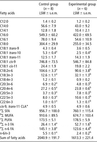
Fatty acid composition (mg/100 g) of longissimus muscle of German Holstein bulls fed with different diets

CLA = conjugated linoleic acid; SFA = saturated fatty acid; MUFA = monounsaturated fatty acid; PUFA = polyunsaturated fatty acid; FA =fatty acid.
a,bindicates significant effect of the diet at P ⩽ 0.05.
* indicates coeluation with C18:2 trans-7, cis-9 and C18:2 trans-8, cis-10.
aΣ SFA = C10:0 + C11:0 + C12:0 + C13:0 + C14:0 + C15:0 + C16:0 + C17:0 + C18:0 + C20:0 + C21:0 + C22:0 + C23:0 + C24:0.
bΣ MUFA = C14:1 + C15:1 + C16:1 + C17:1 + C18:1 trans-9 + C18:1 trans-10 + C18:1 trans-11 + C18:1cis-9 + C18:1cis-11 + C22:1 + C24:1.
cΣ PUFA = Σ n-3 fatty acids (FA) + Σ n-6 FA.
dΣ n-3 FA = C20:3n-3 + C22:6n-3 + C22:5n-3 + C20:5n-3 + C18:4n-3 + C18:3n-3.
eΣ n-6 FA = C22:2n-6 + C20:2n-6 + C18:3n-6 + C22:4n-6 + C20:3n-6 + C18:2n-6 + C20:4n-6.
.
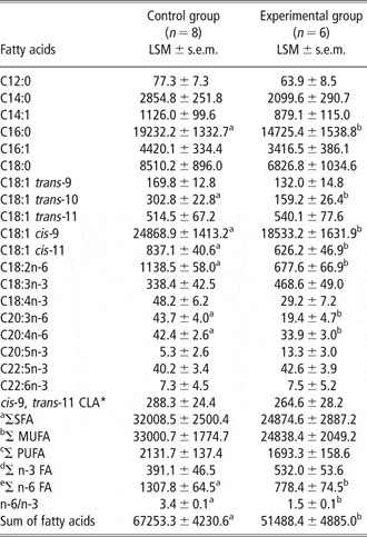
Fatty acid composition (mg/100 g) of subcutaneous adipose tissue of German Holstein bulls fed different diets

CLA = conjugated linoleic acid; SFA = saturated fatty acid; MUFA = monounsaturated fatty acid; PUFA = polyunsaturated fatty acid; FA =fatty acid.
a,bindicates significant effect of the diet at P ⩽ 0.05.
* indicates coeluation with C18:2 trans-7, cis-9 and C18:2 trans-8, cis-10.
aΣ SFA = C10:0 + C11:0 + C12:0 + C13:0 + C14:0 + C15:0 + C16:0 + C17:0 + C18:0 + C20:0 + C21:0 + C22:0 + C23:0 + C24:0.
bΣ MUFA = C14:1 + C15:1 + C16:1 + C17:1 + C18:1 trans-9 + C18:1 trans-10 + C18:1 trans-11 + C18:1cis-9 + C18:1cis-11 + C22:1 + C24:1.
cΣ PUFA = Σ n-3 fatty acids (FA) + Σ n-6 FA.
dΣ n-3 FA = C20:3n-3 + C22:6n-3 + C22:5n-3 + C20:5n-3 + C18:4n-3 + C18:3n-3.
eΣ n-6 FA = C22:2n-6 + C20:2n-6 + C18:3n-6 + C22:4n-6 + C20:3n-6 + C18:2n-6 + C20:4n-6.
Statistical analysis
Data were analysed by the least-squares method using the general linear model procedures of SAS® (2009) with the fixed factor feeding. P ⩽ 0.05 was considered statistically significant.
Results
Fatty acid composition of longissimus muscle and subcutaneous adipose tissue
The fatty acid composition of longissimus muscle and subcutaneous adipose tissue is shown in Tables 3 and 4 and the results are expressed as mg/100 g tissue.
No differences were observed in the content of individual and total SFA and MUFA, except for C18:1 trans-10, which was reduced in experimental group compared with control group (P < 0.001). In grass silage fed group we have also observed a tendency for reduction in C18:1 cis-9 (P = 0.09). The sum of n-3 PUFA in the grass silage fed group (P < 0.001) was significantly increased (by two-fold) because of an increase in the level of all individual n-3 PUFA, except for C18:4n-3. The sum of total and individual n-6 PUFA was significantly decreased in muscles of the experimental animals (P = 0.003). As the result of the opposite changes in n-3 and n-6 PUFA, the muscle n-6/n-3 ratio was significantly lower in experimental group (P < 0.001) and the content of total PUFA did not change significantly (P = 0.534). As there was no significant effect of experimental diet on total SFA, MUFA and PUFA content, the total fatty acids content in muscle (i.e. sum of fatty acids) was not affected either.
In subcutaneous adipose tissue the sum of fatty acids (mg/100 g) was significantly reduced with the experimental diet. This reduction was mainly caused by a lower content of the SFAs (C14:0, C16:0 and C18:0), C18:1 cis-9, C18:1 cis11 and C18:1 trans-10. The adipose tissue of the bulls from experimental group also had a significantly lower content of n-6 PUFA, in particular C18:2n-6 (P < 0.001), C20:3n-6 (P = 0.002) and C20:4n-6 (P = 0.05). The content of n-3 PUFA in adipose tissue was either not affected by the diet (i.e. C22:5n-3 and C22:6n-3; P = 0.65 and P = 0.99, respectively), or there was a trend toward the increase in the level of total (P = 0.07) and individual n-3 fatty acids in experimental group (C18:3n-3 and C20:5n-3; P = 0.07). There was a significant decrease of n-6/n-3 ratio in experimental group (P < 0.001).
Lipogenic enzyme expression
Effect of diet on the lipogenic enzymes protein expression
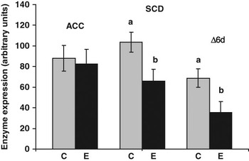
Effects of diet on ACC, SCD and Δ6d protein expression in the longissimus muscle are presented in Figure 1. The expression of ACC protein did not differ significantly between the control and experimental groups (P = 0.71). In contrast to ACC, the experimental diet significantly reduced the expression of SCD protein (by 37%, P = 0.03) and Δ6d protein (by 33%, P = 0.03).
Expression of acetyl-CoA carboxylase (ACC) protein in cytosolic fraction and, stearoyl-CoA desaturase (SCD) and Δ6-desaturase (Δ6d) proteins in microsomal fraction isolated from longissimus muscle. Diets are described in ‘Material and Methods’ section. Bars represent different least squares means for eight animals in control group and six animals in experimental group. The error bars represent standard error for means. The measurements for each animal were performed in duplicate. Bars with different superscripts differ significantly for a given enzyme (P < 0.05). C = control group; E = experimental group.

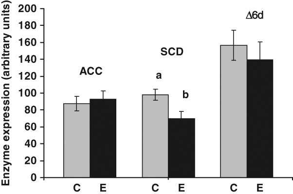
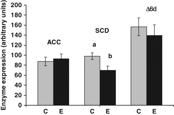
In subcutaneous adipose tissue the expression of SCD in experimental group was decreased by about 29% when compared with control group (P = 0.02). The experimental diet did not have significant effects on ACC and Δ6d protein expression (P = 0.71 and P = 0.55, Figure 2).
Expression of acetyl-CoA carboxylase (ACC) protein in cytosolic fraction and stearoyl-CoA desaturase (SCD) and Δ6-desaturase (Δ6d) proteins in microsomal fraction isolated from subcutaneous adipose tissue. Diets are described in ‘Material and Methods’ section. Bars represent different least square means for eight animals in control group and six animals in experimental group. The error bars represent standard error for means. The measurements for each animal were performed in duplicate. Bars with different superscripts differ significantly for a given enzyme (P < 0.05). C = control group; E = experimental group.
Tissue distribution of lipogenic enzymes
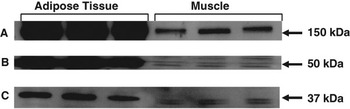
Figure 3a–c shows representative blots for ACC, SCD and Δ6d proteins for three randomly selected animals. The blots clearly demonstrate large between-tissue differences. The presence of an ACC immunoreactive protein was observed in both, muscle and subcutaneous adipose tissue (Figure 3a). The size of the band is consistent with the molecular weight of ACC reported for other species (Tanabe et al., Reference Tanabe, Wada, Okazaki and Numa1975). The intensity of ACC signal was much higher in subcutaneous adipose tissue when compared with muscles.
A–C Representative blots of relative expression of lipogenic enzymes in subcutaneous adipose tissue and in longissimus muscle. (A) Acetyl-CoA carboxylase protein expression in isolated cytosolic fraction. (B) Δ6-desaturase protein expression in isolated micosomal fraction. (C) Stearoyl-CoA desaturase protein expression in isolated microsomal fraction. Each signal represents an individual animal.

Similarly to ACC, the presence of Δ6d-immunoreactive proteins was also observed in both tissues (Figure 3b), and the signal was much higher in subcutaneous adipose tissue when compared with the muscle. The bands’ size was approximately of 50 kDa, which is consistent with the molecular weight of mouse and human Δ6d (Cho et al., Reference Cho, Nakamura and Clarke1999).
Examples of SCD-immunoreactive protein expression are presented in Figure 3c. The size of the immunoreactive proteins was about 37 kDa, which is consistent with the molecular weight of SCD protein reported in rats (Moreau et al., Reference Moreau, Froment, Tosca, Moreau and Dupont2006). The intensity of the SCD-immunoreactive band was higher in the adipose tissue when compared with the muscle.
Discussion
It is well known that fatty acid composition of meat, and hence its nutritional value, can be manipulated by dietary means (Enser et al., Reference Enser, Hallett, Hewett, Fursey, Wood and Harrington1998; Dannenberger et al., Reference Dannenberger, Nuernberg, Scollan, Schabbel, Steinhart, Ender and Nuernberg2004). In monogastric animals, the dietary fatty acids can be directly incorporated in animal tissues (Nuernberg et al., Reference Nuernberg, Fischer, Nuernberg, Kuechenmeister, Klosowska, Eliminowska-Wenda, Fiedler and Ender2005). However, in the case of ruminants, direct fatty acid incorporation is restricted because of rumen biohydrogenation. Nevertheless, a number of experiments demonstrated accumulation of n-6 and n-3 fatty acids in cattle tissues in the case of long-term feeding regimes. In this study we have observed significant increase in most of the individual muscle n-3 fatty acids and decrease in n-6 fatty acids in the case of the diet supplemented with linseed oil and rapeseed cake as sources of C18:3n-3, when compared with control group. The above resulted in an improvement (i.e. reduction) of the n-6/n-3 ratio. These results are in agreement with data of the literature (Scollan et al., Reference Scollan, Hocquette, Nuernberg, Dannenberger, Richardson and Moloney2006; Wood et al., Reference Wood, Enser, Fisher, Nute, Sheard, Richardson, Hughes and Whittington2008). A similar picture was observed for subcutaneous adipose tissue n-3 and n-6 fatty acids and adipose tissue n-6/n-3 ratio. The only difference was that in case of subcutaneous adipose tissue, there was only a trend toward an increase in n-3 fatty acids (P = 0.07); whilst in case of muscles such increase was significant. The lack of significance in case of subcutaneous adipose tissue might be related to large between-individual variations.
There might be a number of possible explanations for the above mentioned dietary effects on n-3 and n-6 fatty acids. One possible reason could be dietary modulation of the rate of n-3 and n-6 fatty acids transfer into the tissues. Waters et al. (Reference Waters, Kelly, O′Boyle, Moloney and Kenny2009) described that the balance between n-6 and n-3 PUFA in the diet is important for the regulation of SCD through SREBP-1c in muscle of beef cattle. However, this explanation could be applied to C18:2n-6 and C18:3n-3 fatty acids, but not to longer-chain fatty acids. Changes in the longer-chain fatty acid profile could be related to dietary effects on activity/expression of Δ5 or Δ6-desaturases and this possibility is discussed below.
Our earlier study on pigs demonstrated that diets, including PUFA-supplemented diets, can trigger changes in the expression of lipogenic enzymes, and these changes are related to variations in fatty acid profiles (Doran et al., Reference Doran, Moule, Teye, Whittington, Hallett and Wood2006; Missotten et al., Reference Missotten, De Smet, Raes and Doran2009). Regulatory effects of the dietary fatty acids on enzyme expression have also been demonstrated in experiments on laboratory animals and on human (Jump and Clarke, Reference Jump and Clarke1999; Ntambi, Reference Ntambi1999). However, there is very limited information regarding lipogenic enzymes distribution and regulation in ruminant tissues.
This study reports the presence of ACC, SCD and Δ6d immunoreactive bands in cattle longisimmus muscle and subcutaneous adipose tissue. ACC is the key enzyme controlling de novo biosynthesis of SFAs (Hardie, Reference Hardie1989). SCD is the enzyme catalysing two types of reactions: (i) biosynthesis of MUFA through the insertion of a double bound in SFAs between carbon C9 and C10 and (ii) the tissue biosynthesis of cis-9, trans-11 CLA from trans-vaccenic acid (Enoch et al., Reference Enoch, Catala and Strittmatter1976). Δ6d is involved in a complex process of biosynthesis of longer chain n-3 and n-6 fatty acids through conversion of linoleic and linolenic acids to their products (Stoffel et al., Reference Stoffel, Holz, Jenke, Binczek, Gunter, Kiss, Karakesisoglou, Thevis, Weber, Arnold and Addicks2008). The molecular weights of ACC, SCD and Δ6d immunoreactive bands detected in this study were 150, 37 and 50 kDa respectively, which is consistent with the molecular weights of ACC, SCD and Δ6d proteins reported for other species (Tanabe et al., Reference Tanabe, Wada, Okazaki and Numa1975; Cho et al., Reference Cho, Nakamura and Clarke1999; Moreau et al., Reference Moreau, Froment, Tosca, Moreau and Dupont2006). In a case of the SCD protein expression (in muscle and subcutaneous adipose tissue) and muscle Δ6d, the two closely positioned immunoreactive bands were observed. One possible explanation for the two bands could be existence of more than one SCD (Lengi and Corl, Reference Lengi and Corl2007) and Δ6d isoforms in cattle tissues. More than one isoform of Δ6d were found in Mucor rouxii by Na-Ranong et al. (Reference Na-Ranong, Laoteng, Kittakoop, Tanticharoen and Cheevadhanarak2006) and in rats by Skrzypski et al. (Reference Skrzypski, Bellenger, Bellenger, Sinclair, Poisson, Tessier, Rialland and Narce2009). The presence of several SCD isoforms, their tissue-specific distribution and regulation also has been previously reported in mice and rats (Thiede et al., Reference Thiede, Ozols and Strittmatter1986; Miyazaki et al., Reference Miyazaki, Jacobson, Man, Cohen, Asilmaz, Friedman and Ntambi2003). The number of Δ6d isoforms in ruminants remains unclear.
The intensity of ACC, SCD and Δ6d immunoreactive bands in this study were relatively higher in subcutaneous adipose tissue when compared with the muscles. These results are in agreement with the data of the literature suggesting that subcutaneous adipose tissue is the primary site of fatty acid biosynthesis in cattle (Smith et al., Reference Smith, Chapman, Lunt, Harris and Savell2007).
In this study we analysed expression of ACC, SCD and Δ6d proteins in animals fed either with C18:2n-6 or with C18:3 n-3 supplemented diets to evaluate potential contribution of the above enzymes in the variations in fatty acid composition. We have shown that increase in the individual and total n-3 PUFA in muscles of experimental animals was not accompanied by increase in Δ6d protein expression. Moreover, Δ6d protein level was significantly decreased in experimental group. Therefore Δ6d protein expression does not seem to contribute to the n-3 PUFA variations. It is important to admit that in this study we have only analysed the Δ6d protein expression (not the actual Δ6d activity). It is well known that fatty acid composition might be regulated by direct changes in enzyme activity and further study is required to investigate this possibility.
Our data on the cattle Δ6d are consistent with the reports on inhibitory effects of the dietary n-3 PUFA on expression of Δ6d gene in other species (Theil and Lauridsen, Reference Theil and Lauridsen2007). Interestingly, in our experiment inhibition of Δ6d was observed in cattle muscles but not in subcutaneous adipose tissue. The reason for a tissue-specific response of Δ6d protein expression to the n-3 PUFA-supplemented diet is not clear. It is known that in a number of species the regulation of desaturase gene expression is mediated through the sterol-regulatory element binding protein (SREBP), and that expression of SREPB is tissue specific (Felder et al., Reference Felder, Klein, Patsch and Oberkofler2005). It might be possible that the tissue-specific effect of n-3 PUFA-supplemented diet on Δ6d protein expression in our experiments is related to tissue-specific distribution/regulation of the relevant transcription factors. This suggestion is supported by data of the literature that n-3 PUFA-enriched diet inhibits SREBP-1c in muscle tissue of bulls (Waters et al., Reference Waters, Kelly, O′Boyle, Moloney and Kenny2009).
In respect to the SCD expression, the level of this protein was significantly decreased under the n-3 PUFA-supplemented diet in both, muscle and subcutaneous adipose tissue. This is consistent with the data of the literature regarding inhibitory effects of PUFA on SCD expression in other species (Flowers and Ntambi, Reference Flowers and Ntambi2008). In contrast to Δ6d, we did not observe any tissue-specific effects of diet on the SCD protein expression. This is a particularly interesting observation because according to the literature, the regulation of both, Δ6d and SCD in other species involves the same transcription factors. Different responses of the cattle SCD and Δ6d to the dietary manipulations suggest the existence of tissue-specific mechanisms, and possibly tissue-specific expression of the transcription factors is required for the regulation of these enzymes in ruminants.
Inhibition of SCD protein expression in animals fed with C18:3n-3 supplemented diet was not accompanied by changes in total muscle or subcutaneous adipose tissue MUFA content in this study. However, we observed a significant decrease in one of the products of SCD catalysed reaction, C18:1 cis-9 in subcutaneous adipose tissue. We would also like to mention that the average values for total and individual MUFA were lower in C18:3n-3 fed group when compared with the group receiving C18:2n-6. This was observed for both, muscle and subcutaneous adipose tissue. Lack of significant differences might be related to large between-individual variations within the groups and relatively small number of animals per group.
In this study we have also observed some decrease in individual and total SFAs in muscles and subcutaneous adipose tissue of animals fed the experimental diet. However, this decrease was only significant for subcutaneous adipose tissue and only for C16:0 (by 23.4%). Reason for this decrease is not clear. One explanation could be that the tissue-specific dietary inhibition of the enzymes are involved in SFA biosynthesis. That ACC is diverse regulated in different tissues was shown by Xiao et al. (Reference Xiao, Wood, Huang, Abbe, Gilani, Cooke and Curran2006) in rats. However, the diet supplemented with n-3 PUFA (experimental diet) did not have significant effects on ACC protein expression. This does not rule out a possible direct inhibitory effect of the experimental diet on ACC activity, which could take place without inhibition of ACC protein expression. Further investigation is necessary.
In term of total fatty acids content, this parameter was lower in both muscles and subcutaneous adipose tissue with the experimental diet. However, the differences between the control and experimental diets were statistically significant for subcutaneous adipose tissue only. This might be another indicator of tissue-specific mechanisms regulating fatty acid deposition in bulls.
To summarise, this study established that: (i) a long-term feeding regime with n-3 fatty acids supplementation results in an accumulation of these health-beneficial fatty acids in cattle muscle; (ii) ACC, SCD and Δ6d proteins are expressed in both, cattle muscle and subcutaneous adipose tissue, with relatively higher expression in adipose tissue; (iii) n-3 PUFA-enriched diet tissue-specifically inhibited the expression of bovine SCD and Δ6d proteins and (iv) the lower Δ6d and SCD protein expression in muscles of animals receiving C18:3n-3 supplemented diet was accompanied by a lower n-6 PUFA and C18:1 cis-9 content when compared with the animal fed with C18:2n-6 supplemented diet. It is suggested that dietary manipulation of fatty acid composition in ruminants is mediated, at least partially, through the regulation of lipogenic enzymes expression; and that the regulation of the bovine lipogenic enzymes expression is tissue specific.
Acknowledgements
We would like to thank B. Jentz, H. Rooch and M. Dahm of the Department of Muscle Biology and Growth for their excellent laboratory support. This study was supported by British-German Academic Research Collaboration Programme (Project no 1299) and DAAD Project no D/07/09929.