INTRODUCTORY COMMENTS: THE Gln/Glu (GABA) CYCLE (GGC) AND COMPARTMENTATION OF Glu/GABA SYNTHESIS IN THE CNS
Glutamine (Gln) is a ubiquitous amino acid in mammalian tissues where it is a precursor or product in multiple metabolic pathways, most of which proceed in a similar manner in both the central nervous system (CNS) and in the peripheral tissues. Features of Gln metabolism and function or dysfunction that are common to all mammalian tissues, or specific to tissues other than CNS, will not be dealt with in this review, as relevant accounts can be readily found elsewhere (Hall et al., Reference Hall, Heel and McCauley1996; Alpers, Reference Alpers2006; Corless et al., Reference Corless, Kiely, McClenaghan, Flatt and Newsholme2006; Haussinger and Schliess, Reference Haussinger and Schliess2007; Brasse-Lagnel et al., Reference Brasse-Lagnel, Lavoinne and Husson2009; Szeliga and Obara-Michlewska, Reference Szeliga and Obara-Michlewska2009). This article focuses exclusively on the specific roles of Gln in neurotransmission. Particular attention will be paid to the contribution of Gln as a precursor of the neurotransmitter amino acids: the excitatory amino acids, glutamate (Glu) and aspartate (Asp), and the inhibitory amino acid, γ-amino butyric acid (GABA).
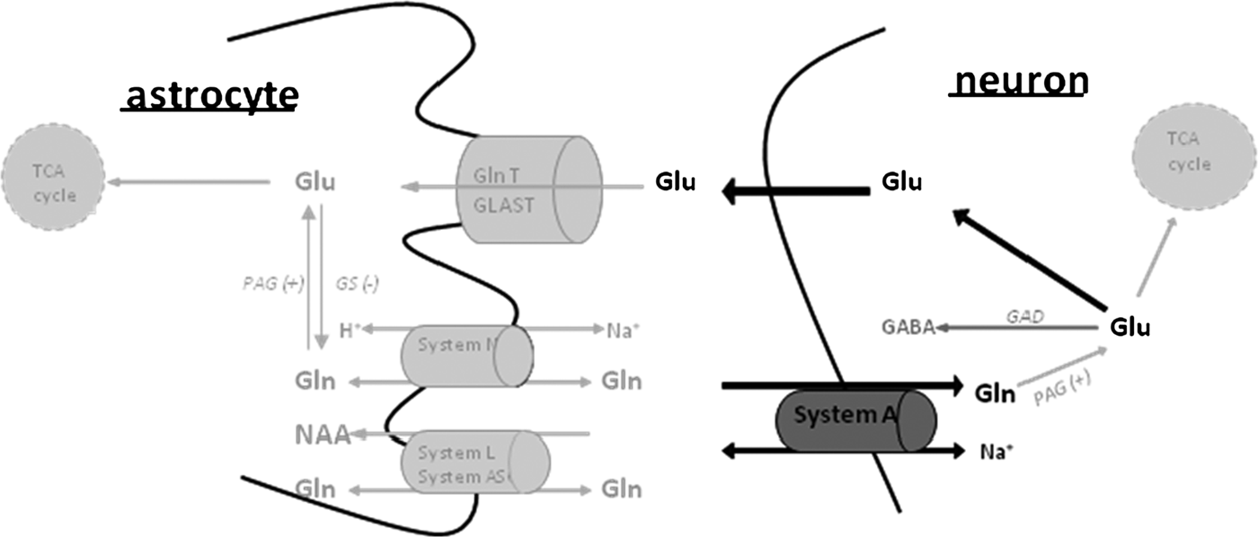
The specific and important features of Gln metabolism in the CNS which enable the controlled channeling of its amino acid products toward their metabolic and neurotransmitter pools are (1) Gln proceeds in the Gln/Glu(GABA) cycle (GGC) and (2) the reactions within the cycle are compartmentalized between astrocytes and neurons. The functional sequence of events constituting the GGC takes place as follows: Gln is synthesized from Glu and ammonia in astrocytes in a reaction catalyzed by Gln synthetase (GS), an enzyme located primarily, if not exclusively, in astrocytes. Newly synthesized Gln is then released from astrocytes, transferred to neurons and hydrolyzed by phosphate-activated glutaminase (PAG), an enzyme found abundantly in neurons, thereby giving rise to Glu, a portion of which may be decarboxylated to GABA or transaminated to Asp (for a review see Bak et al., Reference Bak, Schousboe and Waagepetersen2006). Multiple amino acid transporting proteins in the CNS demonstrate affinity for Gln. Proteins belonging to systems A and N show cell-specific localization, rendering them well-suited for modulating Gln transfer from astrocytes to neurons and, in this way, the GGC (Table 1). The unidirectional system A transporters, SNAT1 and SNAT2, are located in neurons and catalyze neuronal Gln uptake, while the N system transporters, SNAT3 and SNAT5, are located in the astrocytic cell membranes (Chaudhry et al., Reference Chaudhry, Reimer and Edwards2002). N system transporters are bidirectional nature, and one of their major activities in the brain is to mediate the efflux of newly synthesized Gln from astrocytes (Chaudhry et al., Reference Chaudhry, Krizaj, Larsson, Reimer, Wreden and Storm-Mathisen2001, Reference Chaudhry, Reimer and Edwards2002). Most recently, a new system N transporter, SNAT7, has been described. Located specifically in neurons, the exact function of SNAT7 in the GGC remains to be established (Hagglund et al., Reference Hagglund, Sreedharan, Nilsson, Shaik, Almkvist and Backlin2011). The activities of systems A and N transporters are regulated at the transcriptional and translational levels (Palacin et al., Reference Palacin, Estevez, Bertran and Zorzano1998; Boehmer et al., Reference Boehmer, Okur, Setiawan, Broër and Lang2003); the activities of system N transporters are additionally regulated by membrane trafficking (Balkrishna et al., Reference Balkrishna, Broër, Kingsland and Broër2010). Trafficking of SNAT3/SN1 in astrocytic cell membranes is specifically mediated by its phosphorylation involving PKCα and PKCγ (Nissen-Meyer et al., Reference Nissen-Meyer, Popescu, Hamdani el and Chaudhry2011), a process resembling the trafficking of astrocytic Glu transporter 1 (GLT-1) (Kalandadze et al., Reference Kalandadze, Wu and Robinson2002; Gonzalez et al., Reference Gonzalez, Susarla and Robinson2005) and glutamate/aspartate transporter (GLAST) (Guillet et al., Reference Guillet, Velly, Canolle, Masmejean, Nieoullon and Pisano2005). Similar mechanisms underlying the regulation of the transporters mediating astrocytic Glu uptake and Gln efflux may serve to facilitate the coordination of their activities and functional interdependence, as reflected in the control of Gln efflux from astrocytes by neuron-derived Glu (Albrecht, Reference Albrecht1989; Broër et al., Reference Broër, Deitmer and Broër2004).
Distribution and mechanism of action of Gln (GLN)-transporting systems A and N involved in the GLN/GLU cycle in the CNS* (Zielińska et al., Reference Zielińska, Ruszkiewicz, Hilgier, Fresko and Albrecht2011)

* For reviews of Chaudhry et al. (Reference Chaudhry, Reimer and Edwards2002) and Conti and Melone (Reference Conti and Melone2006).
**The term ‘SNAT’ holds for ‘small neutral amino acid transporter’, and the isoforms are designated here as proposed by Mackenzie and Erickson (Reference Mackenzie and Erickson2004).
As will be discussed in more detail in the following sections, the A system-mediated neuronal Gln uptake, which is specifically inhibited by methyl-isoaminobutyric acid (MeAiB) appears critical for maintaining an adequate supply of Gln to glutamatergic terminals as well as for generating neurotransmitter pools of Glu and GABA.
The sequence of events within the GGC has been derived from in vivo and in vitro studies by following the fate of Gln labeled with 14C or with stable isotopes (15N/13C) and is sketched in Fig. 1. Neurons lack the enzymes necessary to perform de novo synthesis of the neurotransmitters, Glu and (GABA), from glucose. One of the pathways proposed for neuronal neurotransmitter repletion is the GGC cycle (Van den Berg, Reference Van den Berg, Balazs and Cremer1972; Martinez-Hernandez et al., Reference Martinez-Hernandez, Bell and Norenberg1977; Kvamme and Lenda, Reference Kvamme and Lenda1982; Szerb and O'Regan, Reference Szerb and O'Regan1985; Schousboe et al., Reference Schousboe, Westergaard, Sonnewald, Petersen, Huang and Peng1993; Sonnewald et al., Reference Sonnewald, Westergaard, Schousboe, Svendsen, Unsgard and Petersen1993; Martin, Reference Martin, Kettenmann and Ransom1995; Kanamori and Ross, Reference Kanamori and Ross1995). The ability to measure the GGC has important implications in the study of brain function in both normal and pathological conditions.
Schematic representation showing the interdependence of the Gln/Glu (GABA) transport and metabolism cycle between astrocytes and neurons. Glu released from presynaptic terminals is transported to the astrocytes via Glu carriers (GLAST and GlnT-1), where it is converted to Gln by the GS. In turn, Gln transporters mediate its release into the extracellular space and its transfer into the neurons, where Glu is regenerated via phosphate-dependent glutaminase (PAG). Glu may be subsequently converted into GABA via decarboxylation by GAD. In both astrocytes and neurons a portion of Glu is utilized for the synthesis of alpha-ketoglutaric acid (α KG), a substrate for the TCA cycle, by oxidative deamination mediated by Glu dehydrogenase.

Studies using L-[U-14C]Glu have demonstrated that cultured primary astrocytes produce Gln from Glu and export this amino acid to the extracellular space (Waniewski and Martin, Reference Waniewski and Martin1986). In vivo studies have revealed that brain labeling with [U-14C]Gln or [3H]Gln results in the labeling of cellular and extracellular Glu and GABA (Ward et al., Reference Ward, Thanki and Bradford1983). Additional in vivo studies have shown that the application of 6-diazo-5-oxo-L-norleucine, a potent and selective phosphate-dependent glutaminase (PAG) inhibitor, to the rat cerebral cortex causes the rapid (<30 min) loss of Glu immunoreactivity in pyramidal cell bodies and dendrites (Conti and Minelli, Reference Conti and Minelli1994). Moreover, there is evidence that Gln serves as a carbon source for the repletion of Glu and GABA (Shank and Campbell, Reference Shank and Campbell1984).
Nuclear magnetic resonance (NMR) spectroscopy permits the detection of 13C incorporation from 13C- and 15N-labeled precursors into the various carbon positions of small metabolites, such as intermediates of the tricarboxylic acid cycle (TCA cycle) and amino acids (e.g. reviews by Westergaard et al., Reference Westergaard, Sonnewald and Schousboe1995; Bachelard, Reference Bachelard1998; Cruz and Cerdan, Reference Cruz and Cerdán1999; Rothman et al., Reference Rothman, Sibson, Hyder, Shen, Behar and Shulman1999). This method represents an important tool for studying brain energy metabolism and substrate trafficking between neurons and astrocytes both in vivo and in vitro. Therefore, 13C and 15N NMR are excellent tools for studying Glu/Gln (GABA) cycling between neurons and astrocytes. The NMR method has confirmed the existence of the GGC in vivo (Sibson et al., Reference Sibson, Dhankhar, Mason, Behar, Rothman and Shulman1997; Lebon et al., Reference Lebon, Petersen, Cline, Shen, Mason and Dufour2002; Petroff et al., Reference Petroff, Errante, Rothman, Kim and Spencer2002). Studies in rodents as well as in humans have indicated that Gln/Glu (GABA) cycling flux is necessary to replenish Glu and GABA and accounts for >80% of the net Gln synthesis. Moreover, studies using in vivo microdialysis and NMR methods have shown that neuronal Glu is the precursor for 80–90% of astrocytic Gln synthesis through Gln/Glu (GABA) cycling (Kanamori et al., Reference Kanamori, Ross and Kondrat2002). Gln labeling from [1-13C] glucose has revealed that the GGC is highly active in the rat cerebral cortex and in the human occipital/parietal cortex (Gruetter et al., Reference Gruetter, Novotny, Boulware, Mason, Rothman and Shulman1994, Reference Gruetter, Seaquist, Kim and Ugurbil1998; Shen et al., Reference Shen, Petersen, Behar, Brown, Nixon and Mason1999). Simultaneous measurements from [1-13C] glucose into the Glu and Gln pools have demonstrated that the rate of glucose oxidation increases in a 1:1 molar stoichiometric relationship with the Glu/Gln cycle in the rat cerebral cortex, suggesting a common mechanism for these fluxes (Pellerin and Magistretti, Reference Pellerin and Magistretti1994; Sibson et al., Reference Sibson, Shen, Mason, Rothman, Behar and Shulman1998). Similarly, Gln/Glu (GABA) cycling was found to demonstrate major metabolic flux in the human occipital/parietal cortex with a rate approximately 80% of glucose oxidation (Shen et al., Reference Shen, Petersen, Behar, Brown, Nixon and Mason1999). These observations indicate that Gln/Glu (GABA) cycling is not only responsible for amino acid transmission, but that such cycling also ensures normal brain energetic function.
Gln AS A PRECURSOR OF THE NEUROTRANSMITTER POOLS OF AMINO ACIDS Glu, GABA AND Asp
Stimulus-driven release of Gln-derived Glu and GABA from different CNS preparations
In the late 1970s and early 1980s, several laboratories reported that the depolarization of slices of different brain regions preloaded with radiolabeled Gln evokes the release of radiolabeled Glu (Reubi et al., Reference Reubi, Van Der Berg and Cuenod1978; Hamberger et al., Reference Hamberger, Chiang, Sandoval and Cotman1979a, Reference Hamberger, Chiang, Nylen, Scheff and Cotmanb) and/or GABA (Reubi et al., Reference Reubi, Van Der Berg and Cuenod1978; Tapia and Gonzalez, Reference Tapia and Gonzalez1978). Glu release occurred readily in the presence of calcium, a condition mimicking the brain ‘at work’ (Hamberger et al., Reference Hamberger, Chiang, Nylen, Scheff and Cotman1979b), and more of the released Glu originated from Gln than from glucose (Hamberger et al., Reference Hamberger, Chiang, Sandoval and Cotman1979a). Depolarization per se acted as a factor stimulating Gln uptake and its conversion to Glu (Hamberger et al., Reference Hamberger, Chiang, Sandoval and Cotman1979a) or GABA (Szerb, Reference Szerb1984). The inhibition of PAG, which catalyzes the generation of Glu from Gln in neurons, reduced the synaptosomal pools of Glu and Asp and decreased the depolarization-dependent release of Glu (Bradford et al., Reference Bradford, Ward and Foley1989). The in vitro findings have been largely confirmed in in vivo experiments with radiolabeled Gln being infused via implanted cannulae (Gauchy et al., Reference Gauchy, Kemel, Glowinski and Besson1980; Thanki et al., Reference Thanki, Sugden, Thomas and Bradford1983). Gallo et al. (Reference Gallo, Ciotti, Coletti, Aloisi and Levi1982) who studied the development of the profile of the CGC in culture demonstrated that the ability of these cells to release endogenous and Gln-derived Glu develop in parallel during the course of cell maturation.
Convincing evidence that Gln plays a substantial role in feeding the releasable pools of Glu or GABA was derived from NMR spectroscopy studies using 13C- or 15N-labeled Gln or its precursors/products in vivo (Patel et al., Reference Patel, Rothman, Cline and Behar2001; Kanamori and Ross, Reference Kanamori and Ross2004; Wang et al., Reference Wang, Maher and Wurtman2007), or in vitro in cultured CGC (Waagepetersen et al., Reference Waagepetersen, Qu, Sonnewald, Shimamoto and Schousboe2005), GABA-ergic neurons (Waagepetersen et al., Reference Waagepetersen, Sonnewald, Gegelashvili, Larsson and Schousboe2001), or brain tissue slices and prisms (Rae et al., Reference Rae, Hare, Bubb, McEwan, Broër and McQuillan2003).
Immunocytochemical evidence for Gln transfer from astrocytes to neurons and its conversion to synaptic Glu and GABA
The above described metabolic studies convincingly demonstrate that Gln is a precursor of the Glu or GABA pools which reside in neurons and are released by depolarization. The question of whether these releasable pools are derived from astrocytic Gln and eventually find their way to the synaptic vesicles to undergo exocytosis, has been addressed in studies using the immunogold technique to label amino acids in the different CNS compartments. In cultured hippocampal slices, a major proportion of tissue Gln is located in glial cells, while Glu predominates in the nerve terminals (Laake et al., Reference Laake, Slyngstad, Haug and Ottersen1995). The inhibition of Gln synthesis with the GS inhibitor, methionine sulfoximine (MSO), reduced the Glu immunoreactivity in neurons and increased both Glu and Gln in glial cells (Laake et al., Reference Laake, Slyngstad, Haug and Ottersen1995). The addition of Gln to the MSO-treated slices restored Glu accumulation in the nerve terminals (Laake et al., Reference Laake, Slyngstad, Haug and Ottersen1995). In an identical hippocampal slice preparation, Gln prevented the depolarization-induced loss not only of Glu but also of Asp (Gundersen et al., Reference Gundersen, Chaudhry, Bjaalie, Fonnum, Ottersen and Storm-Mathisen1998); the latter finding supported the existence of a Gln-derived neurotransmitter pool of Asp.
Several studies have emphasized the role of the PAG-mediated degradation of Gln in the synthesis of synaptic Glu or GABA. Gln has been shown to prevent the depolarization-induced decrease of Glu immunoreactivity in synaptic endings in hippocampal slices (Storm-Mathisen et al., Reference Storm-Mathisen, Ottersen, Fu-Long, Gundersen, Laake and Nordbo1986), the redistribution of Glu from parallel-fiber terminals to glial cells, and the movement of GABA from Golgi fiber terminals to glial cells in depolarized cerebellar slices (Holten and Gundersen, Reference Holten and Gundersen2008). Additional studies have demonstrated that these outcomes are abrogated by the inhibition of PAG with diazonorleucine (DON) (Holten and Gundersen, Reference Holten and Gundersen2008). Furthermore, the topical application of DON to the rat sensorimotor cortex in vivo has been shown to abolish Glu immunoreactivity in neurons in this brain structure (Conti and Minelli, Reference Conti and Minelli1994). Consistent with the conclusions derived from immunocytochemical observations, severe impairment of glutamatergic transmission has also been recorded in the respiratory network of glutaminase-null mice (Masson et al., Reference Masson, Darmon, Conjard, Chuhma, Ropert and Thoby-Brisson2006).
Contribution of astroglia-derived Gln to the electrophysiological manifestations of glutamatergic and GABA-ergic transmission
Bacci (Bacci et al., Reference Bacci, Sancini, Verderio, Armano, Pravettoni and Fesce2002) have found that spontaneous epileptiform discharges reflecting increased glutamatergic transmission in neurons of primary hippocampal cultures and hippocampal slices are suppressed upon the addition of MeAiB or MSO to the bathing medium, and Hülsmann (Hülsmann et al., Reference Hülsmann, Oku, Zhang and Richter2000) reported that either of these two drugs inhibits the activity of the respiratory network in the mouse spinal cord. In both instances, the inhibition of Glu-ergic transmission was alleviated by the addition of exogenous Gln. MSO and MeAiB also served as tools to demonstrate the role of Gln in GABA-ergic transmission. In two independent studies, either compound reduced the inhibitory postsynaptic currents and miniature inhibitory postsynaptic currents in rat hippocampal pyramidal neurons in the CA1 region, indicating that Gln is required for filling the synaptic vesicles in GABA-ergic synapses with GABA (Liang et al., Reference Liang, Carlson and Coulter2006; Fricke et al., Reference Fricke, Jones-Davis and Mathews2007).
Direct demonstration of the roles of system A transporting proteins SNAT1 and SNAT2 in routing Gln toward replenishment of synaptic Glu and GABA
The ability of the system A inhibitor, MeAIB, to attenuate various aspects of glutamatergic and GABA-ergic transmission (see above) indicates that system A may play a rate-limiting role in supplying the glutamatergic synapse with Gln. In support of this notion, both of the two system A proteins present in brain, SNAT2 (SAT2) and SNAT1 (SAT 1) (Table 1), have been shown to be distinctly involved in Gln delivery to glutamatergic or GABA-ergic synapses. Immunocytochemically, SNAT2 (SAT2) expression in many brain regions is primarily confined to the somatodendritic compartments of glutamatergic neurons (Jenstad et al., Reference Jenstad, Quazi, Zilberter, Haglerod, Berghuis and Saddique2009). This specific localization of SNAT2 appears to be functional, as indicated by studies which demonstrated that (1) SNAT2 (SAT2)-containing dendrites efficiently accumulate Gln; (2) in activated dendritic segments, electrical stimulation in vivo and depolarization in vitro trigger Gln conversion to Glu; and (3) MeAIB reduces the neuronal uptake of Gln with the concomitant reduction of intracellular Glu concentrations (Jenstad et al., Reference Jenstad, Quazi, Zilberter, Haglerod, Berghuis and Saddique2009). In turn, the SNAT1 (SAT1) transporter has been shown to be highly enriched in GABA-ergic neurons in several brain regions that also harbor the two other GABA-ergic markers: the GABA-synthesizing enzyme, glutamic acid decarboxylase (GAD) 67 isoform and and/or the GABA-ergic neuron-specific Ca2+-binding protein parvalbumin (Solbu et al., Reference Solbu, Bjorkmo, Berghuis, Harkany and Chaudhry2010). However, it is important to note that a few studies did not support the ubiquitous role of SNAT1 and SNAT2 in the synaptic Gln uptake (Rae et al., Reference Rae, Hare, Bubb, McEwan, Broër and McQuillan2003; Conti and Melone, Reference Conti and Melone2006; Tani et al., Reference Tani, Dulla, Huguenard and Reimer2010). These discrepant findings may reflect the following factors or combination thereof: (i) the contribution of some as yet unidentified transport proteins which may not conform to system A characteristics; (ii) regional differences in the localization of Gln-transporting proteins; (iii) their different subcellular localization, a possibility brought up by Jenstad et al. (Reference Jenstad, Quazi, Zilberter, Haglerod, Berghuis and Saddique2009) with regard to SNAT1 (see above). The regional distribution of system N proteins is not overlapping either. SNAT3 tends to abound in the plasma membrane of astrocytes in the vicinity of GABA-ergic nerve terminals (Boulland et al., Reference Boulland, Osen, Levy, Danbolt, Edwards, Storm-Mathisen and Chaudhry2002), whereas relatively more SNAT5 is located in astrocytes adjacent to glutamatergic nerve terminals and in astrocytic end-feet (Cubelos et al., Reference Cubelos, Gonzalez-Gonzalez, Gimenez and Zafra2005). Irrespective of the nature of the Gln-transporting proteins involved (one of which could be the newly discovered synaptic N system transporter, SNAT7) (Hagglund et al., Reference Hagglund, Sreedharan, Nilsson, Shaik, Almkvist and Backlin2011), functional evidence favors the role of neuronal Gln uptake as a factor in controlling the supply of the synaptic pools of Glu and GABA.
Does system N-mediated Gln efflux from astrocytes stimulate glutamatergic or GABA-ergic transmission?
The system N transporters, SN1 (SNAT3) (Chaudhry et al., Reference Chaudhry, Krizaj, Larsson, Reimer, Wreden and Storm-Mathisen2001) and SN2 (SNAT5) (Cubelos et al., Reference Cubelos, Gonzalez-Gonzalez, Gimenez and Zafra2005), promote Gln efflux from astrocytes and are primarily involved in astrocytic processes covering the glutamatergic terminals. The features of the two transporters make them well-suited to participate in glutamatergic or GABA-ergic transmission. However, the degree to which the N-system-mediated Gln efflux from astrocytes contributes to the synthesis of functional pools of Glu is not entirely clear. In contrast to the almost exclusive involvement of system A in Gln uptake to neurons, Gln efflux from astrocytes appears to engage an array of different sodium dependent and independent transporters (Deitmer et al., Reference Deitmer, Broër and Broër2003). Distinguishing between the relative roles of the different systems is hampered by the unavailability of a specific system N inhibitor. His, the amino acid most commonly used to block system N (Table 1), was found to limit the Gln-dependent synthesis of neurotransmitter Glu and GABA in brain slices or prisms and in cultured CNS cells (Rae et al., Reference Rae, Hare, Bubb, McEwan, Broër and McQuillan2003), and it attenuated the electrical activity of pharmacologically disinhibited neocortical slices (Tani et al., Reference Tani, Dulla, Huguenard and Reimer2010). However, His shares most cell membrane transport routes with Gln (Palacin et al., Reference Palacin, Estevez, Bertran and Zorzano1998; Broër et al., Reference Broër, Friedrich, Wagner, Fillon, Ganapathy and Lang2001). Accordingly, in this study (Tani et al., Reference Tani, Dulla, Huguenard and Reimer2010), an effect similar to His was attained with a nonmetabolized neutral amino acid analog, α-amino-isobutyric acid (AIB), a substrate of a wide spectrum of amino acid transporters. Thus, it is impossible to judge whether and to what extent AIB actually inhibited astrocytic release or the neuronal uptake of Gln, respectively.
Direct versus TCA cycle-mediated contributions of Gln to the neurotransmitter pools of Glu and GABA
While the studies described above demonstrate that Gln does indeed enrich the synaptic pool of Glu, the extent to which glutamatergic or GABA-ergic transmission actually depends on the activity of the GGC has not yet been determined. Analogous to the cells outside the brain, neurons synthesize Glu and GABA through the TCA cycle (Hertz et al., Reference Hertz, Dringen, Schousboe and Robinson1999), and evidence from CNS cell culture studies suggests that some of the TCA-cycle-derived Glu or GABA feeds the neurotransmitter pools, whereby a portion of the TCA intermediates is derived from Gln. In cultured neurons, the amount of Glu released from CGC or the amount of GABA released from cerebral cortical neurons that stem directly from Gln hydrolysis is roughly equal to that derived from Gln metabolites produced in the tricarboxylic cycle (Waagepetersen et al., Reference Waagepetersen, Sonnewald, Gegelashvili, Larsson and Schousboe2001, Reference Waagepetersen, Qu, Sonnewald, Shimamoto and Schousboe2005). A recent electrophysiological study revealed that synaptic transmission in hippocampal slices or cultured hippocampal neurons can remain active for a long period of time notwithstanding the interruption of the Gln supply that followed the inhibition of its astrocytic synthesis or uptake by neurons (Kam and Nicoll, Reference Kam and Nicoll2007). Interestingly, in contrast to the short-term effects of Gln described in the previous section, this particular study demonstrated that transmission was augmented upon the prolonged exposure of the neurons to Gln (Kam and Nicoll, Reference Kam and Nicoll2007). In the authors' interpretation, these results reflect a relatively efficient production of the neurotransmitter pool of Glu in the TCA cycle and/or its long-term storage in the vesicles. In this scenario, Gln serves as a source of TCA intermediates for synaptic Glu synthesis. The relative roles of Gln hydrolysis versus its cycling through the TCA cycle may increase with increased brain activity, considering that the pyruvate carboxylase-mediated anaplerotic replenishment of the TCA cycle precursors of Glu is considerably higher in alert as opposed to anesthetized rats (Oz et al., Reference Oz, Berkich, Henry, Xu, LaNoue and Hutson2004). Also, it is reasonable to expect the occurrence of regional variations in the Gln dependence of neurotransmitter Glu synthesis. Such variations may be attributed to the heterogeneity of mitochondria with regard to their PAG content (Laake et al., Reference Laake, Takumi, Eidet, Torgner, Roberg and Kvamme1999), but also to the above discussed differences in the regional and/or subcellular localization of Gln transporters.
Gln distribution in the CNS favors its neurotransmitter precursor role
Gln distribution between the different brain regions is relatively even, and its content in the brain is very similar to, or at least of the same order of magnitude, as the contents of the neuroactive amino acids, Glu, Asp and taurine (Tossman et al., Reference Tossman, Delin, Eriksson and Ungerstedt1987). Among the different rat brain regions, Gln content ranges from ~5 nmol mg−1 in the hippocampus to ~7 nmol mg−1 in the cerebellum. Among the rabbit brain regions, Gln content ranges from ~4 nmol mg−1 in the frontal cortex to ~ 8 nmol mg−1 in the hippocampus, whereas Glu and taurine contents in the different regions are in the ranges of ~4 to ~20, and ~8 to ~40 nmol mg−1, respectively (Nitsch et al., Reference Nitsch, Schmude and Haug1983; Eloqayli et al., Reference Eloqayli, Dahl, Gotestam, Unsgard and Sonnewald2004). However, Gln content in the extracellular fluid (microdialysates) (ECF) or in the cerebrospinal fluid (CSF) (~0.5–1 mM in either compartment) exceeds, by at least one order of magnitude, the extracellular contents of other amino acids in these compartments (Hamberger and Nystrom, Reference Hamberger and Nystrom1984; Kanamori and Ross, Reference Kanamori and Ross2004). An additional unique feature of Gln is that its arterial blood concentration is very close to its concentration in the ECF or CSF (Xu et al., Reference Xu, McAdoo, Hughes, Robak and de Castro1998). These small differences between Gln concentrations in the different compartments of the CNS implicate flat Gln gradients across the cell membranes, facilitating the operation of transporters that deliver Gln synthesized in astrocytes to the nerve endings and its ‘spillover’ to the periphery (for discussion see Broër and Brookes, Reference Bröer and Brookes2001).
Gln AS A NEUROTRANSMITTER AND NEUROMODULATOR
Direct interaction of Gln with Glu receptors (GluRs): a critical evaluation
By classic definition, neurotransmitters are (1) stored in the synaptic vesicles; (2) released by exocytosis; and (3) bind to receptors on the postsynaptic cellular membranes. While Gln certainly does not meet criteria (1) and (2), its ability to bind to the NMDA receptor has, for a long time, been considered a likely possibility; this question, however, remains unresolved. While an early ligand binding study suggested that Gln is an agonist of the glycine site of the N-methyl-d-aspartate (NMDA) receptor (Stumpo et al., Reference Stumpo, Pullan and Salama1989), further studies in this direction were discouraged by reports that the effects ascribed to Gln may arise from the contamination of Gln preparation by Glu (Yamada and Rothman, Reference Yamada and Rothman1989). Recently, however, this concept has received renewed interest following a study that showed that Gln, added at a milimolar concentration to primary cultured rat hippocampal neurons, is able to evoke dose-dependent transmembrane inward currents which are not shared by other amino acids, are sensitive to inhibitors of ionotropic GluRs and display some features typical for a current evoked by NMDA application (Kolbaev and Draguhn, Reference Kolbaev and Draguhn2008). Notably, the Gln-evoked responses differed from the responses evoked by NMDA in their antagonist sensitivity and kinetics in a sufficient degree to consider them to be Gln specific (Kolbaev and Draguhn, Reference Kolbaev and Draguhn2008). Clearly, this single report deserves further confirmation.
Currents activated by a variety of ion-coupled amino acid transporters depolarize or hyperpolarize cell membranes, as is the case for some of the Gln transporting proteins which mediate Gln-coupled charge transfer or uncoupled conductance. The system A transporter, SNAT1 (ATA1, GlnT), mediates a sodium-dependent, electrogenic transport, coupled to inward charge movement (Mackenzie et al., Reference Mackenzie, Schafer, Erickson, Hediger, Weihe and Varoqui2003; Kanamori and Ross, Reference Kanamori and Ross2004). The other system A transporter, SNAT2, inserted in HEK293 T cells catalyzes a leak anion conductance which is inhibited by the binding of the transported substrates to the transporter (Zhang and Grewer, Reference Zhang and Grewer2007). The anion conductance could be a possible mechanism by which glial cells could influence neuronal activity through the SNAT2 transporter. High extracellular amino acid concentrations inhibit the SNAT2 anion conductance, leading to the increased excitability of neurons (Zhang and Grewer, Reference Zhang and Grewer2007). The N system transporter, SNAT3 (SN1), although not directly electrogenic, elicits Gln-dependent uncoupled proton conductance (Chaudhry et al., Reference Chaudhry, Krizaj, Larsson, Reimer, Wreden and Storm-Mathisen2001; Schneider et al., Reference Schneider, Broër, Broër and Deitmer2007), and, in this way, may modulate membrane potential. The ASC transporter family member, ASCT2, mediates electroneutral obligatory antiport but requires Na+ for its proper function. The substrate activation of the ASCT2 transporter results in amino acid transport and a Na+/substrate-dependent anion conductance (Broër et al., Reference Broër, Wagner, Lang and Broër2000). The above data indicate that Gln carriers, similar to some other amino acid transporter classes (Ryan and Vandenberg, Reference Ryan and Vandenberg2002), can function in ways similar to ion channels, thereby contributing to neuronal signaling.
Modulation of the nitric oxide (NO)/cyclic GMP pathway by Gln
The activation of NMDA receptors triggers intracellular signaling via the synthesis of NO, which generates cyclic guanosine monophosphate (cGMP), a marker of NO synthesis and a downstream carrier of its signal. Data from a recent study using microdialysis showed that pharmacologically elevated extracellular Gln in the brain causes a decrease of NO and cGMP synthesis (Hilgier et al., Reference Hilgier, Fresko, Klemenska, Beresewicz, Oja and Saransaari2009). This phenomenon was achieved by limiting the transport of the NO precursor, arginine, by the y+LAT2 transport system for which Gln and arginine compete (Broër et al., Reference Broër, Wagner, Lang and Broër2000). y+LAT2 is a member of y+L system and is distinct from other systems transporting Gln in that it recognizes cationic amino acids in the absence of Na+ and neutral amino acids in the presence of Na+ (Dye et al., Reference Dye, Vause, Johnston, Clark, Firth and D'Souza2004). y+LAT2 is expressed in the rat brain and in cultured astrocytes and cortical neurons (Broër et al., Reference Broër, Wagner, Lang and Broër2000). However, there are no data allocating this transporter in the brain in situ to a given cell type (astrocytes/neurons); hence, the physiological importance of the modulation of the NO/cGMP pathway by Gln remains to be investigated. In acute hyperammonemia, ammonia induces the NMDA receptor-mediated activation of the NO/cGMP pathway, a major source of nitrogen radicals and peroxynitrates in this condition (Kosenko et al., Reference Kosenko, Kaminsky, Lopata, Muravyov, Kaminsky and Hermenegildo1998; Hilgier et al., Reference Hilgier, Wegrzynowicz, Maczewski, Beresewicz, Oja and Saransaari2008). In astrocytes, where this pathway is also detected (Schliess et al., Reference Schliess, Gorg, Fischer, Desjardins, Bidmon and Herrmann2002), Gln, by reducing NO synthesis, might counteract oxidative/nitrosative stress and, consequently, astrocytic swelling and dysfunction.
IMPAIRMENT OF THE Gln/Glu CYCLE: A POTENTIAL CAUSE OF ALTERED NEUROTRANSMISSION IN DIFFERENT BRAIN PATHOLOGIES
Epilepsy
Epilepsy is a neurological disorder characterized by sudden and temporary bursts (synchronized discharges) of a group of neurons (partial or focal epilepsy) or of the ‘whole’ cortex (general epilepsy). Its widely accepted definition is ‘a condition characterized by recurrent (two or more) epileptic seizures, unprovoked by any immediate identified cause’ (Hauser and Kurland, Reference Hauser and Kurland1975). There is a wealth of evidence indicating that epileptic discharges occur due to the imbalance between glutamatergic and the GABA-ergic transmission, in favor of the former. Epileptogenic activity in the hippocampi of patients with medial temporal lobe epilepsy (MTLE) is associated with the excessive extracellular accumulation of Glu (During and Spencer, Reference During and Spencer1993), accompanied by a decreased extracellular Gln/Glu ratio (Cavus et al., Reference Cavus, Kasoff, Cassaday, Jacob, Gueorguieva and Sherwin2005). These changes are evident in subjects in whom epileptogenic activity could be simultaneously recorded. The role of Gln as a trigger of epileptiform discharges was first described in a study by Bacci (Bacci et al., Reference Bacci, Sancini, Verderio, Armano, Pravettoni and Fesce2002), which was performed in hippocampal slices and cultures; as mentioned earlier, the ability of MeAiB and MSO to inhibit the Gln-induced potentials shown in this study was the first demonstration of a role for Gln in glutamatergic transmission. In later studies, slices obtained from the ‘undercut’ cortex, which is the rat model of traumatic epilepsy, were shown to be more susceptible to the induction of epileptiform activity by Gln than were slices from control rats. Furthermore, this increased susceptibility was found to be related to increased system A-mediated Gln transport (Tani et al., Reference Tani, Bandrowski, Parada, Wynn, Huguenard and Prince2007). In the pharmacologically disinhibited neocortical slice preparation, where the intensity of the stimulus is directly and proportionally translated to the electrically evoked activity, Gln was shown to stimulate the activity and Glu release from the slices simultaneously (Tani et al., Reference Tani, Dulla, Huguenard and Reimer2010). Entorhinal cortex hippocampal slices from pilocarpine-treated chronic epileptic rats responded to the presence of physiologically relevant Gln concentrations in the medium (0.5–2 mM) with ionotropic Glu receptor AMPA-mediated recurrent epileptiform discharges; no such an activity could be elicited in control slices (Sandow, Reference Sandow2009).
The increased glutamatergic activity in MTLE was found to be well-correlated with Gln/Glu cycle dysfunction. In the hippocampus of MTLE patients, the decreased rate of Glu/Gln cycling, as revealed by 13C NMR spectroscopy (Petroff et al., Reference Petroff, Errante, Rothman, Kim and Spencer2002), is accompanied by a correlated decrease of GS expression and activity (Eid et al., Reference Eid, Thomas, Spencer, Runden-Pran, Lai and Malthankar2004) as well as by increased PAG immunoreactivity (Eid et al., Reference Eid, Hammer, Runden-Pran, Roberg, Thomas and Osen2007). GS expression was also found to be lower in the brains of glioma patients presenting symptoms of epilepsy than it was in patients in whom the symptoms had not yet developed (Rosati et al., Reference Rosati, Marconi, Pollo, Tomassini, Lovato and Maderna2009). In rats with pentylentetrazole-induced epilepsy, decreased GS activity is not due to its decreased expression, but is associated with the increased nitration of tyrosine residues in the enzyme (Bidmon et al., Reference Bidmon, Gorg, Palomero-Gallagher, Schleicher, Haussinger and Speckmann2008). However, the involvement of GS may not be a universal occurrence, as no changes in the activities of this enzyme were detected in the brain tissue of frontal cortical epilepsy patients (Steffens et al., Reference Steffens, Huppertz, Zentner, Chauzit and Feuerstein2005).
In the scenario encompassing all the epilepsy-associated changes in Gln metabolism and transport, increased system A-mediated Gln transport to neurons in association with increased PAG activity would contribute to excessive extracellular Glu accumulation in the epilepsy-vulnerable region, thereby resulting in overexcitation. In cases in which GS activity is decreased, the neutralization of Glu in astrocytes would be further impaired. However, even under conditions of decreased Gln synthesis, the normally high extracellular Gln reserves would be sufficient to maintain its supply to neurons (Alvestad et al., Reference Alvestad, Hammer, Eyjolfsson, Qu, Ottersen and Sonnewald2008; Kanamori and Ross, Reference Kanamori and Ross2011) (Fig. 2).
A schematic view of epilepsy-associated changes in the Gln/Glu shuttle which lead to overactivation of glutamatergic transmission. Gln transport to neurons is increased in association with the upregulation of system A activity. Excessive Gln accumulation in neurons and its increased degradation by phosphate-dependent glutaminase (PAG), collectively lead to the excessive release of Glu and its extracellular accumulation. The tone may be further increased by Glu back-flux from astrocytes resulting from its impaired conversion to Gln in astrocytes, which is due to decreased GS activity, as well as, possibly, by increased Gln degradation by upregulated PAG.

As mentioned earlier, Gln has been suspected to play an important role in the inhibitory GABA-ergic transmission. This concept found strong support in a recent study in which reactive astrocytosis was selectively induced in mouse CA1 region of hippocampus to mimic epilepsy-associated sclerotic changes (Ortinski et al., Reference Ortinski, Dong, Mungenast, Yue, Takano and Watson2010). In this experimental setting, astrocytosis was associated with failure of the GGC cycle (inhibition of Gln synthesis), and promoted erosion of inhibitory GABA-ergic currents coupled with hyperexcitation in hippocampal currents, which could be reversed by exogenous Gln administration. Impaired feeding of the neurotransmitter pool of GABA by Gln is likely to be involved in a spectrum of diseases of CNS where reactive astrocytes are involved in the pathogenetic chain. Further experiments in this direction are clearly warranted.
Ammonia neurotoxicity
A high concentration of ammonia in the blood and subsequently in the brain is a causative factor of the group of neurological disorders collectively defined as hyperammonemic encephalopathies (Prakash and Mullen, Reference Prakash and Mullen2010). Among these, the most common is hepatic encephalopathy (HE), a condition which is associated with acute or chronic liver dysfunction (Albrecht and Jones, Reference Albrecht and Jones1999; Prakash and Mullen, Reference Prakash and Mullen2010). Acute HE develops as a consequence of viral or toxic liver damage (fulminant hepatic failure: FHF, also named acute liver failure: ALF) and rapidly progresses to coma and death. Brain edema, increased intracranial pressure and herniation are the immediate causes of death for patients with ALF. These disease conditions occur primarily as the results of astrocytic swelling (Blei, Reference Blei2007). Chronic HE is a complex neuropsychiatric syndrome developed due to liver cirrhosis, and it presents a broad range of abnormalities of varying severity. HE patients demonstrate psychomotor, motor, intellectual, cognitive, emotional and behavioral alterations (Prakash and Mullen, Reference Prakash and Mullen2010). The neurological alterations in HE manifest as consequences of an imbalance between the excitatory glutamatergic and inhibitory GABA-ergic neurotransmission (Felipo and Butterworth, Reference Felipo and Butterworth2002). Alterations in glutamatergic neurotransmission impair intellectual and motor function, as well as coordination and circadian rhythms, all of which are frequently observed in HE patients (Felipo, Reference Felipo2006).
For many decades, the prevailing view has been that the key HE symptoms are due to the direct neurotoxic action of ammonia on CNS cells. Indeed, the increase of the blood ammonia level, along with EEG abnormalities and the impairment of the psychometric score, constitute a basic triad for the parametric evaluation of the HE grade (Conn and Bircher, Reference Conn and Bircher1994). However, research conducted in the last two decades has documented the role of Gln as a mediator of ammonia neurotoxicity. The excessive accumulation of Gln in the brains of animals with hyperammonemia or HE as well as in CNS tissues exposed to ammonia in vitro, has been documented in numerous studies and has been repeatedly reviewed (for a recent review, see Albrecht et al., Reference Albrecht, Zielinska and Norenberg2010). Notably, the Gln content in the brain, distinctly high when compared to other amino acids in control animals (see the section on ‘Gln distribution in the CNS’), is further increased in animal models of hyperammonemia and HE (Vogels et al., Reference Vogels, van Steynen, Maas, Jorning and Chamuleau1997; Hilgier et al., Reference Hilgier, Zielinska, Borkowska, Gadamski, Walski and Oja1999) as well as in HE patients, where it may reach a concentration as high as ~10 mM (Tofteng et al., Reference Tofteng, Hauerberg, Hansen, Pedersen, Jorgensen and Larsen2006). The two, interrelated pathogenical events which pertain to the Gln-associated impairment of amino acidergic neurotransmission are brain edema and NMDA receptor dysfunction; each is discussed below.
In the ammonia-exposed brain, Gln causes astrocytic swelling by a combination of osmotic swelling and mitochondrial dysfunction, both of which can be prevented and attenuated upon the inhibition of Gln synthesis with MSO (Albrecht et al., Reference Albrecht, Zielinska and Norenberg2010). The prevailing interpretation of this phenomenon is encompassed in the ‘Trojan Horse’ hypothesis, according to which Gln carries and unloads excess ammonia in mitochondria, thus behaving like a ‘Trojan horse’. Consequently, the Gln-derived ammonia triggers mitochondrial impairment and the ensuing chain of deleterious reactions leading to astrocytic dysfunction, swelling and brain edema (Albrecht and Norenberg, Reference Albrecht and Norenberg2006). Astrocytic swelling is thought to affect neurotransmission primarily by increasing the perisynaptic concentrations of neuroactive compounds. Indeed, hyperammonemia is associated with increased extracellular concentrations of Glu (Michalak et al., Reference Michalak, Rose, Butterworth and Butterworth1996; Vogels et al., Reference Vogels, van Steynen, Maas, Jorning and Chamuleau1997; Hilgier et al., Reference Hilgier, Zielinska, Borkowska, Gadamski, Walski and Oja1999) and potassium ions (Sugimoto et al., Reference Sugimoto, Koehler, Wilson, Brusilow and Traystman1997), both of which lead to the overexcitation and increase of the glutamatergic tone. Gln, but not ammonia, is responsible for the decreased expression of the inward rectifying potassium channel, Kir 4.1, in the brains of HE rats or in ammonia-treated astrocytes (Obara-Michlewska et al., Reference Obara-Michlewska, Pannicke, Karl, Bringmann, Reichenbach and Szeliga2011). In astrocytes exposed to Gln, the decrease of Kir4.1 expression has been shown to be associated with impaired cell membrane potassium transport and cell swelling (Obara-Michlewska et al., Reference Obara-Michlewska, Pannicke, Karl, Bringmann, Reichenbach and Szeliga2011), changes that inevitably affect glutamatergic or GABA-ergic signaling.
As mentioned above, prolonged exposure to ammonia leads to the desensitization or inactivation of NMDA receptors, resulting in the decrease of cGMP, which contributes to the neuropsychological symptoms inherent to chronic HE. Inhibition of the excitatory neurotransmission contributes to the cognitive impairment associated with the decrease of the NO/cGMP pathway. In the brains of hyperammonemic rats, an increased Gln level limits the availability of arginine for NO synthesis and decreases the generation of cGMP, by a mechanism consisting of the enhanced y + LAT2-mediated exchange of extracellular Gln for intracellular Arg, a process which is associated with the increased expression and activity of this transporter (Hilgier et al., Reference Hilgier, Fresko, Klemenska, Beresewicz, Oja and Saransaari2009; Zielinska et al., 2011).
Manganese (Mn) neurotoxicity
Mn is a naturally occurring trace metal commonly found in the environment. It is essential for maintaining the proper function and regulation of many biochemical and cellular reactions (Erikson et al., Reference Erikson, Syversen, Aschner and Aschner2005). Despite its essentiality, Mn is a common environmental contaminant and, at excessive levels, is toxic to the CNS. Mn has been identified as an occupational health hazard for miners, ferroalloy workers, battery manufacturers and automotive repair technicians (Cotzias, Reference Cotzias1958; Roth and Garrick, Reference Roth and Garrick2003; Weiss, Reference Weiss2006). The excessive accumulation of Mn in specific brain areas, such as the substantia nigra, the globus pallidus and the striatum, produces neurotoxicity resulting in a neurological brain disorder, referred to as manganism. This disorder is characterized by early psychotic symptoms and is frequently followed by chronic symptoms similar to idiopathic Parkinson's disease (Lucchini et al., Reference Lucchini, Martin and Doney2009). Although the underlying mechanisms of manganism are not completely understood, the disturbance in neurotransmitters metabolism is a key mediator in its etiology (Plaitakis and Shashidharan, Reference Plaitakis and Shashidharan2000).
Mn downregulates Glu uptake by astrocytes and reduces the expression of the astrocytic GLAST, leading to excessive extracellular Glu levels (Erikson and Aschner, Reference Erikson and Aschner2002; Fitsanakis et al., Reference Fitsanakis, Au, Erikson and Aschner2006; Lee et al., Reference Lee, Sidoryk, Jiang, Yin and Aschner2009). Study has shown that Chinese hamster ovary cells transfected with GLAST or GLT-1 fail to transport Glu after Mn exposure (Mutkus et al., Reference Mutkus, Aschner, Fitsanakis and Aschner2005). Other finding showed that nonhuman primate brain responds to long-term airborne Mn by downregulation of GLAST and GLT-1, both at mRNA and protein levels (Erikson et al., Reference Erikson, Dorman, Lash and Aschner2008). Additionally, Mn exposure was found to promote excitotoxicity through the activation of GluRs. Other studies have shown that exposure to Mn increases sensitivity of postsynaptic receptors to Glu, leading to the increased vulnerability of pallidal neurons (Spadoni et al., Reference Spadoni, Stefani, Morello, Lavaroni, Giacomini and Sancesario2000). Hyperactivity of GluRs was found to be associated with inhibition of mitochondrial activity (Castilho et al., Reference Castilho, Ward and Nicholls1999), thus energy deprivation could be involved in Mn-induced impairment of glutamatergic neurotransmission.
Mn was found to be involved in the attenuation of GABA clearance/uptake in striatal synaptosomes (Anderson et al., Reference Anderson, Cooney and Erikson2007). Additionally, a microdialysis study has shown that Mn increases extracellular GABA concentrations and decreases GABA transporter dopamine transporter protein and mRNA levels in a rat model (Fordahl et al., Reference Fordahl, Anderson, Cooney, Weaver, Colyer and Erikson2010). Cultured astrocytes possess a greater ability to accumulate Mn than do neurons, and an excess of brain Mn primarily affects the functioning of glial cells (Aschner et al., Reference Aschner, Gannon and Kimelberg1992). Taken together, the imbalance between excitatory glutamatergic and inhibitory GABA-ergic neurotransmission and the targeting of the majority of this toxic compound to astrocytes, constitute the events common to ammonia- and Mn-induced neurotoxicity.
Immunocytochemical analysis has revealed a selective Mn-mediated decrease of GS in the globus pallidus (Erikson et al., 2008). Such a decrease could alter the synaptic levels of Glu in astrocytes. It has been proposed that the downregulation of GS is promoted by the potential increase of oxidative stress after chronic Mn exposure (Erikson et al., 2008). Other studies have demonstrated that oxidative stress mediates Mn toxicity in astrocytes (Milatovic et al., Reference Milatovic, Yin, Gupta, Sidoryk, Albrecht and Aschner2007).
More recent findings have shown that the disruption of Gln turnover, and thus the impairment of the GGC, in astrocytes may also contribute to Mn toxicity (Sidoryk-Wegrzynowicz et al., Reference Sidoryk-Wegrzynowicz, Lee, Albrecht and Aschner2009). Rat neonatal cortical primary astrocytes treated with Mn displayed a significant decrease in Gln uptake mediated by the principal Gln transporting systems, N and ASC. Moreover, systems N, ASC and L were less efficient in Gln export after Mn exposure. Mn treatment caused a significant reduction of mRNA expression and protein levels of SNAT3, SNAT2 and LAT2 and also lowered the protein levels of ASCT2, while no effects were observed for transporters SNAT5 and LAT1, which are less specific to Gln. Additional studies have established that the SNAT3 transporter, which plays a prominent role in the uptake and release of Gln in astrocytes (Table 1), is the most sensitive to Mn exposure among all investigated Gln transporters. This transporter was found to be degraded upon a relatively short time of Mn exposure (Sidoryk-Wegrzynowicz et al., Reference Sidoryk-Wegrzynowicz, Lee, Albrecht and Aschner2009). A more recent finding has shown that the ubiquitin-mediated proteolytic system may be involved in the Mn-mediated downregulation of SNAT3 (Sidoryk-Wegrzynowicz et al., Reference Sidoryk-Wegrzynowicz, Lee, Ni and Aschner2010). Mn exposure has also been found to induce ubiquitin B mRNA overexpression and to increase both free ubiquitin levels and ubiquitinated proteins in primary cultures of astrocytes. Additionally, this study demonstrated a selective interaction of SNAT3 with the ubiquitin ligase, Nedd4-2 (neuronal precursor cell expressed, developmentally downregulated 4-2) as well as an increase of Nedd4-2 expression after Mn exposure in astrocytes (Sidoryk-Wegrzynowicz et al., Reference Sidoryk-Wegrzynowicz, Lee, Ni and Aschner2010). Nedd4-2 was found to be involved in the posttranslational regulation of proteins via the covalent addition of one or more ubiquitin molecules, subsequently leading to degradation by the proteasome (Jentsch and Schlenker, Reference Jentsch and Schlenker1995). Most recently, Nedd4-2 has been described as a mediator of the downregulation of numerous proteins, negatively controlled by the serum glucocorticoid-inducible kinase, SGK1 (Bhalla et al., Reference Bhalla, Daidie, Li, Pao, LaGrange and Wang2005; Zhou et al., Reference Zhou, Patel and Snyder2007; Kabra et al., Reference Kabra, Knight, Zhou and Snyder2008). Several laboratories have reported that Nedd4-2/SGK1 signaling is involved in the regulation of membrane channels, transporter expression and function by promoting their ubiquitination (Debonneville et al., Reference Debonneville, Flores, Kamynina, Plant, Tauxe and Thomas2001; Boehmer et al., Reference Boehmer, Okur, Setiawan, Broër and Lang2003, Reference Boehmer, Laufer, Jeyaraj, Klaus, Lindner and Lang2008; Lang et al., Reference Lang, Henke, Embark, Waldegger, Palmada and Bohmer2003). It has also been shown that Mn exposure decreases SGK1 expression and phosphorylation. Additionally, it was established, in a Xenopus laevis oocyte expression system, that SNAT3 is downregulated by Nedd4-2 and that this effect is restored by SGK1 (Boehmer et al., Reference Boehmer, Okur, Setiawan, Broër and Lang2003). These findings, concomitant with evidence that chronic Mn exposure alters the expression of genes associated with the ubiquitin/proteasome system (Guilarte et al., Reference Guilarte, Burton, Verina, Prabhu, Becker and Syversen2008), suggest that Nedd4-2/SGK1 signaling is involved in the Mn-mediated disruption of the SNAT3 transporter (Sidoryk-Wegrzynowicz et al., Reference Sidoryk-Wegrzynowicz, Lee, Ni and Aschner2010). The mechanism(s) responsible for the Mn-dependent downregulation of ASCT2, SNAT2 and LAT2 expression and function in primary astrocytes remains to be determined.
In general, the Mn-mediated disruption of Gln/Glu(GABA) cycling, a pathway in which intact neuronal–astrocytic interactions are present, leads to dysfunction in both cell types. The deregulation of Gln homeostasis may consequently diminish the availability of Glu to neurons. These effects are posited to lead to the impairment of Glu-ergic and GABA-ergic neurotransmissions, which is one of the most prominent pathological effects of Mn neurotoxicity.
CONCLUDING COMMENTS
The available experimental evidence strongly supports the views that (1) astroglia-derived Gln gives rise to neuronal Glu and, via Glu, to GABA, and that (2) significant proportions of each of the amino acids assume neurotransmitter functions. Glu entering the synaptic ending may either derive directly from the PAG-mediated hydrolysis of Gln or may originate from a portion of the Gln that was metabolized in the tricarboxylic cycle. The relative contribution of either of the two routes to the neurotransmitter pools of amino acids may vary in different brain regions, but may also depend on neuronal activity at a given moment, reflecting the response of the actual demands for the transmitters. To date, the functioning of the GGC in the brain, and thus the precursor–product relationship between Gln and the synaptic Glu/GABA, has only been analyzed in rodents. It is to be expected that further refinement of noninvasive brain imaging techniques in situ will extend our knowledge of the regulation of amino acid neurotransmitter synthesis in humans. Experiments in situ should also provide the means for definitively confirming or denying the direct interaction of Gln with the NMDA receptors.
Disturbances of Gln delivery to neurons and/or its metabolism to Glu or GABA likely contribute to neurotransmission imbalance in different pathological conditions of the brain. In the authors' opinion, the role of Gln in the three CNS pathologies discussed here, i.e. epilepsy and encephalopathies related to ammonia and Mn neurotoxicity, has been examined in great enough detail to be considered in this review, but additional, more in-depth analyses and reviews may be pursued. However, it is important to note that direct evidence demonstrating that derangements in Gln transport and metabolism are translated into electrophysiologically manifested neurotransmission impairment has only been obtained in the case of epilepsy. Regarding cases of hyperammonemic- or Mn-induced encephalopathy, the causal nexus can be rather safely deduced from relevant metabolic studies; however, the desired electrophysiological evidence is absent.
At least two other possibilities by which Gln may interfere with amino acidergic neurotransmission appear worth investigating. The first derives from observations that, in non-CNS cells, Gln controls the metabolism by modulating gene transcription (reviewed by Rhoads, Reference Rhoads1999; Brasse-Lagnel et al., Reference Brasse-Lagnel, Lavoinne, Loeber, Fairand, Bôle-Feysot, Deniel and Husson2003). In the context of the present study, it is worth noting that, in the pancreas, Gln exerts transcriptional control of the NMDA receptor (Corless et al., Reference Corless, Kiely, McClenaghan, Flatt and Newsholme2006). Thus, it is reasonable to propose that, by a similar mechanism, Gln may modulate NMDA-receptor-mediated transmission in the CNS. The latter mechanism might gain even greater importance under prolonged hyperammonemic conditions. The second possibility may be deduced from the observation that, in an astroglia-derived cell-line (C6), long-term Gln deprivation has been shown to lead to complex rearrangements of the Gln transporting systems (Dolinska et al., Reference Dolinska, Dybel, Hilgier, Zielinska, Zablocka and Buzanska2001). Taken together, Gln dyshomeostasis contributes to changes in Glu-ergic or GABA-ergic transmission associated with neuropathological conditions as best recognized in epilepsy, HE and Mn encephalopathy.
ACKNOWLEDGEMENTS
This work was supported in part by grants from the National Institute of Environmental Health Sciences ES R01 10653 and ES R01 07731 and from the Ministry of Science and Education of Poland, N N401 130839 and N N401 037839.