Introduction
Alzheimer's disease (AD) accounts for 50–60% of all dementia (Blennow, de Leon, & Zetterberg, Reference Blennow, de Leon and Zetterberg2006), with incidence rates doubling every 5 years after the age of 65. It is estimated that half of the population above 80 years old may have symptomatic AD, and that this number will grow rapidly as life expectancy increases. As a prodromal stage of AD, mild cognitive impairment (MCI) refers to the clinical condition between the normal aging and AD. MCI patients usually experience memory loss to a greater extent than one would expect for age, while they do not meet the criteria for AD (Petersen et al., Reference Petersen, Smith, Waring, Ivnik, Tangalos and Kokmen1999). As a subtype of MCI, it is reported that amnestic MCI (aMCI) have a high risk for AD progression and nearly 10–15% of them will convert to AD (Petersen et al., Reference Petersen, Doody, Kurz, Mohs, Morris, Rabins and Winblad2001). Early diagnosis and treatment is preferred to reduce burdens of patients’ families, since the psychological and financial cost of AD is tremendous and rapidly rising. It is thus necessary and urgent to find out easy diagnostic and prognostic biomarkers of aMCI, which provides opportunities for relatively early diagnosis of AD.
Resting-state functional MRI (rs-fMRI) is an imaging method that reflects synaptic activity through changes in blood flow and the oxyhemoglobin:deoxyhemoglobin ratio (De Luca, Beckmann, De Stefano, Matthews, & Smith, Reference De Luca, Beckmann, De Stefano, Matthews and Smith2006; Scholvinck, Maier, Ye, Duyn, & Leopold, Reference Scholvinck, Maier, Ye, Duyn and Leopold2010). Through measuring functional connectivity (FC) between spatially distinct brain regions, rs-fMRI can be used to evaluate brain function (Biswal, Yetkin, Haughton, & Hyde, Reference Biswal, Yetkin, Haughton and Hyde1995; Cordes et al., Reference Cordes, Haughton, Arfanakis, Carew, Turski, Moritz and Meyerand2001; Mantini, Perrucci, Del Gratta, Romani, & Corbetta, Reference Mantini, Perrucci, Del Gratta, Romani and Corbetta2007). It is characterized by its economy and easy application. One important functional connectivity network is referred as “default model network (DMN),” which is active during episodical and autobiographical memory retrieval but shows a decreased activity during the performance of cognitive tasks that demand attention to external stimuli (Greicius, Krasnow, Reiss, & Menon, Reference Greicius, Krasnow, Reiss and Menon2003; He et al., Reference He, Wang, Zang, Tian, Zhang, Li and Jiang2007; Raichle et al., Reference Raichle, MacLeod, Snyder, Powers, Gusnard and Shulman2001). Previous rs-fMRI studies have shown a disruption in FC between structures that are part of the DMN at an early stage of AD and aMCI (He et al., Reference He, Wang, Zang, Tian, Zhang, Li and Jiang2007; Raichle et al., Reference Raichle, MacLeod, Snyder, Powers, Gusnard and Shulman2001; Sorg et al., Reference Sorg, Riedl, Muhlau, Calhoun, Eichele, Laer and Wohlschlager2007; Wang et al., Reference Wang, Zang, He, Liang, Zhang, Tian and Li2006). Rombouts et al. used a visual encoding task and a nonspatial working memory task to investigate the deactivations in DMN for normal subjects, aMCI subjects, and AD subjects (Rombouts, Barkhof, Goekoop, Stam, & Scheltens, Reference Rombouts, Barkhof, Goekoop, Stam and Scheltens2005). The deactivation in anterior medial frontal cortex significantly distinguishes aMCI from normal older patients. Sorg et al. investigated resting-state fMRI networks of normal older and aMCI groups and found that functional connectivity between hippocampus and PCC was absent in aMCI during the resting state (Sorg et al., Reference Sorg, Riedl, Muhlau, Calhoun, Eichele, Laer and Wohlschlager2007).
However, recent studies have shown that, in addition to functional connectivity, effective connectivity between different regions are both important and essential in detailing working mechanisms of the brain's functional architecture. The resultant model primarily concerns with the directions of neural interactions and how one neural system exerts influence over another. Granger causality has been highlighted in recent years, and is proved suitable for the study of directionality in neuronal interactions through assessment on neurophysiologic data in both the frequency and time domains (Demirci et al., Reference Demirci, Stevens, Andreasen, Michael, Liu, White and Calhoun2009; Dhamala, Rangarajan, & Ding, Reference Dhamala, Rangarajan and Ding2008; Ding, Chen, & Bressler, Reference Ding, Chen and Bressler2006; Rombouts et al., Reference Rombouts, Barkhof, Goekoop, Stam and Scheltens2005; Wang et al., Reference Wang, Zang, He, Liang, Zhang, Tian and Li2006). It is assumed that the autoregressive prediction of the first time series at present time could be improved by including the past information of the second time series if the second time series has a causal influence on the first (Granger, Reference Granger1969). In the present study, we attempted to explore changes of the causal interaction connectivity within the DMN in the aMCI patients. We would further compare the results with that of normal controls, providing diagnostic references for clinical treatment.
Material And Methods
Subjects
A total of 36 right-handed subjects were recruited in the present study. Eighteen aMCI patients were enrolled from Beijing Tiantan Hospital. Eighteen age-matched normal elderly were randomly selected from a community investigation of epidemiological research. This epidemiologic research was specifically designed to investigate the relation between older persons’ health status and their social integration and social networks (family, children, friends, and community). The study population comprised people who lived at home with a total number of 1154. The criteria for identification and diagnosis of MCI patients were as follows (Cordes et al., Reference Cordes, Haughton, Arfanakis, Carew, Turski, Moritz and Meyerand2001): (a) showing objective memory impairment by verbal memory delayed-recall test; (b) demonstrating a memory complaint corroborated by an informant; (c) having generally normal cognitive function [Mini-Mental State Examination (MMSE) score >24] and intact activities of daily living; (d) an overall Clinical Dementia Rating (CDR) rating of 0.5, with at least a 0.5 in the memory subcategory; (e) lacking in diagnosed dementia according to the DSM-IV (Diagnostic and Statistical Manual of Mental Disorders, 4rd edition, revised). The criteria for inclusion of healthy elderly controls were as follows: (a) matching with MCI patients by the gender, age and education years; (b) displaying no memory complaints, psychiatric and neurological illness; (c) MMSE score of 28 or higher. All the experimental protocols were approved by the Medical Research Ethics Committee of Tiantan Hospital. The experiment was also conducted in accordance with the Declaration of Helsinki. All subjects provided informed consent before the initiation of the study.
Neuropsychological Test
In the current study, the Chinese version of the Rey Auditory Verbal Learning Test (AVLT) was adopted and administered by two certified psychologists in standard manner (Vakil, & Blachstein, Reference Vakil and Blachstein1997). The AVLT consisted of one learning trial in which a list of 15 Chinese double-character words was read and the subject was asked to immediately recall as many items as possible. After that, the trial was repeated four times and the Immediate Recall score was the average accurate recall of the five times. After a 20-min delay, each subject was asked to recall the words from the initial list (AVLT-Delay Recall). Then subjects were told to identify the 15 studied words mixed with 15 novel words (AVLT-Recognition). In addition, the clock drawing test was administered on a blank piece of paper and the patients were instructed to draw the face of a clock with all the numbers on it and set the time to 10 after 11. The test was scored according to Shulman (Reference Shulman2000), since this scoring method has been better at predicting AD compared to other scoring methods (Buchhave, Stomrud, Warkentin, Blennow, & Minthon, Reference Buchhave, Stomrud, Warkentin, Blennow and Minthon2008). Five points were given for a perfect clock and 0–4 points depending on the severity of the errors.
Image Acquisition
All subjects endured a resting state scanning for 8 min 20 s and were instructed simply to rest with their eyes closed, relaxed, but not to fall asleep. All experiments were performed on a Siemens Trio 3-Tesla whole-body MRI system. A custom-built head holder was used to prevent head movements. Functional MR images were obtained using a gradient echo T2*-weighted pulse sequence with repetition time (TR) = 2000 ms, echo time (TE) = 30 ms, matrix = 64 × 64, field of view (FOV) = 256 mm × 256 mm, and flip angle (FA) = 85°. Then 20 slices (6 mm thickness, no gap) oriented parallel to the AC-PC line were collected to cover the whole brain. After the functional run, a high-resolution T1-weighted three-dimensional MRI sequence with the following parameters was used: 1 mm thickness, no gap, TR = 2100 ms, TE = 3.25 ms, FOV = 230 mm × 230 mm, and FA = 10°.
Data Preprocessing
The first five volumes were discarded to eliminate equilibrium effects of magnetization. After that, all the remaining images were pre-processed using statistical parametric mapping (SPM5, http://www.fil.ion.ucl.ac.uk/spm/). First, the image data underwent realignment for head motions using least-squares minimization. None of subjects had head movements exceeding 2 mm on any axis and head rotation greater than one degree. A mean image created from the realigned volumes was coregistered with the subject's individual structural T1-weighted volume image. Then, the standard MNI template provided by SPM5 was used in spatial normalization with resampling at 2 mm × 2 mm × 2 mm. Then these data were filtered by using a bandpass filter (0.01–0.08 Hz) to reduce the effect of low-frequency drift and high-frequency noise. In the end, the functional images were spatially smoothed with an isotropic Gaussian kernel (FWHM = 6 mm).
ICA Analysis and the Definition of the Regions of Interest
Group spatial ICA was conducted for all the preprocessed data using the fMRI toolbox (GIFT, http://icatb.sourceforge.net/), which included principal component analysis (PCA) reduction, application of ICA, and back-reconstruction for each subject. The images were reduced to 40 dimensions using principal component analysis, and the number of independent components (ICs) was estimated to be 25 using the MDL criteria. The mean ICs of all subjects, the corresponding mean time courses and ICs for each subject were obtained from group ICA separation and back-reconstruction. The maps of these ICs across all subjects were generated for a random effect analysis using a one-sample t test (p < .05, correction using the false discovery rate criterion). For each independent component (IC) from ICA analysis, the waveform corresponds to the time course of a specific pattern of brain activity, and the intensity, with which this activity is found across voxels, is expressed by the associated spatial map. The intensity values in each spatial map were converted to Z scores to indicate the voxels that contributed most strongly to a particular IC. Voxels with absolute Z values greater than 1.5 were considered as active voxels of the IC in this study (Liao et al., Reference Liao, Mantini, Zhang, Pan, Ding, Gong and Chen2009). Negative Z scores indicate voxels with BOLD signals modulated opposite to the IC waveform. The independent component, which best matched the default mode network (largest spatial correlations) as previously reported (Sorg et al., Reference Sorg, Riedl, Muhlau, Calhoun, Eichele, Laer and Wohlschlager2007; Wang et al., Reference Wang, Zang, He, Liang, Zhang, Tian and Li2006), were selected from each group. Every template region was spherical with a radius of 5 mm (varying sphere size had no effect on component identification), and the average value of voxels within the template minus that of voxels outside the template was calculated for each component. Finally, the component with the greatest difference was determined to be the best-fit component and was designated as the DMN for each group. All neuroimaging approaches for identifying the default network including task-induced deactivation (Shulman et al., Reference Shulman, Fiez, Corbetta, Miezin, Raichle and Petersen1997), resting state functional connectivity (Greicius et al., Reference Greicius, Krasnow, Reiss and Menon2003), independent component analysis (Mantini et al., Reference Mantini, Perrucci, Del Gratta, Romani and Corbetta2007), and anticorrelations between networks (Fox et al., Reference Fox, Snyder, Vincent, Corbetta, Van Essen and Raichle2005), similarly converge on a consistent set of regions within the default mode network that includes the anteromedial prefrontal cortex (amPFC), posterior cingulate cortex (PCC)/precuneus, inferior parietal lobule (IPL), and lateral temporal cortex (LTC). Other regions, such as the lateral PFC (Greicius et al., Reference Greicius, Krasnow, Reiss and Menon2003; Harrison et al., Reference Harrison, Pujol, López-Solà, Hernández-Ribas, Deus, Ortiz and Cardoner2008) and hippocampal formation (HC), have been thought to belong to this network, yet the role of HC remains controversial (Mantini et al., Reference Mantini, Perrucci, Del Gratta, Romani and Corbetta2007; Greicius, Srivastava, Reiss, & Menon, Reference Greicius, Srivastava, Reiss and Menon2004; Sorg et al., Reference Sorg, Riedl, Muhlau, Calhoun, Eichele, Laer and Wohlschlager2007). In the present study, the ROIs within the default mode network extracted from the ICA included the precuneus and posterior cingulate cortex (PreCN/PCC, BA 7/31), bilateral inferior parietal lobule (IPL, BA 40), right anterior cingulate cortex (ACC, BA 32), bilateral ventral medial PFC (vmPFC, BA 10/11), right lateral prefrontal cortex (LPFC, BA 47), right dorsal MPFC (dMPFC, BA 9/10), right middle temporal gyrus (MTG, BA 21), right fusiform gyrus (FG, BA 37), and left hippocampus (HC, BA 35). To avoid the bias for any group, we selected the ROIs based on the conjunction analysis (Price & Friston, Reference Price and Friston1997) of the default mode network between the aMCI and healthy control. The spherical ROIs were defined as sets of voxels which were included in 6-mm spheres centered on the local maximum activation clusters extracted from ICA. For bilaterally activated regions, we selected both laterals and average the results. Then, the time series were averaged across voxels within each ROI. Finally, the averaged time course across subjects within each group was normalized to form a single vector per ROI and then served as the inputs for mGCA.
MGCA Analysis
The mGCA used the directed transfer function (DTF) (Blinowska, Kus, & Kaminski, Reference Blinowska, Kus and Kaminski2004; Kaminski, Ding, Truccolo, & Bressler, Reference Kaminski, Ding, Truccolo and Bressler2001; Korzeniewska, Manczak, Kaminski, Blinowska, & Kasicki, Reference Korzeniewska, Manczak, Kaminski, Blinowska and Kasicki2003; Kus, Kaminski, & Blinowska, Reference Kus, Kaminski and Blinowska2004), computed from a multivariate autoregressive model of the time series in the selected ROIs. In this study, we also adopted the weighted DTF with partial coherence to emphasize direct connections and inhibit mediated influences. To assess the significance of path weights, a null distribution was obtained by generating 2500 sets of surrogate data and calculating the DTF from these 2500 datasets (Deshpande, LaConte, James, Peltier, & Hu, Reference Deshpande, LaConte, James, Peltier and Hu2009; Kaminski et al., Reference Kaminski, Ding, Truccolo and Bressler2001; Kus et al., Reference Kus, Kaminski and Blinowska2004; Seth, Reference Seth2010; Theiler, Eubank, Galdrikian, & Farmer, Reference Theiler, Eubank, Galdrikian and Farmer1992). For instance, Fourier transform was applied to each regional time series, and the phase of the transformed signal was randomized. Inverse Fourier transform was then applied to generate one instance of surrogate data. Test statistics were then computed by fitting the VAR model to the surrogate data. The DTF value was compared with the null distribution for a one-tailed test of significance with a p-value of .01 [false discovery rate (FDR) corrected for multiple comparisons].
To better extract information on the temporal relations among the regions obtained from mGCA, a node interaction analysis was performed. In-degree of a node in a Granger causal connectivity network means the number of causal in-flow connections to the node from any of the other nodes. Out-degree of a node means the number of causal out-flow connections from the node to any of the other nodes in the network. We calculated “In + Out degree” for every node within the DMN, respectively. The region was identified as the hub in the network if its sum of “In degree” and “Out degree” were at least one standard deviation (SD) greater than the average “In + Out degree” for all regions (i.e., sum > mean + SD). Between-group differences in the causal connectivity graphs were determined as follows. Differences, in terms of difference influence (doi) (i.e.,  $$${{\rm F}_{\rm x \rightarrow \rm y}}\:{\rm{ - }}\:{{\rm F}_{ \rm y \rightarrow \rm x}} $$$), between the two groups were computed, for each link. The links that showed between-group changes in the strength of causal influence were those whose mean difference in the doi term significantly differed from empirically constructed null distributions of doi differences (p < .01, FDR corrected).
Correlation Analysis of MGCA Mapping and Neuropsychological Measures
To identify the association between the interregional causal interactions varying with the disease severity and cognitive performances in aMCI patients, the intensities of the mGCA mapping were extracted and correlated with CDT, AVLT-immediate recall, AVLT-delayed recall, AVLT-recognition, and MMSE score using Pearson's correlative analysis with SPSS software (SPSS, Inc., Chicago, IL). Post hoc multiple comparisons were performed to correct for the number of neuropsychological measures.
Results
Demography and Neuropsychological Test
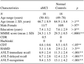
Demographics and neuropsychological findings of aMCI patients and normal elderly were shown in Table 1. Demographics of aMCI patients and normal elderly, including age, sex and education level, were matched between the two groups. However, the groups were significantly different with regard to MMSE (p < .0001), CDT (p < .05), and AVLT scores (p < .001) by using two-sample two-tailed t test.
Demographic information for aMCI and normal controls
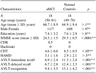
Note. aMCI = amnestic mild cognitive impairment; MMSE = Mini-Mental State Examination; CDR = Clinical Dementia Rating Scale; CDT = Clock Drawing Test; HAMD = Hamilton Depression Scale; AVLT = Auditory Verbal Learning Test.
*Under a Pearson x2 two-tailed test.
**Under a two-sample two-tailed t-test.
Default Mode Network for aMCI and Healthy Controls From ICA Analysis
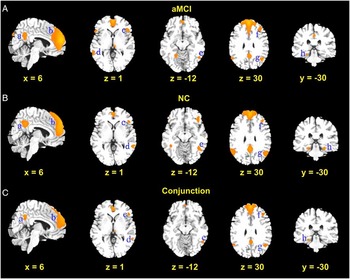
Most spatial locations in DMN were similar between the aMCI patients and healthy elderly (shown in Figure 1) from the conjunction analysis (p < .05, FDR corrected). These regions included the bilateral PreCN/PCC (BA 7/31), bilateral IPL (BA 40), right ACC (BA 32), bilateral vmPFC (BA 11), right LPFC (BA 47), right dMPFC (BA 9/10), right MTG (BA 21), right FG (BA 37), and left HC (BA 35). Results from ICA in aMCI group included the bilateral PreCN/PCC (BA 7/31), bilateral IPL (BA 40), right ACC (BA 32), bilateral vmPFC (BA 10/11), right LPFC (BA 47), right dMPFC (BA 9/10), right MTG (BA 21), right FG (BA 37), and left HC (BA 35). The areas for healthy controls were primarily located in the bilateral PreCN/PCC (BA 7/30/31), bilateral IPL (BA 39/40), right ACC (BA 32), bilateral vmPFC (BA 10), right dMPFC (BA 10/11), right LPFC (BA 47), bilateral MTG (BA21), bilateral FG (BA 37), and bilateral HC (BA 35/36).
Results of the Default mode network (p < .05, FDR corrected). (A) Amnestic mild cognitive impairment (aMCI); (B) Normal controls (NC); (C) Conjunction analysis. a, precuneus/posterior cingulate cortex; b, dorsal medial prefrontal cortex/anterior cingulate cortex; c, ventral medial prefrontal cortex; d, middle temporal gyrus; e, fusiform gyrus; f, lateral prefrontal cortex; g, inferior parietal lobule; h, hippocampus.

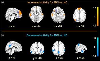
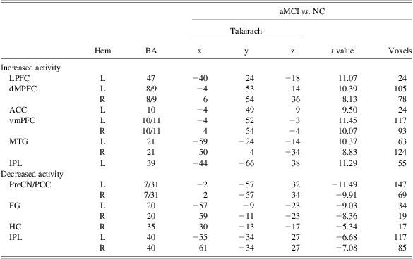
The between-group differences were shown in Figure 2 and Table 2 (p < .05, FWE corrected). Decreased activity in the aMCI patients was presented mainly in the bilateral PreCN/PCC (BA 7/31), bilateral FG (BA 20), right HC (BA 35), and bilateral IPL (BA 40), compared with the healthy controls. Increased activity in the aMCI was primarily observed in the left LPFC (BA 47), bilateral dMPFC (BA 8/9), left ACC (BA 10), bilateral vmPFC (BA 10/11), bilateral MTG (BA 21), and left IPL (BA 39).
Between-group maps of the default mode network for aMCI groups vs. normal controls (NC) (p < .05, FEW corrected, cluster size >15 voxels). Representative color-coded statistical maps exhibited the distribution of foci with significant increase (shown in the spectrum from orange to yellow) and decrease (shown in blue) activities. a, dorsal medial prefrontal cortex/anterior cingulate cortex; b, ventral medial prefrontal cortex; c, middle temporal gyrus; d, lateral prefrontal cortex; e, inferior parietal lobule; f, precuneus/posterior cingulate cortex; g, hippocampus; h, inferior parietal lobule; i, fusiform gyrus.

Talairach coordinates of peak significance of both increased and decreased DMN activity in aMCI patients compared to normal controls
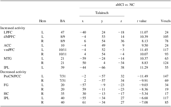
Note. p < .05, FWE corrected, cluster size > 15 voxels.
aMCI = amnestic mild cognitive impairment; NC = normal controls; LPFC = lateral prefrontal cortex; dMPFC = dorsal medial prefrontal cortex; ACC = anterior cingulate cortex; vmPFC = ventral medial prefrontal cortex; MTG = middle temporal gyrus; IPL = inferior parietal lobule; PreCN/PCC = precuneus/posterior cingulate cortex; FG = fusiform gyrus; HC = hippocampus; BA = Brodmann area; Hem = hemisphere.
MGCA Mapping for aMCI and Healthy Controls
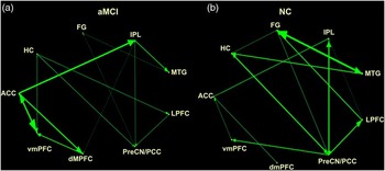
A causal connectivity graph was constructed using the thickness of connecting arrows to indicate the strengths of the causal influences. Only significant effective connectivities (p < .01, corrected for multiple comparisons) were divided into four levels (25%, 50%, 75%, and 100%) relative to the maximum significant dDTF value and presented in the graphs (shown in Figure 3). In normal controls, the mean “In + Out degree” for every region was 3.7 with the SD of 1.2. The PreCN/PCC served as the hub according to the standard, and was a central target. In patients with aMCI, the mean “In + Out degree” for every region was 3.4 with the SD of 1.3. The PreCN/PCC was also identified as the hub in patients with aMCI, not only as the central target but also the central source.
Results of the effective connectivity patterns of patients with amnestic mild cognitive impairment (aMCI) and normal controls (NC) from the multivariate Granger causality analysis (p < .01, corrected for multiple comparisons). The relative causal influence strengths were divided into four levels relative to the maximum significant direct directed transfer function (dDTF) value, and are represented by the thicknesses of the green lines. Abbreviations: FG, fusiform gyrus; IPL, inferior parietal lobule; MTG-middle temporal gyrus; LPFC, lateral prefrontal cortex; PreCN/PCC, precuneus/posterior cingulate cortex; dMPFC, dorsal medial prefrontal cortex; vmPFC, ventral medial prefrontal cortex; ACC, anterior cingulate cortex; HC-hippocampus.

As a whole, it was a trend that the causal interactions between the PreCN/PCC and HC, IPL, and LPFC were attenuated in the patients with aMCI in comparison to the normal controls. It was also noted that significant decreased causal influence in aMCI patients were particularly occurred between the PreCN/PCC and HC, PreCN/PCC and IPL, MTG and HC, MTG and FG, as well as FG and HC in terms of intensity (p < .01; two-sample t test). On the other hand, the aMCI showed saliently increased interregional causal interactions within the frontal cortices and between the frontal and parietal cortices. Results showed that strong bi-directional causal influence primarily emerged among the ACC, vmPFC, and dmPFC in the patient with aMCI, which presented significant differences in the dDTF values between these two groups (p < .01, FDR corrected). In addition, the increased causal influence from the ACC to the IPL were also exhibited in the patient with aMCI (p < .01, FDR corrected), compared with the normal controls.
Correlations Between Interregional Causal Interactions and Neuropsychological Scores
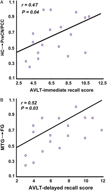
We also found causal connectivity patterns correlated with memory performance scores in patients with aMCI (Figure 4). AVLT-immediate scores were significantly positively related with the causal influence strength from the HC to the PreCN/PCC (r = 0.47; p < .05). AVLT-delayed scores were significantly positively related with the causal influence strength from the MTG to the FG (r = 0.52; p < .05). Other correlations were not significant (p > .09).
Correlation of mGCA mapping and neuropsychological scores. A. Causal influence from the hippocampus (HC) to precuneus/posterior cingulate cortex (PreCN/PCC) and auditory verbal learning test (AVLT)-immediate recall scores (r = 0.47; p < .05); B. Causal influence from the middle temporal gyrus (MTG) to fusiform gyrus (FG) and auditory verbal learning test (AVLT)-delayed recall scores (r = 0.52; p < .05).

Discussion
The present study examined causal interactions within the DMN for the aMCI patients, in contrast to the normal controls. It aimed to illustrate the working mechanism of the inter-region interactions within the DMN, and locate the specific dysfunctional effective connectivity of the resting brain network underlying the aMCI. Results from mGCA powerfully demonstrated the dynamic reorganization of interregional influences in aMCI and the normal control group: aMCI patients exhibited a trend toward impaired information flow between the PreCN/PCC and HC, PreCN/PCC and IPL, MTG, and HC, MTG and FG, as well as FG and HC; meanwhile, significantly increased interregional causal interactions in aMCI were particularly found among the frontal cortices and between the frontal and parietal cortices. We reiterate here that the effective connectivity patterns within the DMN can reflect the cognitive decline in aMCI.
Previous neuroimaging studies have focused on the abnormal functional connectivity for aMCI in the resting state. However, methodological limitations made it difficult to directly compare how the distributed networks were operated in processing the information underlying aMCI. Nowadays, the identification of the causal relationship between relevant regions offers a reliable choice. With the improvement of fMRI's sensitivity to detect systematic temporal relationship between regions, promising evidence has supported that the temporal relationship between fMRI hemodynamic measures can track the flow of neural signaling. Granger causality mapping is one of the proper candidates and provides us a way to understand interregional interactions among these spatially defined brain regions. It concerns about how multiple levels of regional activities jointly cause the abnormal signal processing underlying the aMCI.
The PreCN/PCC is the site of early metabolic abnormalities (Minoshima et al., Reference Minoshima, Giordani, Berent, Frey, Foster and Kuhl1997). As the hub of the DMN, its altered resting-state activity seems to be a meaningful functional hallmark to distinguish aMCI from normal controls. In our study, GCA revealed a reduced causal influence from the medial temporal lobe (HC) to the PreCN/PCC observed in the patient with aMCI, in comparison with normal controls. This result is consistent with previous studies that damaged connectivity between the MTL and PreCN/PCC, as a result of MTL lesions, is thought to be responsible for PreCN/PCC abnormalities (Wang et al., Reference Wang, Zang, He, Liang, Zhang, Tian and Li2006). We also observed a significant correlation between cognitive performance and the causal influence from the HC to the PreCN/PCC. It demonstrated that this abnormal effective connectivity may be an index for cognitive decline in the aMCI patients. Moreover, we also noted that the PreCN/PCC became the central hub in both aMCI and normal control group due to their “In + Out degree” of causal interactions. However, the PreCN/PCC was driven by other nodes, working as the terminal receiver of causal influences within the DMN in normal controls. In contrast, it served as both central target and source in aMCI group. Since previous study has already proved that Granger causal influences can be used to predict BOLD activity levels (Jiao et al., Reference Jiao, Lu, Zhang, Zhong, Wang, Guo and Liu2011), the abnormal signal flows may provide a potential cause of the decreased activity in the PreCN/PCC of DMN.
Meanwhile, our results also indicated that the bi-directional casual interactions between MTG and HC, and MTG and FG became weakened in aMCI than the normal controls. One hypothesis from the histological finding has pointed out that the pathological origin of AD is in the MTG (Braak & Braak, Reference Braak and Braak1991). Even though reduced PCC activity is consistently observed in prior aMCI studies of the DMN, reduced MTG has never been identified. Profiting from the multivariate Granger causality analysis, we found the coexistence of attenuated casual interactions between MTG and HC as well as MTG and FG. Along this line, we speculated that the abnormal activity in MTG can further trigger a cascade of abnormalities in HC and FG. Although the exact functional meaning of FG with fMRI BOLD activity is debated, there is consensus on its importance in memory-related function in recognition, particularly in face recognition (McCarthy, Puce, Gore, & Allison, Reference McCarthy, Puce, Gore and Allison1997). Anatomically, FG can feed information into the parahippocampal cortex (posterior) and perirhinal cortex (anterior), which are connected to the HC through the entorhinal cortex (Preston & Wagner, Reference Preston and Wagner2007). It should be no surprise that FG serves as a part of the integrated system of memory processes during resting states (Bookheimer et al., Reference Bookheimer, Strojwas, Cohen, Saunders, Pericak-Vance, Mazziotta and Small2000; Buckner, Reference Buckner2004). In consideration to the positive relation between the AVLT-delayed recall scores and the causal interactions from the MTG to the FG, we postulate that such attenuated causal interactions between MTG and FG may be an “accessory” indicator of impaired memory function associated with aMCI.
Apart from the impaired mechanism, causal interaction maps also indicated increased causal interregional influences in aMCI patients compared with the normal group. Our results showed that increased causal interregional interactions were mainly distributed among the ACC, dmPFC, and vmPFC, as well as between the frontal and parietal cortices. It is already found that an increased recruitment of the frontal and parietal lobes is observed to compensate in AD patients in memory task (Anderson et al., Reference Anderson, Iidaka, Cabeza, Kapur, McIntosh and Craik2000; Cabeza, Anderson, Locantore, & McIntosh, Reference Cabeza, Anderson, Locantore and McIntosh2002; Gould et al., Reference Gould, Arroyo, Brown, Owen, Bullmore and Howard2006; Grady, Bernstein, Beig, & Siegenthaler, Reference Grady, Bernstein, Beig and Siegenthaler2002; Logan, Sanders, Snyder, Morris, & Buckner, Reference Logan, Sanders, Snyder, Morris and Buckner2002; Pariente et al., Reference Pariente, Cole, Henson, Clare, Kennedy, Rossor and Frackowiak2005). Recent reports also show increased fMRI responses in the MTL, as well as a trend toward increased frontal activation, compared to controls (Hamalainen et al., Reference Hamalainen, Pihlajamaki, Tanila, Hanninen, Niskanen, Tervo and Soininen2007).The present study may suggest that aMCI patients could recruit network resources, primarily from the frontal-parietal regions, to maintain memory functions after reduced MTL activity.
Our study has some limitations. First, we instructed all the participants to keep their eyes closed during the scanning run. However, one recent research from Van Dijk et al. has demonstrated that functional connectivity strength of brain network is influenced by tasks such that fixation and eyes open rest yielded stronger correlations in the networks examined against eyes closed rest or a continuous semantic classification task (Van Dijk et al., Reference Van Dijk, Hedden, Venkataraman, Evans, Lazar and Buckner2010). Therefore, instructing subjects simply to keep their eyes open is a practical condition in studies where setting up a display is difficult. Contrasts of resting-state brain networks of aMCI, AD and normal subjects with eyes open could be our further research interests. Another limitation is participants were asked to not fall asleep during scanning. We just checked that participants were not sleeping by an oral report after scanning. In future, a better way of adopting eye-tracking for direct monitoring is suggested.
To conclude, we combined an effective connectivity based analysis of multivariate Granger causality approach with ICA to explore the causal interactions within the DMN involving both aMCI patients and normal age-matched controls. Research results presented an impaired effective connectivity between the PreCN/PCC- parietal-temporal areas as well as within the temporal lobe, which was strongly correlated with the cognitive declines in the aMCI patients. We also found an enhanced effective connectivity within the frontal cortex, which may maintain memory functions after reduced DMN activity. The findings highlight the importance of effective connectivity changes in the pathophysiology of aMCI. These criteria may serve as an imaging-based biomarker for the diagnosis and monitoring of aMCI patients.
Acknowledgments
This work was supported by Program for New Century Excellent Talents in University of Ministry of Education of China (grant number NCET-08-0801); Key Program of the “11th Five Year” Plan of National Education Science of China (grant number DBA070070); the Fundamental Research Funds for the Central University; the National Natural Science Foundation of China (grant numbers 81071217); the Beijing Nova program (grant number Z111101054511116); and Beijing Natural Science Foundation (grant number 4122082). There are no conflicts of interest. Drs. Yan, and Zhang contributed equally to this work.