Inferior displacement of the septal and posterior leaflets of the tricuspid valve characterises Ebstein anomaly. Associated genetic disorders and cardiac malformations can significantly exacerbate the clinical management and outcome of these infants. In this report, we describe an infant with both Ebstein anomaly and Williams–Beuren syndrome.
Case report
The infant, born at full term, was a male weighing 3 kilograms. Maternal sickle cell trait and a history of genital herpes simplex virus complicated the pregnancy. Moderate meconium fluid staining complicated the delivery, which was via caesarean section secondary to non-reassuring foetal cardiac tones. The infant’s apgar scores were as follows: three at 1 minute, six at 5 minutes, and seven at 10 minutes. Resuscitation at delivery included stimulation, oral suctioning, and face mask ventilation. The infant was intubated at 1 hour of life secondary to continued cyanosis. His arterial blood gas showed a pH of 7.21, with a partial pressure of oxygen of 53 torr. The infant was started on prostaglandins, with saturations increasing into the low 70s. The infant was transferred to a tertiary medical facility. Upon arrival, the repeat arterial blood gas showed a pH of 7.43 and a partial pressure of oxygen of 21 torr.
On general evaluation, the infant was noted to have periorbital fullness, broad nasal base, long philtrum, and full lips with a wide mouth. Cardiac examination revealed a right ventricular lift, normal first cardiac sound, single second cardiac sound, and a 3/6 holosystolic murmur at the left lower sternal border. The lungs were clear, with shallow respirations and mild intercostal retractions. Pulses were equal and full in all extremities. There was no hepatomegaly.
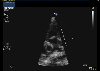
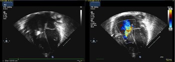
A chest radiograph revealed an enlarged cardiac silhouette with no airspace disease but with mildly prominent interstitial markings. Echocardiography showed functional pulmonary atresia with normal-appearing valve morphology and severe branch pulmonary artery stenosis (Fig 1). There was a large reverse angle patent arterial duct with left-to-right shunting. The tricuspid valve had a mild Ebstein anomaly with apical displacement of the posterior and septal leaflets with severe tricuspid valve insufficiency (Fig 2). The infant’s body surface area was 0.2 metres squared with an apical displacement of the septal leaflet of 5 millimetres, which resulted in a displacement index of the tricuspid valve of 25 millimetres per metre squared. The anterior leaflet of the tricuspid valve was elongated and appeared calcified. A moderately sized pericardial effusion was also seen.
Parasternal short-axis view showing severe branch pulmonary artery stenosis.

Apical four-chamber view showing apical displacement of the septal leaflet in two-dimensional imaging and severe tricuspid valve regurgitation by colour flow Doppler.

Secondary to concerns for pulmonary valve atresia, the infant underwent cardiac catheterisation for possible valvuloplasty. The right ventricular angiogram showed severe tricuspid valve insufficiency, a normal-appearing right ventricle, a normal-appearing right ventricular outflow tract, and a normal-appearing and patent pulmonary valve. The angiogram confirmed severe main pulmonary artery stenosis, branch pulmonary artery stenosis, and Ebstein malformation with severe tricuspid valve regurgitation. After cardiac catheterisation, the prostaglandins were discontinued.
Other issues during the infant’s hospitalisation included the following: respiratory distress requiring intubation until day 13 of life, a right renal artery cyst, and mild cholestasis. His ionised calcium and urine calcium to creatinine ratio remained normal throughout the hospitalisation. His blood tested positive for Williams–Beuren syndrome via the fluorescence in situ hybridisation study. On day 36 of life, he was discharged home.
Currently, this 5-month-old infant continues to be asymptomatic with weight at less than 5%. His most recent echocardiogram continues to show Ebstein anomaly of the tricuspid valve with severe regurgitation, severe branch pulmonary artery stenosis, and normal biventricular function. He does not require cardiac medications at this time. Williams-Beuren syndrome has contributed to his failure to thrive, and he is currently receiving palliative care treatment.
Discussion
It was J.C.P. Williams and then A.J. Beuren who first described what is known as the Williams–Beuren syndrome in the 1960s.Reference Williams, Barratt-Boyes and Lowe1, Reference Beuren, Apitz and Harmjanz2 It affects approximately 1/10,000 individuals including all racial and ethnic groups.Reference Stromme, Bjornstad and Ramstad3 The genetic cause is a micro-deletion at chromosome 7q11.23, which creates a disruption of the elastin gene.Reference Ewart, Morris and Atkinson4 It is a multi-system disorder affecting the cardiovascular system as well as having distinctive facial features, endocrine abnormalities, and intellectual handicaps.
The elastin gene defect results in arteriopathy with stenotic lesions of the medium- and large-sized arteries. The most common cardiovascular defect is supravalvular aortic stenosis. Other defects may also include diffuse aortic hypoplasia, coarctation of the aorta, aortic valve disease, pulmonary valve stenosis, stenosis of the branch pulmonary arteries, coronary arterial lesions, and mitral valve disease.Reference Bruno, Rossi, Thuer, Cordoba and Alday5 In our review of the literature, this is the first patient with Ebstein malformation of the tricuspid valve with documented Williams–Beuren syndrome.
Ebstein anomaly was initially described by Wilhelm Ebstein in 1866. It occurs in one to five per 2,00,000 births, representing less than 1% of all congenital cardiac diseases.Reference Dearani and Danielson6 There is an equal distribution between genders and most cases are sporadic. Embryologically, Ebstein anomaly represents the failure of leaflet delamination of the posterior and septal leaflets of the tricuspid valve.
The morphology of the tricuspid valve in Ebstein anomaly is variable. There is some degree of apical displacement of the septal and posterior leaflets. This apical displacement can be measured in the apical four-chamber view and indexed to body surface area resulting in the displacement index.Reference Shiina, Seward, Edwards, Hagler and Tajik7 The anterior leaflet is usually elongated and sail-like. The proximal portion of the right ventricle between the true annulus and the displaced valve leaflets is the atrialised portion of the right ventricle. This atrialised portion of the right ventricle is very thin, while the more distal right ventricle is usually normal in thickness. Ebstein anomaly is known to be associated with the following cardiac defects: atrial septal defects, patent oval foramen, pulmonary valve stenosis, ventricular septal defects, pulmonary atresia with intact septum, atrioventricular canal defects, tetralogy of Fallot, coarctation of the aorta, and congenitally corrected transposition of the great vessels.
Ebstein anomaly has been reported in association with the following genetic conditions: Down syndrome, Ellis–van Creveld syndrome, and Noonan syndrome.Reference Johnson, Ortiz-Colom, Sainz de la Pena and Barroso8–Reference Wright, Summitt and Ainger10 Most cases of Ebstein anomaly are sporadic and familial cases are rarely reported. Although it is well recognised that Ebstein anomaly is associated with extracardiac anomalies, in our review of the literature we were unable to find other reported patients with both Williams–Beuren syndrome and Ebstein anomaly. Clinicians must remain aware of genetic disorders, which may alter the clinical management of patients with congenitally malformed hearts.