I. INTRODUCTION
Several metals and alloys such as silver and copper exhibit antibacterial properties because they release metal ions Reference Zhao and Stevens1,Reference Galeano, Korff and Nicholson2 and reactive oxygen species (ROS) Reference Chatterjee, Chakraborty and Basu3 in aqueous environments. Certain metallic nanoparticles that release no harmful diffusive species (e.g., metal ions and ROS) also exhibit antibacterial activity (AA), which is due to their incorporation into the cytoplasm of bacteria, and subsequent deterioration of cytoplasmic proteins such as ribosomes. Reference Sadhasivam, Shanmugam, Veerapandian, Subbiah and Yun4–Reference Hernández-Sierra, Ruiz, Pena, Martínez-Gutiérrez, Martínez, Guillén, Tapia-Pérez and Castañón7
Nanoporous metals are nanostructured materials with open porous structures containing nanometer-scale pores and ligaments. Reference Erlebacher, Aziz, Karma, Dimitrov and Sieradzki8 The first electron microscopy observations of nanoporous metals were reported in 1979–1980 by Forty and Durkin, Reference Forty9,Reference Forty and Durkin10 and nanoporous metals have since received much interest from researchers in various fields. Nanoporous gold (NPG) has been intensively investigated because it is easily fabricated by the dealloying of gold–silver alloys. Lattice distortion occurs at the surface of the nanosized ligaments because of their large curvature. This results in an interesting catalysis behavior of NPG in organic reaction that is not observed in bulk gold. Reference Kameoka and Tsai11–Reference Hakamada, Hirashima and Mabuchi15 NPG reportedly exhibits catalytic activity in the oxidation of carbon monoxide Reference Kameoka and Tsai11–Reference Xu, Su, Xu, Liu, Zhao, Tian and Ding13 and methanol Reference Wittstock, Zielasek, Biener, Friend and Bäumer14 in contrast to bulk gold. NPG also catalytically decomposes methyl orange (MO, an organic azo dye), whereas bulk gold does not. Reference Hakamada, Hirashima and Mabuchi15 The decomposition kinetics of MO was not simply proportional to the surface area of NPG, which suggests that active sites due to lattice distortion are important in the catalysis by NPG. Thus, NPG and its catalytic properties strongly affect the stability of the surrounding organic matter.
On the other hand, NPG has been also investigated as a substrate for mammalian cells. Reference Seker, Berdichevsky, Staley and Yarmush16–Reference Chapman, Chen, Stamou, Biener, Biener, Lein and Seker18 These studies demonstrated that NPG generally does not cause severe deactivation of cells, although individual processes such as adhesion and growth are sensitive to the porosity of NPG. To our knowledge, however, there have been few reports on the effect of NPG on the activity of bacteria, Reference Santos, de Santi Ferrara, Zhao, Rodrigues and Shih19 which have a different composition and structure from those of mammalian cells.
The activity of bacteria is generally sensitive to the surface morphology of nanomaterials. Reference Anselme, Davidson, Popa, Giazzon, Liley and Ploux20 Rizzello et al. reported that the fimbriae of bacteria disappeared on a gold substrate containing nanoscale surface features, whereas fimbriae were still observed on a flat gold (FG) substrate. Reference Rizzello, Sorce, Sabella, Vecchio, Galeone, Brunetti, Cingolani and Pompa21 The effects of nanostructured substrates on the activity of adherent bacteria have been widely investigated, although the underlying mechanism is still in debate.
Bacteria are systematically and functionally assembled organic matters. NPG may, therefore, catalytically decompose organic materials within the components of bacteria, thus suppressing the bacterial activity because NPG strongly affects the stability of the surrounding organic matter as described in the second paragraph. This led us to investigate the antibacterial properties of NPG against Escherichia coli (E. coli) and Staphylococcus epidermidis (S. epidermidis). We found that NPG can catalytically damage these bacteria without exploiting diffusive bacteria-killing species such as metal ions and ROS.
II. EXPERIMENTAL METHODS
A. Preparation of NPG and FG substrates
Gold–silver films sputtered on glass substrates were dealloyed in concentrated nitric acid at 253 and 298 K for 24 h, to fabricate NPG substrates with different ligament sizes. A radio-frequency sputtering apparatus (SVC-700RF, Sanyu Electron Co., Ltd., Tokyo, Japan) was used to fabricate the gold–silver alloy thin film, which was then dealloyed to form NPG. A 50-nm-thick pure gold film (>99.9 mass%) was sputtered on a glass substrate of length × width × thickness of 50 × 50 × 1.2 mm. A 50-nm-thick Au0.47Ag0.53 alloy was then sputtered on the thin gold film. NPG was synthesized by dealloying (free corrosion) of the alloy at 253 or 298 K for 24 h, in 69 mass% HNO3. The microstructures of NPG and FG were observed using scanning electron microscopy (SEM; SU-6600, Hitachi High-Technologies Corporation, Tokyo, Japan) equipped with an energy-dispersive X-ray spectroscopy (EDXS) apparatus. For comparison, a polyethylene (PE) film and 50-nm-thick FG sputtered on glass were also prepared.
B. Bacterial strain
Type strains of E. coli (K-12, NBRC 3301) and S. epidermidis (NBRC 100911) were obtained from the National Institute of Technology and Evaluation (Tokyo, Japan). We incubated bacteria in Luria–Bertani (LB) medium at 308 K for 48 h, before treating in antimicrobial tests. Casein-peptone glucose yeast extract agar (Wako Pure Chemical Industries Ltd., Osaka, Japan) was used for the incubation.
C. Tests of antibacterial activities
The antibacterial properties of NPG, FG, and control PE were tested, essentially according to the Japanese Industrial Standard “Antibacterial products—Test for antibacterial activity and efficacy.” 22 Control PE was necessarily used for the confirmation of validness of each test, although its physical and chemical states of the surface differ from those of gold. First, one quantity of platinum loop of bacteria incubated in the medium was removed from the colony and placed in 5 mL of 1/500 nutrient broth, followed by vortex mixing. Second, 400 µL of the bacterial suspension was dropped onto the samples and then the PE film covered the bacterial suspension. In this way, bacterial suspensions were incubated on samples for a given period (up to 24 h), in humidity-controlled incubators at 308 K. Finally, the incubated bacteria were recovered and diluted in LB medium, to make a 10-fold dilution series of agar pour plates. These were then incubated at 308 K for 48 h. The number of colonies in the agar pour plates was then counted. The identical tests were repeated 4 times. Viable bacteria counts (VBCs) were statistically analyzed by the one-way analysis of variance followed by a post-hoc test. All results were expressed as mean ± standard deviation. Differences were considered statistically significant at P-value < 0.05.
D. Observation of bacteria
The size and morphology of the bacteria incubated for 24 h on the NPG and FG substrates were examined by SEM. Quantitative information on the three-dimensional geometry of E. coli was also collected by scanning probe microscope (SPM; NanoWizard III, JPK instruments AG, Berlin, Germany). Prior to SEM and SPM analyses, bacteria on the samples were fixed with 2% glutaraldehyde solution for 2 h, which was then removed by rinsing with phosphate-buffered saline (PBS), after which specimens were dehydrated in a series of ethanol solutions (50, 60, 70, 80, 90, 99, and 100 vol%) and in tert-butyl alcohol. The samples were then freeze-dried from tert-butyl alcohol and coated with Pt–Pd thickness of 10 nm.
E. Transcriptomic analyses
The expressions of two different genes (fimA and lrhA) were investigated for E. coli cultured in the suspension dropped on NPG and FG (24 h, RH of 60%) by real-time polymerase chain reaction (PCR). Reference Rizzello, Sorce, Sabella, Vecchio, Galeone, Brunetti, Cingolani and Pompa21 The gapA gene, encoding D-glyceraldehyde-3-phosphate dehydrogenase A, was used as an independent internal control. After incubation on substrates (three independent biological replicates), the surfaces were washed out with a mixture of RNAprotect Bacteria Reagent (1000 µL) + PBS (500 µL) (pH 7.5), which stripped the bacteria from the substrates. The total Ribonucleic acid (RNA) was extracted from the bacterial cells from the NPG and FG samples, using tris-ethylenediaminetetraacetic acid (Tris-EDTA) buffer solution containing lysozyme. The PCR was carried out against the total RNA.
The expression of approximately 5000 genes was investigated for E. coli cultured on NPG and FG (24 h, RH of 60%) by microarray analyses. Reference Cui, Zhao, Tian, Zhang, Lü and Jiang5 E. coli were collected from four independent biological replicates by centrifugation at 8000 rpm for 5 min, for the microarray analyses. Samples were hybridized for 16 h on Affymetrix GeneChip E. coli Genome 2.0 Array containing approximately 10,000 probe sets for all 20,366 genes. We analyzed the microarray data by Significance Analysis of Microarrays using Genechip Operating Software ver1.4 (Affymetrix, Santa Clara, California). Genes were considered as being regulated with greater than or equal to 2-fold change, meanwhile the P-value was required to be below 0.05. We performed DAVID (Database for Annotation, Visualization and Integrated Discovery) analysis 23 for comparison of the expression data. We imported lists of regulated genes to generate the gene ontology (GO) enrichment lists.
III. RESULTS
A. Synthesis and characterization of NPG
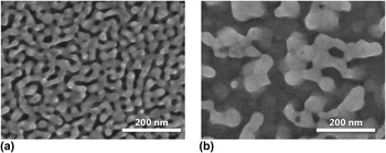
SEM images of the surface microstructure of the synthesized NPG are shown in Fig. 1. A nanoporous structure with average ligament and pore sizes of approximately 20 nm was observed for the NPG fabricated by dealloying at 253 K. The NPG fabricated by dealloying at 298 K had larger ligament and pore sizes of approximately 50 nm. EDXS analysis was carried out during SEM observations. EDXS revealed that the residual Ag content was very small, being below the 1 at.% detection limit (see Supplementary Material for EDXS results). On the other hand, control PE film and FG sputtered on glass did not possess nanoporous structures (see Supplementary Material for SEM image of FG). Water wettability of NPG and FG was similar during our experimental handling, although a strict evaluation such as contact angle measurement was not conducted.
SEM images of NPG. Samples were fabricated by dealloying at (a) 253 K and (b) 298 K, in 69 mass% HNO3. The lower dealloying temperature led to smaller pore and ligament sizes.

B. Antibacterial properties of NPG
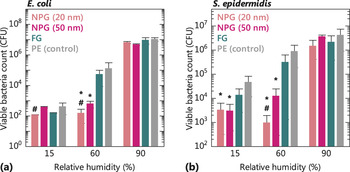
Figure 2 shows VBCs of E. coli and S. epidermidis on NPG and FG after incubation for 24 h at various RHs. At an intermediate RH of 60%, the VBCs on NPG were much lower than those on FG. FG showed no significant decrease in the VBCs of E. coli and S. epidermidis on its surface, compared with the control PE film, which was devoid of antibacterial properties. These results indicated that at an intermediate RH, NPG possessed antibacterial properties against E. coli and S. epidermidis, whereas FG did not. NPG with a pore size of 20 nm reduced the VBCs of both bacteria more efficiently than NPG with a pore size of 50 nm. The VBCs on NPG at a high RH of 90% showed no significant difference to those on FG and the control PE film. The VBCs on NPG at a low RH of 15% were also slightly lower than those on FG and the control PE film. Irrespective of the substrate, a lower RH significantly decreased the VBCs and a higher RH increased the VBCs. This was attributed to drying and is explained later. These results suggested that the antibacterial properties of NPG were significant at an intermediate RH, which may have practical importance. Reference Møretrø and Langsrud24–Reference Møretrø, Høiby-Pettersen, Halvorsen and Langsrud26
Viable bacterial counts (VBCs) on NPG, FG, and control PE films as a function of relative humidity (RH), for 24 h incubation of (a) E. coli and (b) S. epidermidis. * denotes significant difference between NPG and FG with P < 0.05, while # denotes significant difference between NPG (20 nm) and NPG (50 nm) with P < 0.05. An intermediate RH of 60% differentiated the VBCs on NPG from those on FG, while lower and higher RHs did not.

The AA of nanoporous Au can be expressed by
 $${\rm{AA}} = {\log _{10}}\left( {{{{N_0}} \mathord{\left/ {\vphantom {{{N_0}} N}} \right. \kern-\nulldelimiterspace} N}} \right)\quad ,$$
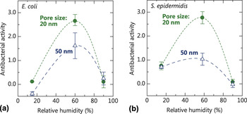
where N 0 and N are the VBCs on FG (as a control sample) and NPG, respectively. When the AA is >2.0, i.e., when the reduction in VBC by the sample is >99%, then the material is generally regarded as antibacterial. 22 The relationships between the AA of NPG and RH with respect to E. coli and S. epidermidis are shown in Figs. 3(a) and 3(b), respectively. NPG exhibited the best antibacterial performance at an intermediate RH of 60%. The antibacterial properties diminished at lower or higher RH. NPG with a pore size of 20 nm exhibited better antibacterial performance than that with a pore size of 50 nm. This suggested that the small pores and ligaments were the source of the antibacterial properties in NPG. Figure 3(b) shows that the AA of NPG against S. epidermidis was approximately 0.7 at low RH, whereas that of NPG against E. coli was approximately zero. The slightly higher AA of NPG against S. epidermidis was due to the fact that gram-positive S. epidermidis is more resistant toward dryness than E. coli because of a thicker cell wall, and that N 0 of S. epidermidis was somewhat higher than N [see Fig. 2(b)]. Thus, S. epidermidis can live to some extent at low RH. However, NPG still affected the VBC of S. epidermidis at the low RH.
Antibacterial activities (AAs) of NPG as a function of RH, for 24 h incubation of (a) E. coli and (b) S. epidermidis. The maximum AA against both bacteria was observed at the intermediate RH.

Subsequent investigations were carried out using the NPG with the pore size of 20 nm, because of its higher AA. Figure S3 in Supplementary Material shows the variation in VBCs of E. coli on NPG and FG with incubation time, at a RH of 60%. The VBC on NPG was lower than that on FG at every incubation time. The VBCs on NPG incubated for 12–24 h were significantly lower than those on FG, at the corresponding incubation times. The VBCs on NPG and FG were similar for incubation times of 0–12 h. Thus, antibacterial properties of NPG were evident after incubation for 12 h at an RH of 60%. This behavior is quite different from the AA by metal ions, which typically begin decreasing the VBC immediately after commencing incubation. Reference Zhao and Stevens1
C. Morphological observations of treated bacteria
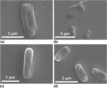
SEM images of E. coli on NPG (pore size of 20 nm) and FG are shown in Fig. 4. The shape of E. coli on NPG was significantly distorted, with a dimple observed in the center of the cell [Fig. 4(a)]. Leakage of the cytoplasm was also apparent, as shown by arrows in Fig. 4(b). The cell membrane of E. coli cultured on FG was not distorted [Fig. 4(c)] as an undented rod shape was observed. No fimbria were observed in the SEM images of E. coli on both NPG and FG, Reference Rizzello, Sorce, Sabella, Vecchio, Galeone, Brunetti, Cingolani and Pompa21 perhaps because of the intermediate RH.
SEM images of E. coli cultured on NPG and FG at RH = 60%. (a) The center of the cell of E. coli on NPG appeared dimpled. (b) Cytoplasm leaked from E. coli on NPG (arrows). (c and d) No dimpling and cytoplasm leakage were observed in E. coli on FG.

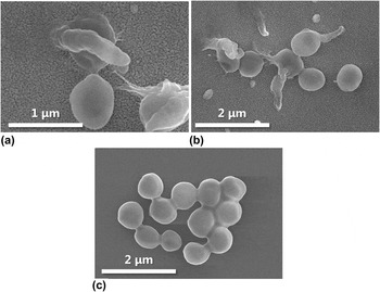
Similar trends were observed for S. epidermidis, as shown in Fig. 5. The membrane of S. epidermidis on NPG was damaged, and its morphology was aspherical. Isolated bacteria were also frequently observed. The cell wall of S. epidermidis cultured on FG was not damaged, and several bacteria formed grape-shaped clusters, which is typical of active Staphylococcus bacteria.
SEM images of S. epidermidis cultured on NPG and FG at RH = 60%. (a) The cell wall of S. epidermidis on NPG was significantly distorted. (b) Isolated S. epidermidis cells were frequently observed on NPG. (c) The cell wall of S. epidermidis on FG was not damaged, and bacteria tended to form grape-shaped clusters, which is typical of active Staphylococcus bacteria.

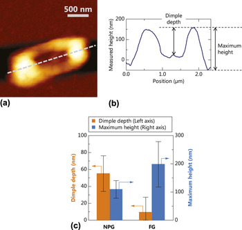
SPM was used to quantify the shape distortion of E. coli on NPG, as shown in Fig. 6. Figure 6(a) shows the morphology of E. coli on NPG, as observed by SPM. The bacteria appeared dented, similarly to the SEM observations. The height profile of the E. coli cell collected along its long axis [Fig. 6(b)] showed an obvious concave structure at the cell center. The dimple depth and maximum height of the E. coli cell are collected from the SPM observations of E. coli on NPG. The dimple depth is the height difference between the highest point and the lowest point of the dimple [Fig. 6(b)]. The maximum height of the E. coli cell is the height difference between the highest point and the base of the E. coli cell [Fig. 6(b)]. Average values of dimple depth and maximum height, as determined from measurements of more than 20 individual E. coli cells, are summarized in Fig. 6(d). The average dimple depth of E. coli on NPG was 55 nm, which was much larger than that of E. coli on FG (9 nm). The average maximum height of E. coli on NPG was 109 nm, which was smaller than that of E. coli on FG (198 nm). These data indicated that the E. coli cell was severely flattened and dimpled on NPG. The SPM measurements indicated the detrimental effect of the NPG structure on the shape of E. coli cells. This correlated with the AA of NPG.
SPM images of E. coli cultured on NPG at RH = 60%. (a) The center of the cell of E. coli on NPG was dimpled. (b) Height profile of the E. coli cell along the dashed line in (a). (c) Dimple depth and maximum height for E. coli cells on NPG and FG. The dimple was deeper in the E. coli cell on NPG than on FG. The maximum height was greater in the E. coli cell on FG than on NPG. The shape distortion was more severe in the E. coli cell on NPG than on FG.

D. Transcriptomic analysis
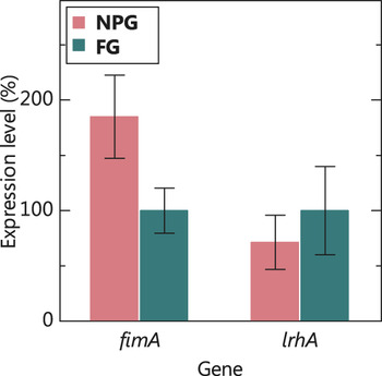
The gene expression related to the fimbriae evolution of E. coli was differentiated in a previous study. Reference Rizzello, Sorce, Sabella, Vecchio, Galeone, Brunetti, Cingolani and Pompa21 To compare the gene expression of E. coli on the present gold samples with that reported previously, the expression levels of two genes related to fimbria evolution were analyzed by real-time PCR. The results are summarized in Fig. 7. Two genes of fimA, which encodes the major subunit of type-1 fimbriae, and lrhA, which encodes the repressor of the fim operon, were investigated. The mRNA expression level for fimA in E. coli grown on NPG was clearly higher than that in E. coli grown on FG. By contrast, lrhA in E. coli grown on NPG was expressed at a slightly lower level than that of E. coli grown on FG. The P-values of expression levels of fimA and lrhA were 0.000087 and 0.54, respectively. Thus, there was a clear difference in the expression level of fimA between NPG and FG, but not for lrhA.
Expression levels of lrhA and fimA in E. coli on NPG compared with E. coli on FG. RH during culturing was 60%. fimA was expressed more in E. coli on NPG. lrhA in E. coli on NPG was expressed at a similar level to that in E. coli on FG.

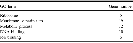
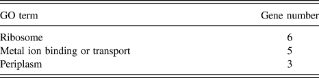
To comprehensively investigate the effects of the antibacterial NPG on E. coli, mRNA expression was analyzed by gene microarray hybridization. Across the GO databases, differently expressed genes in several classifications, including physiological process, cellular processes, catalytic activity, metabolism, binding, transporter activity, protein complex and localization, were identified. According to filtering criteria, 99 genes were differentially expressed in E. coli cultured in the medium on NPG, among which 24 were up-regulated and 75 were down-regulated. Based on the biological process, molecular function, and cellular component classification, these up- or down-regulated genes were classified to different GO terms. We investigated three classifications related to up-regulated and down-regulated genes by GO enrichment analysis, as summarized in Tables I and II. Stricter filtering criteria with fold changes of >3 and P-values of <0.01 detected the significant down-regulation of c0412, cpxP, ytfQ, and htpX, which are related to the membrane system in the cell envelope of E. coli. Reference Danese and Silhavy27–Reference Shimohata, Chiba, Saikawa, Ito and Akiyama34
Significantly down-regulated biological processes in E. coli on NPG.

Significantly up-regulated biological processes in E. coli on NPG.

IV. DISCUSSION
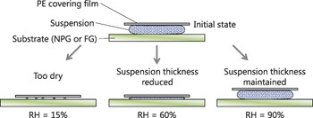
We observed that the thickness of the bacterial suspension on samples reduced because of evaporation at RH values of 15 and 60%. The suspension volume was largely unchanged after incubation for 24 h at RH of 90%. This suggested that the effect of RH on the antibacterial properties of NPG was related to the thickness of the suspension, as illustrated in Fig. 8. A low RH of 15% caused significant drying and subsequent bacterial death on NPG and FG. In this case, the AA of NPG was not overly high because N 0 and N in Eq. (1) were similar. A high RH of 90% minimized evaporation of the suspension during incubation. Bacteria in the suspension could live and proliferate far from the substrate surface without contacting the NPG substrate, resulting in low AA. The intermediate RH of 60% apparently resulted in reduced residual bacterial solution to force the bacteria to contact with the NPG substrate. Hence, the direct contact of bacteria with the NPG substrate was necessary for the antibacterial efficacy of NPG.
Schematic illustration of humidity dependence of antibacterial activity (AA) of NPG. The evaporation of bacterial suspension was related to the high AA at an intermediate relative humidity (RH). This suggested that the direct contact of bacteria with the catalytic NPG substrate was necessary for AA.

The time variation in the VBC cultured in suspension on NPG (Fig. S3) also supported the necessity of the direct contact between the bacteria and NPG. The suspension thickness gradually decreased during incubation for 24 h at a RH of 60%. The VBC on NPG significantly decreased only after 12 h of incubation, where the dryness of samples caused the decrease in suspension volume and bacteria are forced to touch NPG substrates. These results strongly imply that the antibacterial properties of NPG are attributed to the catalytic nature of NPG, and not to the metallic ion and ROS released into the solution from substrates. (If these diffusible species were responsible for the antibacterial properties of NPG, the AA at RH = 90% would be higher due to the diffusion of these species.) We also confirmed that metal ions (silver and gold) and ROS were essentially absent in the culturing solution, as described in Supplementary Material. Thus, NPG itself directly and catalytically deteriorated the bacteria. The range of the catalytic effect of NPG was thought to be limited to within a few nanometers from the surface. Reference Nel, Mädler, Velegol, Xia, Hoek, Somasundaran, Klaessig, Castranova and Thompson35 Hence, a direct contact between the bacteria and NPG was necessary for the AA. The AA of NPG with a smaller pore size was stronger than that of NPG with a larger pore size. A smaller ligament size provided a higher concentration of catalytically active sites on the ligament surface. This resulted from the more pronounced lattice distortion and higher concentration of atomic defects at the larger curvature ligaments.
The AA of NPG depended on the direct contact of the bacteria with the NPG surface. SEM and SPM observations indicated severe distortion in the bacterial shape on NPG. Thus, the nanosized ligaments of NPG directly influenced the physical and/or chemical stability of the bacterial cell wall and membrane, resulting in their death.
In a previous study, the fimbriae of E. coli on nanorough gold synthesized by electroless deposition disappeared, while the fimbriae of E. coli on FG could be observed. Reference Rizzello, Sorce, Sabella, Vecchio, Galeone, Brunetti, Cingolani and Pompa21 Real-time PCR analyses indicated that the mRNA expression level for fimA in E. coli cultured on nanorough gold was lower than that in E. coli cultured on FG, and that the mRNA expression level for lrhA in E. coli cultured on nanorough gold was higher than that in E. coli cultured on FG. This indicated that nanorough gold resulted in the fimbriae of E. coli disappearing from a transcriptomic point of view. In the present study, the mRNA expression level for fimA in E. coli cultured on NPG was higher than that in E. coli cultured on FG. In other words, the real-time PCR result in the present study was opposite to that in the previous study. This difference suggested that small changes in the characteristics of nanostructure and lattice strain caused a significant transcriptomic differentiation.
Microarray analyses of E. coli on NPG showed the significant down-regulation of c0412, cpxP, ytfQ, and htpX. The c0412 gene encodes the transcriptional regulator protein YcjZ controlling the recycling of peptidoglycan, which structurally supports the body of E. coli in its cell envelope. Reference Shimada, Yamazaki and Ishihama36 The CpxP protein governs the Cpx pathway of periplasmic stress response, operating as a proteolytic adapter to facilitate degradation of misfolded proteins in the cell envelope. Reference Thede, Arthur, Edwards, Buelow, Wong, Raivio and Glover28 The YtfQ protein promotes and supports the importing of nutritious substances in the periplasm. Reference Horler, Müller, Williamson, Potts, Wilson and Thomas31 The htpX protein induces an endoproteolytic cleavage, to facilitate dislocating misfolded membrane proteins out of the membrane. Reference Akiyama32–Reference Shimohata, Chiba, Saikawa, Ito and Akiyama34 The down-regulation of these genes suggested that NPG directly (not depending on diffusive species such as metal ions and ROS) and catalytically induced the collapse of the cell envelope system of E. coli. This inhibited the normal exchange of intracellular and extracellular substances through the cell membrane, inducing protein misfolding and subsequent cell death.
The catalytic degradation of nutritious components in the culture media by NPG was not responsible for the present antibacterial properties. If it were, then NPG would exhibit antibacterial properties even at an RH of 90%, where nutritious components could degrade at the catalytic NPG substrate. Hence, the direct contact of NPG with the bacterial cell envelope was necessary for killing bacteria, perhaps inducing the catalytic decomposition of envelope-structuring components such as peptidoglycan and membrane proteins. The physical situation may be similar to that of nanostructured artificial viruses with AA, which bypass the need for membrane-induced folding and destroy bacterial membrane on the direct contact. Reference Castelletto, de Santis, Alkassem, Lamarre, Noble, Ray, Bella, Burns, Hoogenboomb and Ryadnov37 However, it is not currently clear what part of the envelope was catalytically degraded by NPG. This is because even detailed chemical analyses of products from AA tests cannot determine the substance directly affected by NPG. The conformation and subsequent stability of proteins in the cell membrane may be altered by NPG, Reference Hakamada, Takahashi and Mabuchi38,Reference Shulga, Jefferson, Khan, D’Souza, Liu, Demchenko and Stine39 as is observed for other porous materials. Reference Hudson, Cooney and Magner40 Several approaches including investigating purified components such as peptidoglycan and synthesized cell membrane proteins are required for a critical elucidation.
V. CONCLUSIONS
NPG affected biological processes operating in the membrane system of bacteria, resulting in bacterial death under intermediate RH. The antibacterial properties of NPG differed from those of typical antibacterial metals and alloys because metal ions and ROS were not responsible for bacterial death. The monolithic nanostructure of NPG also differentiated its antibacterial properties from those of metallic nanoparticles, the latter of which typically penetrate the cell envelope, enter, and disturb the cytoplasm of the bacteria. Transcriptomic analyses suggested that NPG degraded the cell envelope of E. coli, which correlated with the distorted bacterial shape observed by microscopy. These phenomena may be related to the catalytic properties of NPG, although more evidences will be necessary for further elucidation.
Supplementary Material
To view supplementary material for this article, please visit https://doi.org/10.1557/jmr.2017.157.
ACKNOWLEDGMENT
M.H. acknowledges financial support by JSPS KAKENHI Grant No. JP15H05547.