Congenital cardiac defects are among the most prevalent congenital anomalies affecting newborns. Ventricular septal defect represents the most common type of congenital cardiac defect and accounts for 32% of all cardiac defects diagnosed during the first year of life.Reference Anderson, Macartney, Shinebourne and Tynan1, Reference Ferencz, Rubin and Loffredo2 The postnatal prevalence of isolated ventricular septal defects ranges from 0.34 to 2.68 per 1000 live births. Most isolated ventricular septal defects at birth close spontaneously in the first year of life, and some close up to the age of 5 years.Reference Hoffman and Rudolph3, Reference Alpert, Mellitis and Rowe4 There exist some reports about the natural intra-uterine course of isolated ventricular septal defects,Reference Bahtiyar, Dulay, Weeks, Friedman and Copel5–Reference Axt-Fliedner, Schwarze, Smrcek, Germer, Krapp and Gembruch7 but few data are available about their relevance to foetal echocardiographic features. We aimed to describe the natural history of ventricular septal defect, from intra-uterine diagnosis up to the age of 3 years or until documented echocardiographic closure of the defect.
Methods
Between January, 2004 and December, 2006, 268 (1.8%) cases of congenital cardiac defect were detected in 14,993 pregnancies referred to our hospital for routine foetal echocardiography. The mean gestational age at diagnosis was 29.3 weeks, ranging from 24 to 40 weeks; 125 of the 268 cases (46.6%) were isolated ventricular septal defect. Women underwent ultrasonography every 2 weeks from diagnosis until the ventricular septal defects closed or until 3 years postnatally.
Philips ultrasound (Philips 7500, the Netherlands) was used for cardiac examinations. We obtained four-chamber, left ventricular outflow tract, right ventricular outflow tract, and short-axis views of the aortic root in foetal ventricular septal defect. The following data were retrieved: site – muscular or perimembranous – and size of the defect; mean velocity of left-to-right and right-to-left flow across the defect; pregnancy outcome; neonatal follow-up; and spontaneous closure time of ventricular septal defects. The dimensions of the defect during diastole at diagnosis and during follow-up were calculated.
The closure period of the ventricular septal defects was divided into I – from the foetal period to 12 months postnatally; II – from 13 to 24 months postnatally; and III – from 25 to 36 months postnatally.
Statistical analysis
Statistical analysis involved SPSS 10.0 for Windows XP (SPSS Inc., Chicago, Illinois, United States of America). Data are presented as mean plus or minus standard deviation. The level of statistical significance was set at p equal to 0.05. The differences in spontaneous closure rate and time by defect site and size were analysed using chi-square test. Owing to the fact that ventricular septal defect size and velocity of flow do not obey the normal distribution, they were converted into logarithms. The correlation between defect size, velocity of flow, and the closure rate period was analysed using the Pearson correlation coefficient.
Results
Of the 125 cases with isolated ventricular septal defects, 25 pregnancies were terminated and four resulted in neonatal death, with no intra-uterine foetal death. The remaining 96 infants are alive and thriving. For the remaining 96 continuing pregnancies, two defects closed spontaneously in utero, 55 closed at a mean age of 13.7 months, ranging from 3 days to 32 months, 17 were treated with surgery, nine remained patent, and 13 cases were lost to follow-up. For the 81 cases who were under regular follow-up, all the postnatal diagnoses were consistent with those made prenatally.
For the 17 cases treated by the surgery, the ventricular septal defect size was between 3.0 and 6.0 millimetres (4.5 plus or minus 0.9 millimetre). The age of the children ranged from 3 to 32 months at the time of surgery (17.0 plus or minus 0.9 month). For all of them, the left ventricle had enlarged before surgery.
We selected 57 ventricular septal defects that closed spontaneously prenatally and postnatally and nine that remained unclosed for further investigation.
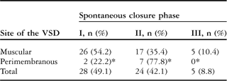
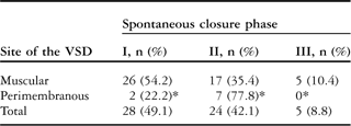
The closure rates of ventricular septal defects by site are presented in Table 1. Only 7.7% (4 out of 52) of the muscular ventricular septal defects remained unclosed as compared with 35.7% (5 out of 14) of perimembranous ventricular septal defects (p = 0.007). Most muscular ventricular septal defects, that is, 26 out of 48 (54.2%), closed in period I (p less than 0.05; Table 2). Most perimembranous ventricular septal defects, that is, 7 out of 9 (77.8%), closed in period II.
Closure of VSD by site.

VSD = ventricular septal defect
*p is 0.007 – muscular VSD versus perimembranous VSD
Spontaneous closure time by VSD site.
VSD = ventricular septal defect
I – from foetal period to 12 months postnatally; II – from 13 to 24 months postnatally; and III – from 25 to 36 months postnatally. *p < 0.05 – muscular VSD versus perimembranous VSD
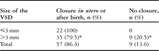
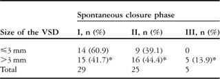
The size of the ventricular septal defect affected the spontaneous closure rate and time: all the ventricular septal defects less than or equal to 3 millimetres closed, whereas only 79.5% (35 out of 44) of the defects greater than 3 millimetres closed before the age of 3 years (Table 3); 60.9% (14 out of 25) of the defects less than or equal to 3 millimetres closed before the age of 1 year as compared with 41.7% (15 out of 36) of the defects greater than 3 millimetres. All defects that closed after the age of 2 years were greater than 3 millimetres (Table 4). After logarithmic conversion, the size of the ventricular septal defects was negatively correlated with closure rate and positively with closure period (Tables 5 and 6).
Closure rate by VSD size.
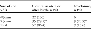
VSD = ventricular septal defect
*p < 0.05 – VSD ⩽ 3 mm versus VSD > 3 mm
Closure time by VSD size.
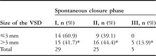
VSD = ventricular septal defect
I – from foetal period to 12 months postnatally; II – from 13 to 24 months postnatally; and III – from 25 to 36 months postnatally. *p > 0.05 – VSD ⩽ 3 mm versus VSD > 3 mm
Relationship of diameter and velocity of shunts to VSD closure rate.

VSD = ventricular septal defect
Data are mean ± standard deviation. *p < 0.05
Relationship of VSD diameter and velocity of shunt to spontaneous closure period.

Data are mean ± standard deviation. I – from foetal period to 12 months postnatally; II – from 13 to 24 months postnatally; and III – from 25 to 36 months postnatally. *p < 0.05
After logarithmic conversion, velocity of right-to-left flow was negatively correlated with closure rate (Table 5) but had no correlation with closure period (Table 6).
Discussion
The field of foetal cardiology has experienced considerable progress in the last few years, with new data on prognosis and prognostic factors and developments in intra-uterine interventions. Our report of 125 foetuses with ventricular septal defect represents an observational study of the natural history of ventricular septal defect diagnosed in utero and the factors related to spontaneous closure of ventricular septal defect. Ventricular septal defects closed in utero or during the postnatal period. Both the size and site of the ventricular septal defect play a significant role in its natural history, with the closure rate increased and the closure period early for small and muscular ventricular septal defects. Finally, the velocity of right-to-left flow across the ventricular septal defect affected the spontaneous closure rate and time.
Early studies have shown that the spontaneous closure of ventricular septal defect occurs mainly during the first postnatal year. Some recent studies have verified that ventricular septal defects can close in utero and that this event is not rare.Reference Nir, Driscoll and Edwards8, Reference Paladini, Palmieri, Lamberti, Teodoro, Martineni and Nappi9 The overall intra-uterine closure rate reached 32.5%.Reference Paladini, Palmieri, Lamberti, Teodoro, Martineni and Nappi9 However, only 3.0% of our cases (2 out of 66) closed in utero. The possible explanation for this discrepancy was the time at which foetal echocardiography was conducted. The mean gestational age at diagnosis was 29.3 weeks, ranging from 24 to 40 weeks, which was later than in the Paladini et alReference Paladini, Palmieri, Lamberti, Teodoro, Martineni and Nappi9 study (mean of 24.8 weeks). In our unit, the first foetal ultrasonography usually began after a gestational age of 20 weeks. Thus, many ventricular septal defects, especially small ventricular septal defects, could not be detected because they had closed at the time of examination. In addition, the Paladini et alReference Paladini, Palmieri, Lamberti, Teodoro, Martineni and Nappi9 analysis had some false-positive diagnoses.
Our study showed that ventricular septal defect closure significantly correlated with its size, site, and flow velocity from right to left. All ventricular septal defects less than or equal to 3 millimetres closed before the age of 3 years. This information may be useful during prenatal counselling to reassure parents of foetuses with a ventricular septal defect less than or equal to 3 millimetres as these ventricular septal defects tend to close spontaneously in most instances.
In contrast with perimembranous defects, muscular defects had a relatively higher postnatal closure rate and earlier closure time, as demonstrated by earlier research.Reference Alpert, Cook, Varghese and Rowe10 In general, the mechanism of closure involves apposition of peripheral tissues. For muscular ventricular septal defects, the muscle grows more quickly to benefit from spontaneous closure. Furthermore, for the mechanism of late intra-uterine spontaneous closure, some authorsReference Paladini, Palmieri, Lamberti, Teodoro, Martineni and Nappi9 speculated that this might be a delay in the physiological development of the heart.
Ventricular septal defect flow in the foetus is bidirectional.Reference Lethor, Marcon and de Moor11 However, no reports exist of the relationship between the velocity of flow across the ventricular septal defect and the natural history. In general, in postnatal life, the velocity of right-to-left flow implies the resistance of pulmonary circulation. Therefore, if the pulmonary resistance remains low in the postnatal period, the ventricular septal defect outcome may be better.
Our study has shown that isolated ventricular septal defects in foetuses can undergo spontaneous closure during intra-uterine and postnatal life. The spontaneous closure rate and time are significantly correlated with the size and site of ventricular septal defects and the velocity of right-to-left flow across the defect.
Acknowledgements
This study was supported by the Echocardiogram Room affiliated to the Shandong Provincial Hospital. Mei Zhu advised on the foetal echocardiogram.