Hepatitis C virus (HCV) is an important global cause of bloodborne infection. Infection is asymptomatic in 80% of infected persons, and 75% of the infected will progress to chronic HCV infection, with a 15%–30% risk of progression to hepatic cirrhosis and hepatocellular carcinoma within 20-30 years.Reference Hajarizadeh, Grebely and Dore 1 Since nucleic acid amplification screening for HCV in blood donors became routine in 2002, nosocomial HCV transmissions have been largely confined to hemodialysis units due to environmental contamination with HCV-positive blood, suboptimal compliance with standard precautions, unsafe handling of multidose heparin and anesthetic vials, and possible reuse of contaminated needles.Reference Fabrizi and Messa 2 – Reference Egro, Nwaiwu, Smith, Harper and Spiess 9 In a case-control study conducted in a skilled nursing facility with an outbreak of acute HCV infections, podiatry and international normalized (prothrombin time) ratio monitoring by phlebotomy were also noted to be significantly associated with case statusReference Calles, Collier and Khudyakov 10 ; however, the exact procedure of phlebotomy and the mechanism of HCV transmission were not identified in this study. Percutaneous exposure from a known HCV-positive patient carries a transmission risk of 1%–3%.Reference Yazdanpanah, De Carli and Migueres 11 The introduction of vacuum extraction tube system as a closed system for phlebotomy markedly reduced the risk of needlestick injury among healthcare workers. The vacuum extraction tube system contains a double-end needle with a patient end (ie, skin-needle) and non–patient end covered by a rubber sleeve (ie, sleeved-needle) that is screwed onto a blood collection tube holder. The tube holder facilitates the fitting of the sleeved-needle with the vacuum-specimen tube in place and protects healthcare workers from direct contact with the collected blood. Moreover, the system allows multiple samples to be taken from a single venipuncture. The best practice is to discard the double-end needle and tube holder as a single unit into a sharps container to minimize the risk of needlestick injury. The World Health Organization (WHO) 12 and the Occupational Safety and Health Administration (OSHA) recommend the use of a single-use disposable tube holder to reduce the risk of needlestick injury. 13 , 14 Manufacturers of tube holders either recommend using such devices as single use or performing disinfection with bleach after each use as reusable device. 15 , 16 Such tube holders are still available in the market and reusing tube holder without disinfection between patients has been a common practice in our public hospitals since its introduction. Disinfection was only advised if there was visible blood in the tube holder. Here, we report an outbreak of nosocomial transmission of HCV and our investigation of the reusable tube holder as a nosocomial source of infection because previous surveillance data in Hong Kong indicated a low incidence of HCV positivity among new blood donors.
Methods
Epidemiological investigation
An investigation of HCV infection in the liver transplant unit (a mixed-gender ward of 28 beds) at Queen Mary Hospital, Hong Kong, was conducted when a case of HCV genotype 6a infection was identified 14 months post liver transplant. The liver donor and all used blood products after the transplant were HCV negative, which excluded the liver donor and blood products as the source.
Since the incidence of HCV antibody positivity among the new blood donor were 0.04% (17 HCV new cases per 48,769 blood donor) and 0.06% (21 HCV new cases per 35,848 blood donor) in 1991 and 2016, respectively, 17 an outbreak was defined as 1 or more patients with newly diagnosed nosocomially acquired HCV (index case) linked epidemiologically to a known HCV carrier (potential source patient) associated with an environmental source during the incubation period, as described in our previous outbreak investigations.Reference Cheng, Chan and Ngan 18 – Reference Cheng, Sridhar and Wong 21 A case of nosocomially acquired HCV was defined as (1) baseline anti-HCV antibody and HCV RNA negativity, followed by (2) anti-HCV antibody seroconversion or a detectable HCV RNA. The index case was interviewed by infection control nurse for personal risk factors for community acquisition of HCV, such as body piercing, acupuncture, tattoo, unprotected sexual contact, and intravenous drug abuse. Moreover, 24 ward-based medical, nursing, and supporting staff in the unit were interviewed and observed by an infection control nurse for recent changes in clinical practice, patient care procedures and staff-to-patient ratio retrospectively. Ethics approval was obtained from the institutional review board of the University of Hong Kong/Hospital Authority Hong Kong West Cluster.
Medical record review and contact tracing
The medical record of the index case was reviewed for all patient-care episodes with risks of percutaneous or mucosal transmission: phlebotomy, blood transfusion, finger pricking for glucose sticks, endoscopy, peripheral and central intravenous catheter insertion, and intravenous injections of medications, including multiple-dose drug vials. Contact tracing was conducted to search for all known HCV carriers present in the same ward with the index case during the incubation period, as previously described.Reference Cheng, Chen and So 22 – Reference Cheng, Tai and Ng 24 A contact case was defined as a patient who stayed in the same ward as the index case. All contact cases who screened positive for HCV could be a potential source patient or a secondary case. Archived blood samples, if available, were retrieved in the laboratory for anti-HCV antibody and HCV RNA viral load assays. Contact cases without archived blood samples were called back for HCV testing.
Environmental surveillance
Environmental sites with direct or indirect contact of blood were collected for detection of HCV RNA. The phlebotomy trolley, tray, reusable tube holders of the vacuum extraction tube system for phlebotomy, and blood-glucose monitoring machine were swabbed using rayon-tipped Copan swab applicators (Copan, Brescia, Italy). The swabs in viral transport medium were sent for testing immediately. Environmental samples with detectable HCV RNA by RT-PCR were subjected to Sanger sequencing and phylogenetic analysis along with those of index and source patients.Reference Sridhar, Yip, Chan, To, Cheng and Yuen 25
In vitro demonstrations during simulated phlebotomy
To ascertain the possibility of HCV transmission through contaminated reusable tube holders, several in vitro experiments were performed. To demonstrate HCV transmission through reflux from a contaminated reusable tube holder toward the patient side, an in vitro experiment was performed in triplicate using HCV-contaminated plasma. First, 5 mm of the rubber sleeve capping the sleeved-needle was dipped into plasma from a known HCV-positive patient with an HCV RNA viral load of 1×10⁶ IU/mL. Then, the double-end needle was screwed onto the tube holder. The skin-needle was inserted into a tube fully filled with HCV-negative EDTA blood (tube A) at atmospheric pressure, to mimic the venous side of a patient. An empty EDTA vacuum-specimen tube (tube B) was then plunged onto the sleeved-needle by fitting into the tube holder, thus mimicking the phlebotomy process. An in-and-out motion was performed 3 times to simulate phlebotomy using 3 vacuum-specimen tubes. Blood samples from tubes A and B were tested for HCV viral load.
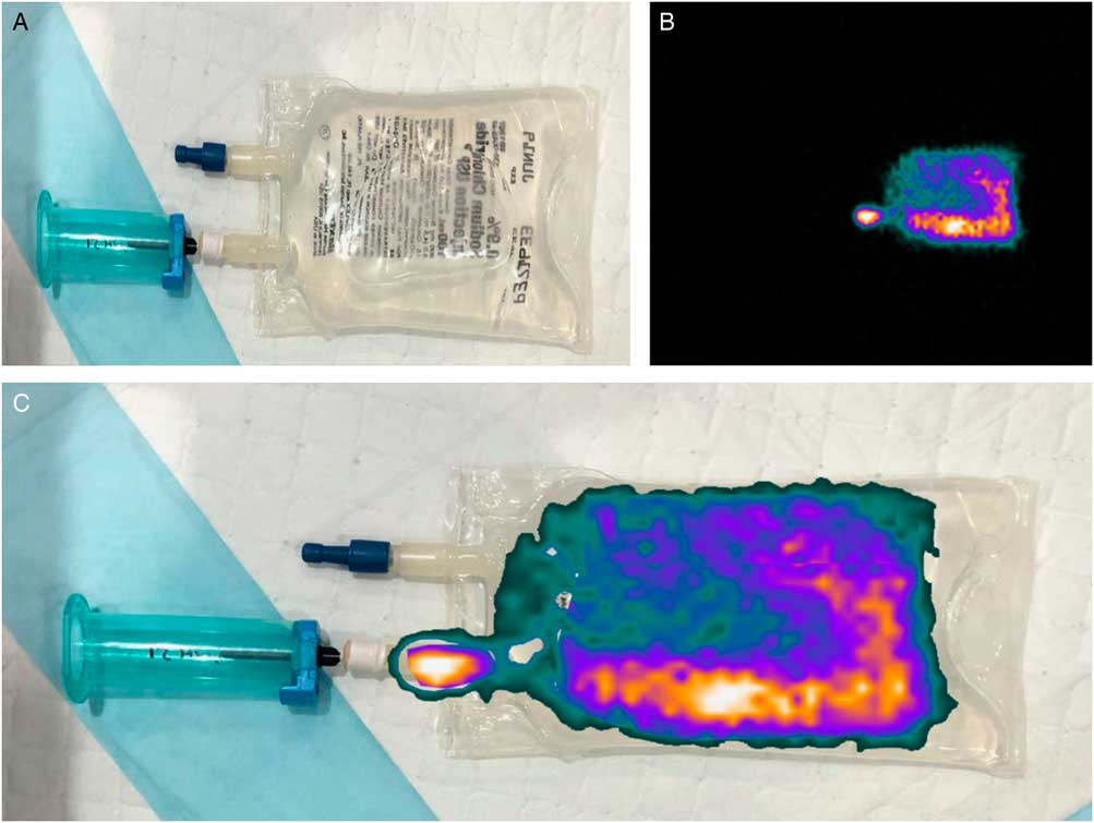
Next, we used radionuclide studies with 99mTc pertechnetate (99mTcO4) to simulate HCV-contaminated blood. This experiment was repeated 3 times by light smearing of the tip of the sleeved-needle with gauze containing a few drops of 99mTcO4. The sleeved-needle was then fitted onto the tube holder between the 2 detector heads of a dual-head SPECT/CT gamma scanner ready for acquisition of radioactivity readings. A saline-prefilled EDTA vacuum-specimen tube (mimicking the engorged vein of patient) was inserted onto the skin-needle, while empty vacuum-specimen tubes were inserted through the tube holder onto the sleeved-needle and then removed 3 times to simulate phlebotomy. Imaging with static acquisition was conducted for 200 seconds. To simulate and image the point of release of the tourniquet, a follow-up experiment was performed by inserting the skin-needle already fitted on the tube holder into a 100-mL saline bag under gentle manual pressure (simulating venous pressure). An EDTA vacuum-specimen tube containing a mixture of 99mTcO4 and saline was plunged into the sleeved-needle inside the tube holder and then withdrawn after 3 seconds to simulate phlebotomy. The manual pressure on the saline bag was released to simulate the release of the tourniquet. Imaging of the saline bag and tube holder was again conducted for 200 seconds.
Finally, we conducted in vivo testing to demonstrate the change in venous blood pressure during the release of a tourniquet at the end of phlebotomy (see online Supplementary File 1).
Data analyses
Patients’ serum samples were tested for the presence of HCV antibody using the ARCHITECT Anti-HCV chemiluminescence immunoassay (Abbott Laboratories, Wiesbaden, Germany) in the Architect i2000SR system (Abbott Diagnostics, Abbott Park, IL) according to manufacturer’s instructions. HCV RNA measurements were performed using the Abbott realtime HCV assay (Abbott Molecular, Des Plaines, IL) using the mSample Preparation System reagents, m2000sp and m2000rt instruments (Abbott Molecular, Des Plaines, IL). The HCV genotypes were determined using the Abbott realtime HCV genotype II assay (Abbott Molecular, Des Plaines, IL). Phylogenetic analysis of the HCV strains were performed using partial E1–E2 sequences obtained by polymerase chain reaction (PCR) using the SuperScript III One-Step RT-PCR System (Invitrogen, Carlsbad, CA) and an ABI 3130xl Genetic Analyzer (Applied Biosystems, Foster City, CA). The sequences were checked manually and trimmed for the construction of phylogenetic tree using the neighbor-joining method with ClustalX version 2.0 software. Detailed laboratory tests are described in Supplementary File 1. SPSS version 23 software (SPSS, Chicago, IL) was used to perform the statistical analyses.
Results
Epidemiological investigation
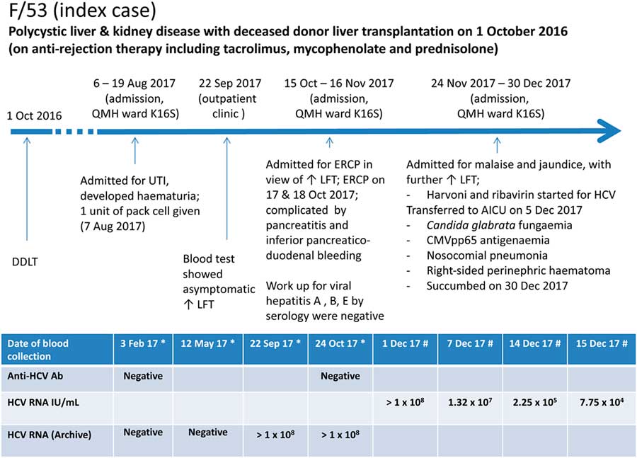
On December 5, 2017, a 53-year-old female (index case) who had received a post-deceased donor liver transplant 14 months previously for polycystic liver and kidney disease was confirmed to have HCV genotype 6a infection with a viral load of >1×10⁸ IU/mL. Archived blood samples were persistently negative for anti-HCV antibodies, while the HCV RNA was retrospectively found to have been positive since September 22, 2017 (Fig. 1). No personal risk factors for HCV acquisition were present. The liver donor and all used blood products after the transplant were confirmed to be HCV negative.
Clinical history of index patient with nosocomial acquisition of hepatitis C virus. *Retrospective analysis of archived blood samples; # prospective analysis of blood samples. Note. LFT, liver function test; DDLT, deceased donor liver transplantation; UTI, urinary tract infection.

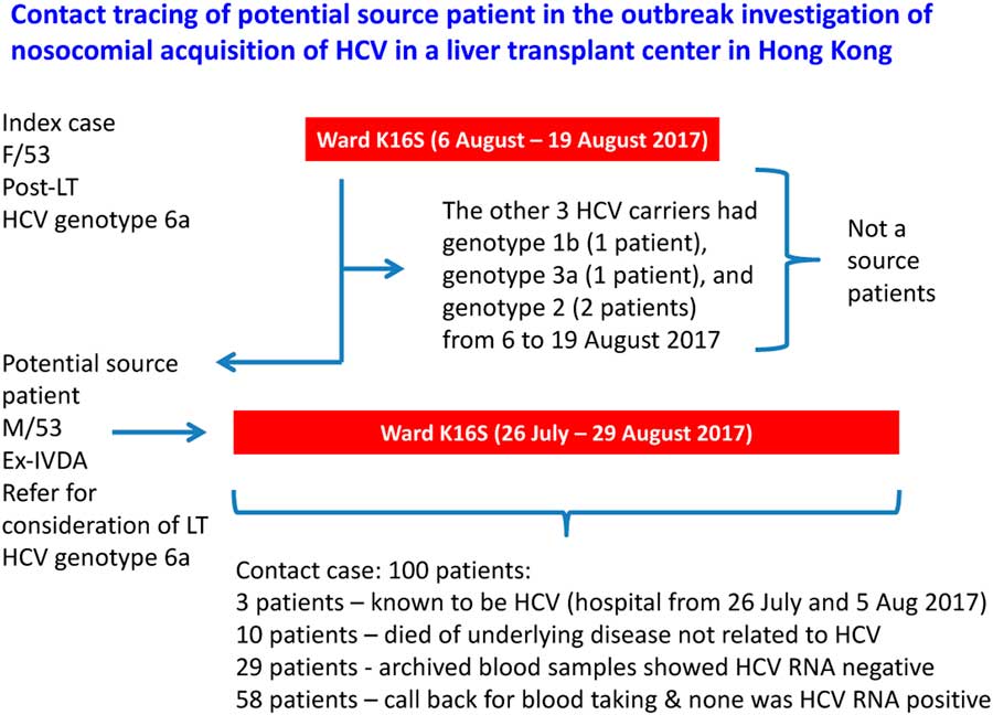
Contact tracing was conducted according to the incubation period of HCV (2 weeks to 6 months before the onset of liver function derangement), and the at-risk period of HCV acquisition for the index case was estimated to be March 22, 2017, to September 8, 2017. The index case had been hospitalized in the liver transplant center between August 6, 2017, and August 19, 2017. Also, 4 known HCV carriers stayed in the same ward during the index case’s hospitalization. Except for 1 patient with history of intravenous drug abuse who had HCV genotype 6a (potential source patient) and a viral load of 2.75×10⁵ IU/mL, the other 3 HCV carriers had HCV genotype 1 (1 patient), 1b (1 patient), and 3a (1 patient). Further contact tracing was performed for patients hospitalized in the same ward as this potential source patient during his stay, which yielded another 100 potential contact cases for HCV testing (Fig. 2). No other secondary cases of HCV infection were found.
Contact tracing of potential source patient in the outbreak investigation of nosocomial acquisition of HCV in a liver transplant center in Hong Kong. Note: IVDA, intravenous drug abuser; LT, liver transplantation.

Interviews with ward staff and observation of patient care practice initially did not reveal any irregularities in practice or change in the staff-to-patient ratio. A review of the time log in the barcoding system in the computerized laboratory information system showed that 14 instances of phlebotomy from the source patient were followed by phlebotomy from the index patient on the same day, whereas the reverse only happened in 5 instances. Further investigation showed that the phlebotomy trolley and the reusable tube holders were the only shared items between the source and index patients with risk of cross infection. Because it was determined that reusable tube holders were not properly disinfected between patients, all reusable tube holders were seized and were replaced with single-use disposable devices. Despite the viral load of HCV RNA decreasing to 7.75×10⁴ IU/mL after 2 weeks of Harvoni (ledipasvir/sofosbuvir) and ribavirin therapy, the index case succumbed with multi-organ failure.
Environmental surveillance
A total of 34 environmental samples were collected, including the inner surface (14 samples) and the outside surface (14 samples) from 14 tube holders, a glucometer tray (1 sample), a tray used for phlebotomy (3 samples), and a phlebotomy trolley (2 samples). HCV RNA (genotype 6a) was detected on the inner surface of a single tube holder.
In vitro demonstrations during simulated phlebotomy
Using HCV-contaminated plasma
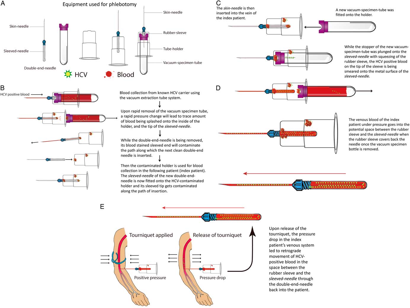
After the simulation test, EDTA tube A (fully filled with HCV-negative blood mimicking a patient’s vein) had a median HCV RNA load of 765 IU/mL (range, 85–2,041 IU/mL). As expected, the EDTA vacuum-specimen tube B (mimicking the blood collection side) had a higher median HCV RNA load of 10,728 IU/mL (range, 5,630–18,127 IU/mL). The possible mechanism of retrograde transmission of HCV-contaminated plasma is presented graphically (Fig. 3, A–E) and photographically (Supplementary Fig. 1).
Postulated mode of transmission of HCV from source to index patient through contaminated reusable tube holder. (A) The double-end needle is fitted onto the tube holder by a screw-on motion, with rubber sleeve capping sleeved-needle being ‘inside’ the tube holder. The skin-needle is then inserted into the patient’s vein for blood collection. The vacuum-specimen tube is plunged into the sleeved-needle by insertion into the tube holder. Blood is drawn automatically by negative pressure into the vacuum tube. (B) From above to below, blood collection from known HCV carrier. Upon removal of the vacuum-specimen tube, a rapid pressure change leads to a trace amount of blood being splashed onto the inside of the holder and the tip of the sleeve capping the sleeved-needle. While the double-end needle is being removed, its blood-stained sleeved tip contaminates the path along which the next clean double-end needle is inserted. The sleeved-needle of the new double-end needle is now fitted onto the HCV-contaminated holder and its sleeve tip gets contaminated by HCV-positive blood along the path of insertion. (C) The skin-needle is then inserted into the vein of the index victim patient. While the stopper of the new vacuum-specimen tube is plunged onto the sleeved-needle with compression of the rubber sleeve, the HCV-positive blood on the tip of the sleeve is being smeared onto the exposed metal surface of the sleeved-needle. (D) On removal of the vacuum-specimen tube, the rubber sleeve cap will recoil to its original position covering the sleeved-needle. The venous blood of the index patient under pressure goes into the potential space between the rubber sleeve and the sleeved-needle. (E) Upon release of the tourniquet, the relatively lower venous pressure above the tourniquet sucks the HCV-contaminated blood collected in the space between the rubber sleeve and the sleeved-needle through the double-end needle back into the patient’s vein.

Radionuclide studies using 99mTcO4
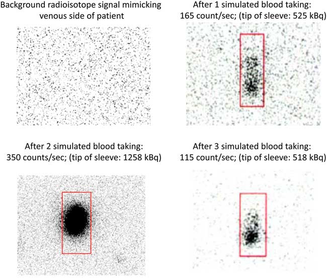
99mTcO4 was detected in all saline pre-filled EDTA tubes suggesting reflux from the sleeved-needle back to the patient side (Fig. 4). Furthermore, the amount of 99mTcO4 activity in EDTA tubes was proportional to the degree of radioactivity contamination on the rubber sleeve tip of the sleeved-needle and markedly above the background counting of the scanner detector (30 counts per second). In the second experiment, scintigraphic images obtained after release of manual pressure from the saline bag (simulating release of tourniquet) clearly showed reflux of radioactive material into the saline bag (simulating the venous patient side) (Fig. 5).
Detection of 99mTc pertechnetate (99mTcO4) in saline pre-filled EDTA tubes, suggesting reflux from the rubber sleeved-needle back to the patient side. Note. Bq, becquerel is the SI derived unit of radioactivity. One becquerel is defined as the activity of a quantity of radioactive material in which 1 nucleus decays per second.

Scintigraphic images obtained after release of manual pressure from the saline bag (simulating release of tourniquet) to demonstrate the reflux of radioactive material into the patient side. (A) Set-up of simulated blood collection experiment to demonstrate reflux of radioisotope from collection side of reusable tube holder to saline bag (signifying patient venous system), manual pressure was used to simulate effect of tourniquet on venous pressure. (B) Scintigram of saline bag following release of pressure showing radioactivity within the bag. (C) Fused scintigraphic image.

In vivo demonstration of change in venous pressure during release of a tourniquet
Upon release of the tourniquet, the mean (±standard deviation) venous pressure drop at sitting and lying positions, respectively, were 11.5±3.21 mmHg and 12.1±3.73 mmHg (right arm). The corresponding values were 12.0±2.4 mmHg and 12.0±2.79 mmHg (left arm), respectively, and the venous pressures were significantly different before and after release of the tourniquet (P<.01).
Phylogenetic analysis of HCV
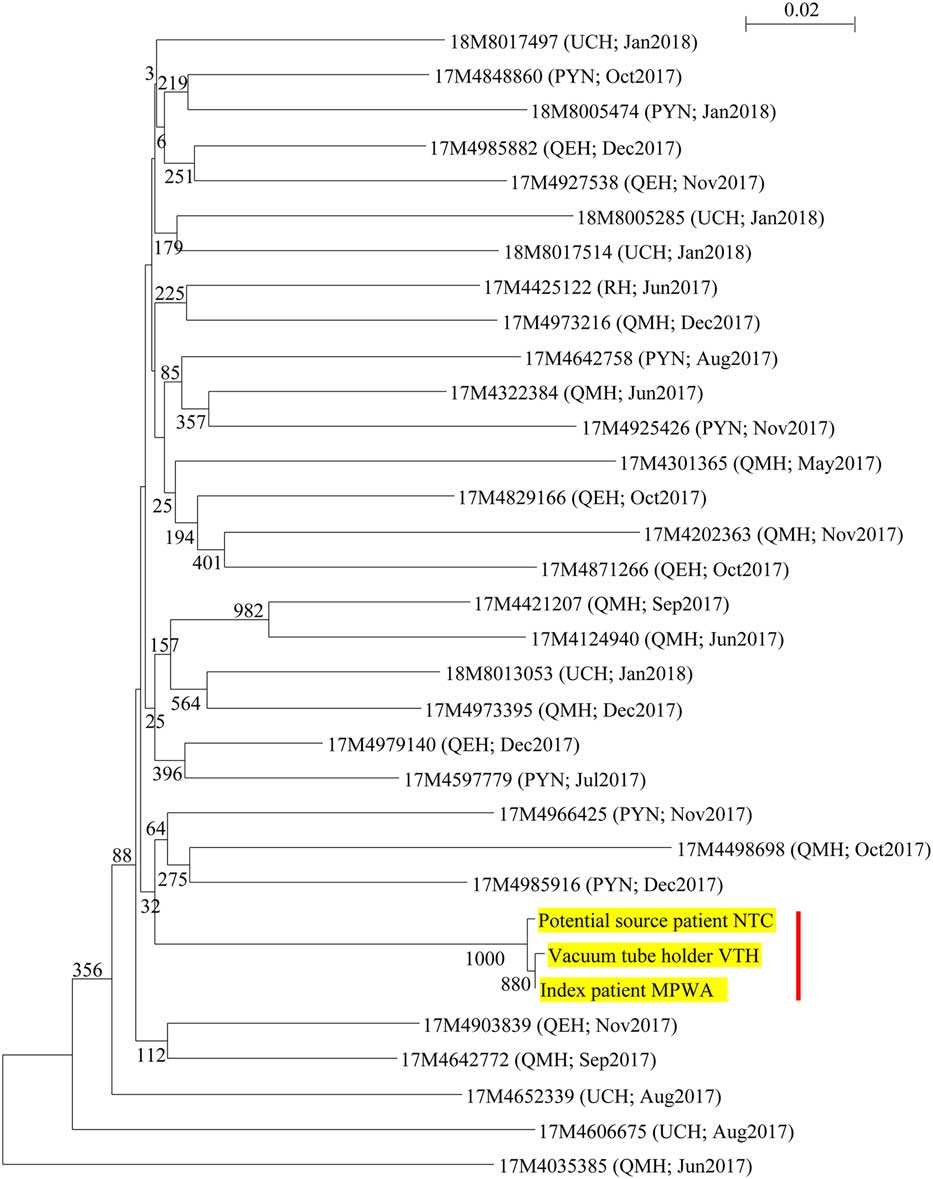
A phylogenetic tree was constructed using a partial envelope gene (E1–E2 with hypervariable region) of 653 nucleotide positions of HCV strains identified from the index patient, the potential source patient, and the environmental sample from the tube holder. The tree suggested clonality between the virus strains infecting the index case, the potential source patient, and the tube holder (Fig. 6).
Phylogenetic relationship of the index patient, potential source patient, and environmental sample using the hypervariable region of HCV partial envelope gene (E1–E2). A phylogenetic tree constructed using partial envelope gene sequences (E1–E2) of HCV genotype 6a strains (detected in the index case, the potential source patient, and the tube holder) and 30 epidemiologically unrelated patient samples collected from patients in Pamela Youde Nethersole Eastern Hospital (PYNEH, 7 patients); Queen Elizabeth Hospital (QEH, 6 patients); Queen Mary Hospital (QMH, 10 patients); Ruttonjee Hospital (RH, 1 patient); and United Christian Hospital (UCH, 6 patients) between May 2017 and January 2018 in this study. The tree was inferred from data using the neighbor-joining method with bootstrap values calculated from 1,000 trees. A total of 653 nucleotide positions in the E1–E2 region were included in the analysis. While the base-pair differences between the index case and the 30 epidemiologically unrelated control samples were 73 to 125, the number of base-pair differences between the index case and the potential source patient was 2, and the base-pair difference between the index case and inner side of tube holder was only 1, suggesting clonality between the virus strains infecting the index case, the potential source patient, and the tube holder (highlighted in yellow and with a red bar). Scale bars indicate estimated number of nucleotide substitutions per 50 nucleotides.

Discussion
Hepatitis C virus is primarily transmitted through percutaneous exposure to blood. However, no risk factors are identified in up to 40% of patients with HCV infection.Reference Manns, Buti and Gane 26 While a previous study noted international normalized (prothrombin time) ratio monitoring by phlebotomy as a risk factor for nosocomial transmission of HCV, the exact mechanism has never been ascertained.Reference Calles, Collier and Khudyakov 10 We postulate that HCV-positive blood can reflux through the double-end needle from the tip of the rubber sleeve that was inadvertently contaminated during needle insertion after the tube holder was used previously on a HCV-positive source patient (Fig. 4). Vacuum extraction tube systems are designed to reduce percutaneous injury during phlebotomy among healthcare workers.Reference Valls, Lozano and Yanez 27 It is generally assumed that blood under pressure from the tourniquet-treated vein always flows from the patient toward the vacuum-specimen tube during phlebotomy. However, our in vitro simulated phlebotomy experiment with HCV-positive plasma and radioisotope suggested that retrograde flow of blood is possible. Notably, the rapid removal of vacuum-specimen tubes from the sleeved-needle is often followed by a fine splash of HCV-positive blood that contaminates the inner surface of tube holders. Moreover, during the removal of this double-end needle, the HCV-contaminated sleeve tip inadvertently contaminates the fitting hole of the tube holder and the inner surface of tube holders. When a clean sleeved-needle is inserted through this HCV-contaminated fitting hole, the tip of this clean sleeve capping will be easily contaminated along the path of insertion, especially when the double-end needle is inserted at a slight angle. When another vacuum-specimen tube is inserted through the tube holder, HCV particles contaminating the tip of the new sleeve will be smeared onto the sleeved-needle by the stopper of the vacuum-specimen tube while compressing the rubber sleeve, thus exposing the sleeved-needle. On withdrawal of the vacuum-specimen tube, the rubber sleeve capping the sleeved-needle will recoil and regain its original position, and the space between the rubber sleeve and the HCV-contaminated sleeved-needle will be filled by venous blood under positive pressure from the tourniquet-treated arm. Once the tourniquet is released, the blood in the vein below the tourniquet will flow back to the heart while sucking the HCV-contaminated blood collected in the space between the sleeve and the sleeved-needle back into the patient’s vein. Our in vivo experiment confirmed that there is a significant drop in venous pressure of at least 11 mmHg during the release of a tourniquet, regardless of sitting or lying position. The environmental stability of HCV facilitates transmission via this route. The HCV remains infective for up to 6 weeks after drying on inanimate surfaces at room temperature,Reference Paintsil, Binka, Patel, Lindenbach and Heimer 28 and HCV infectivity was detectable for up to 5 months in a liquid environment at lower temperatures.Reference Ciesek, Friesland and Steinmann 29 Thermostability tests showed that different HCV genotypes possess comparable environmental stability.Reference Doerrbecker, Meuleman and Kang 30 Also, HCV RNA viral loads showed no significant differences after 10 freeze–thaw cycles in serum and plasma samples.Reference Comert, Aktas and Terzi 31 Therefore, it is not surprising that HCV RNA was detected inside the reusable tube holder in the ward after 3 months.
Our radioisotope experiment illustrated that the HCV inoculum contaminating the sleeve tip capping the sleeved-needle during phlebotomy (and hence, the probability of transmission) may depend on the viral load of the original source HCV-infected patient. Based on a recent study showing the presence of hemoglobin and HCV on surfaces even without visible blood contamination,Reference Girou, Chevaliez and Challine 32 we believe that simply disinfecting with alcoholic wipes or discarding only tube holders with visible blood stains is insufficient to effectively eliminate the risk of HCV transmission via this route. One previous report implicated the reuse of tube holders without disinfection between patients as a possible source of HCV outbreak involving 6 patients in an orthopedic ward, when the number of phlebotomies exceeded tube-holder consumption.Reference Ross, Viazov and Khudyakov 33 However, environmental surveillance was not performed in that study.
In addition, the rubber stopper of the vacuum-specimen tube can be contaminated during contact with the contaminated inner surface of tube holder, further contaminating the environment. Lapses in infection control measures, especially hand hygiene, can carry environmental HCV to other patients during phlebotomy and other percutaneous procedures. However, direct observation of the nursing procedures did not support this route of transmission.
This study has several limitations. It is a retrospective investigation, and we have only inferred what could have occurred at the time of transmission. With only 1 case, a robust case-control study could not be performed. Although there were ≤3 base-pair differences in the sequence of hypervariable region of HCV partial envelope gene (E1–E2) among the index patient, source patient, and tube holder, which suggests clonality, it was not possible to determine the direction or exact mechanism of transmission. However, given that RNA viruses have an extremely high mutation rate, that the blood of the index and source patients, that the environmental sample were collected 4 months apart, and most importantly, the lack of disinfection of the tube holder between patients, we conclude that the tube holder was the most likely vector of HCV transmission.
In summary, a single-use disposable tube holder should be used for phlebotomy to prevent needlestick injury according to OSHA and WHO guidelines, which will also minimize patient-to-patient transmission of bloodborne viruses.
Acknowledgments
The authors thank Dr Hin Chu and Wen Lei for the illustrations used in the figures, and the technical support from the staff at the Department of Microbiology.
Financial support
This work was supported in part by the donations of Mr. Michael Tong, Providence Foundation Ltd (in memory of the late Lui Hac Minh) and the Hong Kong Hainan Commercial Association. Funding was also received from the Consultancy Service for Enhancing Laboratory Surveillance of Emerging Infectious Diseases of the Department of Health, Hong Kong Special Administrative Region and from the Collaborative Innovation Center for Diagnosis and Treatment of Infectious Diseases, the Ministry of Education of China.
Conflicts of interest
All authors report no conflicts of interest relevant to this article.
Supplementary material
To view supplementary material for this article, please visit https://doi.org/10.1017/ice.2018.175