Introduction
Hypoplastic left heart syndrome is a condition which encapsulates a wide spectrum of morphologic variants that result in a single functional ventricle. These patients typically undergo a three-staged surgical repair that includes an operation in the first days of life. Reference Šamánek, Slavík, Zbořilová, Hroboňová, Voříšková and Škovránek1 Alternative approaches to the first stage (Norwood procedure) include a combined surgical and interventional approach, placing bilateral pulmonary artery bands and a stent in the ductus arteriosus. Reference Norwood, Lang, Hansen, Lang and Hansen2,Reference Norwood, Kirklin and Sanders3,Reference Gibbs, Wren, Watterson, Hunter and Hamilton4
A fully percutaneous approach to the first palliative stage was first described in 2006 using modified vascular occlusion devices as pulmonary artery flow regulators and ductus stenting. Reference Chan, Mashburn and Boucek5 This concept has since been adapted to using modified microvascular plugs (MVP, Medtronic Inc., Minneapolis, MN) in which parts of the polytetrafuoroethylene (PTFE) coverings are punctured by scalpel or perforated with a low-temperature fine-tip Bovie cautery tool (Symmetry Surgical, Antioch, TN). Reference Nageotte, Shahanavaz, Eghtesady and Balzer6–Reference Boucek, Mashburn and Chan8
The amount of disruption to the PTFE membrane has been discussed previously; Reference Schranz, Esmaeili and Akintuerk7,Reference Kizilski, Recco and Sperotto9–Reference Khan, Hoskoppal and Kumar12 however, the degree of flow restriction provided by each modification is poorly defined. In this study, we constructed a bench-top model to quantify the flow effect that varying amounts of disruption of PTFE membrane had on a controlled flow, single-ventricle simulation circuit.
Methods and materials
We designed an in vitro experiment using a 2.5:1 scale 3D-printed acrylic phantom to simulate the branch pulmonary arteries in a single-ventricle circulation (Figure 1). The model was printed from the 3D segmentation of CT imaging of a 2.5 kg neonate with hypoplastic left heart syndrome (Figure 2a). The model was incorporated into an open, hydrostatically controlled flow circuit using silicone tubing. Water was used as the flow medium. Flow was driven by connecting this model to a pulsatile flow pump (Harvard Apparatus Pump Model 1423) (Figure 2b). The pump was set to provide a stroke volume of 100 L/min and a beat frequency of 20 beats per minute. Outlets representing the systemic and pulmonary arteries were connected to a hydrostatic reservoir positioned at the same height as the model to maintain consistent pressure conditions. The mean flow to each limb of the circuit was measured using ultrasonic flow sensors (Transonic Systems Inc. H9XL) placed at systemic and pulmonary outflows. Resistance was increased in each limb by tightening a flow regulator valve placed at the distal extreme of the tubing. Flow distributions were then calculated at baseline without the placement of the experimental flow restrictors (MVPs) with pulmonary-to-systemic flow (Qp:Qs) ratios of 2:1, 3:1, and 4:1, aimed to simulate the physiology of neonates with single-ventricle circulation (i.e., Qp:Qs of 2:1 was a ratio of 2 times the amount of flow going through the pulmonary outlets compared to the systemic outlet).
Schematic of the bench top model for in-vitro experiment. A 3D printed model was connected via silicon tubing to a pulsatile flow pump. Resistance was altered in each limb by tightening a flow regulator valve placed at the distal extreme of the tubing. The mean flow to each limb of the circuit was measured using ultrasonic flow sensors placed at systemic and pulmonary outflows.

Figure 1. Long description
Schematic of the bench top model for in-vitro experiment. A 3D printed model was connected via silicon tubing to a pulsatile flow pump. Resistance was altered in each limb by tightening a flow regulator valve placed at the distal extreme of the tubing. The mean flow to each limb of the circuit was measured using ultrasonic flow sensors placed at systemic and pulmonary outflows.
Bench-top model and modified MVP set up. A. A 3D printed phantom model of branch pulmonary arteries was used. B. Flow was driven by connecting the model to a pulsatile flow pump (Harvard Apparatus Pump Model 1423). C. Symmetrical slits were made using a scalpel in the PTFE end caps of each device as follows: 1-slit, 2-slit, 3-slit and 4-slits.

Figure 2. Long description
Bench-top model and modified MVP set up. A. A 3D printed phantom model of branch pulmonary arteries was used. B. Flow was driven by connecting themodel to a pulsatile flow pump (Harvard Apparatus Pump Model 1423). C. Symmetrical slits were made using a scalpel in the PTFE end caps of each device as follows: 1-slit, 2-slit, 3-slit and 4-slits.
To ensure fidelity between the scaled up in vitro model and the neonatal physiology represented, Reynolds and Womersley numbers were calculated to establish pump parameters. Reference Ho, Hribernik and Shillaker13,Reference Medar, Kumar and Choi14 The Reynolds number represents the ratio of inertial to viscous forces, whereas the Womersley number represents the ratio of pulsatile to viscous effects. Reynolds and Womersley numbers are defined using the below equations:
 $Re = \rho QD / \mu A$
 $Wo = D(\omega \rho / \mu)^{1/2}$
where ρ is the density of fluid, Q is the maximum volumetric flow rate at the main pulmonary artery, D is the diameter of the main pulmonary artery, μ is the dynamic viscosity, A is the area of the main pulmonary artery, and ω is the angular frequency. The mean volumetric flow rate was converted to maximum volumetric flow rate (Q) by multiplying the mean volumetric flow rate measured by a factor of 2.65. Physiological blood flow in the normal neonatal circulation can be approximately estimated by a Reynolds number ranging from 1,200 to 3,600 and a Womersley number of 15. Reference Ho, Hribernik and Shillaker13–Reference Kurtz, Alsoufi, Wilkens and Kim15 Using the above formulae, Reynolds numbers of 2640, 3381, 4007, and 4489 for Qp:Qs ratios of 1:1, 2:1, 3:1, and 4:1, respectively, and a constant Womersley number of 15 were used.
The model was run initially without positioning experimental devices to obtain a baseline without restriction. The silicone tubing immediately adjacent to the printed model had an internal diameter of 5 mm, and therefore, two microvascular plug (MVP) 9Q devices were selected to ensure adequate apposition of the device to the internal wall of the silicone tubing to reduce the risk of migration and the likelihood of para-device leak. Two MVP-9Q devices were placed (one in the left and one in the right pulmonary artery equivalent) with the following modifications made in the PTFE membrane of the individual devices: intact (no slit), 1-slit, 2-slits, 3-slits, and 4-slits. A sharp scalpel was used to create a uniform 1 cm slit in the inferior aspect of the MVP device. Each slit was objectively measured and subjectively assessed to be uniform in size and length prior to being placed in the bench-top model (Figure 2c).
Flow rates were measured using a flow sensor (Transonic Systems Inc. H9XL) placed in the circuit limb representing the systemic output and the left and right pulmonary arteries. Data from each vessel was collected for 30 seconds and synchronised based on a reference signal to ensure consistent flow values. Flow volume (L/cycle) was calculated by integrating the obtained flow waveform over the measured cycles and measuring an area under the curve (Supplementary Figure 1). Flow distribution between systemic and pulmonary flow was then calculated as a percentage of the total (%), and then the pulmonary-to-systemic flow ratio (Qp:Qs) was calculated.
Results
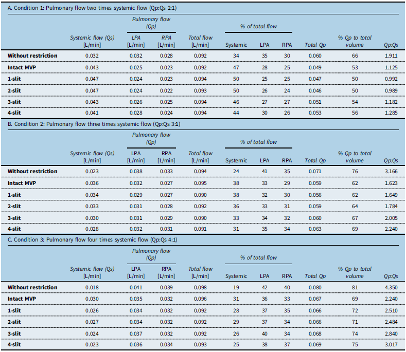
Table 1 illustrates the flow distribution at baseline (without MVP devices) and following insertion of modified MVPs (intact, 1-slit, 2-slit, 3-slit, and 4-slit) in the pulmonary artery limbs of the 3D phantom model. These restriction models were repeated in three shunt conditions (2:1,3:1,4:1).
Flow distribution in three shunt conditions at baseline (without MVPs) and following insertion of modified MVPs in the pulmonary artery limbs of the 3D phantom model

Table 1. Long description
Flow distribution in three shunt conditions at baseline (without restrictor devices) and following insertion of modified MVPs in the pulmonary artery limbs of the 3D phantom model.
Qs = systemic flow; Qp = pulmonary flow; LPA = left pulmonary artery; RPA = right pulmonary artery.
With a model representing a 2:1 Qp:Qs ratio at baseline and devices placed in both pulmonary arteries simultaneously, unmodified MVPs produced a significant decrease in pulmonary blood flow and Qp:Qs decrease to approximately 1:1. A single slit in the covering of the PTFE resulted in a 23% total reduction in pulmonary flow (Qp) while two slits resulted in a 21% reduction (Figure 3). A corresponding increase (directly measured) in systemic flow was seen with placement of modified MVPs in both left and right pulmonary branch limbs of the 3D model (1-slit: 14% increase, 2-slit: 11% increase). Two symmetrical slits in the PTFE membrane resulted in a Qp:Qs ratio closest to 1:1. There was a subsequent increase in pulmonary flow with each subsequent slit (3-slits: 18% reduction and Qp:Qs 1.1:1; 4-slits: 16% reduction and Qp:Qs 1.3:1).
Pulmonary to systemic (Qp:Qs) ratio with varying modified MVPs. Change in the ratio of pulmonary (Qp) to systemic (Qs) flow with each MVP modification in conditions with prescribed baseline Qp:Qs. The red line represents the response with a baseline setting of Qp:Qs= 2:1; the black line with a Qp:Qs= 3:1 and the blue line represents a Qp:Qs= 4:1.

Figure 3. Long description
Pulmonary to systemic (Qp:Qs) ratio with varying modified MVPs. Change in the ratio of pulmonary (Qp) to systemic (Qs) flow with each MVP modification in conditions with prescribed baseline Qp:Qs. The red line represents the response with a baseline setting of Qp:Qs= 2:1; the black line with a Qp:Qs= 3:1 and the blue line represents a Qp:Qs= 4:1.
Higher baseline flow ratio states (Qp:Qs 3:1 and Qp:Qs 4:1) yielded a similar pattern, but the resulting calculated flow distributions with the modified MVPs suggested less flow restriction. Even with intact MVPs, without any modifications to the PTFE membrane, the flow ratio only dropped to the equivalent of a Qp:Qs of 1.6:1 and 2.25:1, respectively.
Discussion
The use of trans-catheter pulmonary artery flow restrictors as an alternative to surgical pulmonary artery banding represents a significant evolution in the palliation of neonates with hypoplastic left heart syndrome and other single-ventricle physiologies. It lowers overall risk by avoiding open-chest surgery and helps avoid cardiopulmonary bypass during the vulnerable neonatal period. Reference Sperotto, Lang and Nathan11,Reference Haddad, Bentham, Adel Hassan, Al Soufi, Jaber, El Rassi and Kasem16 Neonates with multiple risk factors, including prematurity, low birth weight, major comorbidities, ventricular dysfunction, or severe atrioventricular valve regurgitation, may benefit more from this approach. Reference Sperotto, Lang and Nathan11 A fundamental requirement of a completely percutaneous stage I palliation is the ability to reliably and predictably control pulmonary blood flow, achieving a hemodynamic balance between systemic and pulmonary blood flow. Reference Chan, Mashburn and Boucek5,Reference Nageotte, Shahanavaz, Eghtesady and Balzer6 Despite thoughtful approaches towards the modification of MVPs and the use of dynamic angiographic pulmonary artery assessment for device sizing, multiple reports have highlighted difficulties in achieving and maintaining an appropriate hemodynamic balance. This suggests a lack of understanding of the degree of flow restriction provided by current modification strategies. Reference Chan, Mashburn and Boucek5,Reference Boucek, Mashburn and Chan8,Reference Alnoor and Ing10–Reference Khan, Hoskoppal and Kumar12,Reference Haddad, Bentham and Pardo17 This is the first study to use a model that couples progressive modification of microvascular plugs with different baseline Qp:Qs ratios to determine the resultant degree of flow restriction achieved.
Our data supports a conservative approach to MVP modification. Our central finding—that one or two slits yielded an effective reduction in our model of pulmonary flow with a Qp:Qs ratio closest to the physiologically desirable 1:1—has important clinical implications. Although serial increases in the number of slits did not create a directly proportionate increase in flow, 3- and 4-slit models routinely conferred minimal effective flow reduction. This non-linear, progressive correlation between slit number and flow reduction highlights the delicate balance required when modifying microvascular plugs to achieve optimal hemodynamic outcomes. The addition of a third and fourth slit resulted in minimal restriction, especially at higher pulmonary output states, indicating that excessive device modifications may obviate efficacy in controlling pulmonary flow. This is particularly critical in hypoplastic left heart syndrome physiology, where even modest pulmonary over-circulation can compromise systemic perfusion.
With elevated baseline pulmonary flow (Qp:Qs ratios 3:1 and 4:1), even one- or two-slits placed in the devices achieved only modest pulmonary flow reductions. This finding reveals a limitation of current MVP-based flow restriction strategies in patients with markedly excessive pulmonary blood flow or low pulmonary vascular resistance. Additionally, this likely highlights the structural limitation of this device as an effective pulmonary artery flow restrictor. The data recorded with no modification (i.e., an intact MVP) can be explained by several compromises in both the MVP as a vascular flow restrictor and our experimental model. Our use of water as the flow medium may have permitted non-physiologic flow through the PTFE membrane of the device. This may be different using blood as the flow medium, as platelet and fibrin deposition in the MVP after days of implantation would likely further increase the amount of flow restriction. Furthermore, the relatively stiff, completely circular, non-compliant silicone tubing may be less forgiving than vascular walls, leading to flow around the compressed device, not subject to the PTFE membrane, modified, or otherwise (Supplementary Figure 2). The experimental setup for the systemic and pulmonary circuits was also configured to provide identical outlet pressures. Independent control of systemic and pulmonary outlet pressures would likely provide a more physiologically realistic assessment of device performance.
Clinical experience with modified MVP pulmonary artery flow restrictors in patients has revealed significant challenges in precisely regulating pulmonary blood flow, frequently leading to inadequate pulmonary flow restriction. Follow-up catheterisations frequently demonstrated elevated Qp:Qs (>2:1), accompanied by elevated mean distal pulmonary artery pressures despite employing techniques to calibrate the size of the fenestrations. Reference Nageotte, Shahanavaz, Eghtesady and Balzer6 This bench-top study provides valuable insight into how modifying the PTFE membrane of MVPs influences flow dynamics. It also highlights the inherent limitations of this device as a flow restrictor and the inaccuracy of trying to prescribe pulmonary blood flow based on the modifications (e.g., number of slits) made. One hypothesis suggests that continued high pulmonary flow results from flow occurring around the perimeter of the device rather than strictly through the central fenestration. Furthermore, the thin, semi-permeable PTFE membrane may fail to impose enough resistance when subjected to systemic pressures, represented in our model with significant flow still through the MVP with no slits. Future bench-top models may focus on evaluating potential para-device flow paths relative to the size and compliance of the silicon tubing and the physical conformation of the modified MVP to that tubing.
Limitations
Using a 3D-printed pulmonary artery phantom and physiological pulsatile flow conditions, this bench-top model provides a rigorous framework for understanding MVP modification effects on flow. One potential limitation of the bench-top model was the decision to non-dimensionalise the model rather than use a blood analogue in estimating flow. Although non-dimensionalisation and experimental conditions were adjusted to simulate physiological conditions, water’s Newtonian properties and membrane permeability may have resulted in lower resistance to flow and altered pressure-flow relationships, which may have led to an overestimation of pulmonary flow across the pulmonary artery flow restrictors.
Furthermore, patient-specific anatomical variability—particularly in pulmonary artery size and geometry and compliance—was not represented in our model. Although we mitigated the structural limitations of our model as much as possible, we recognise that to make it feasible we had to scale the size up by a factor of 2.5. The model was created by a combination of 3D-printed silicone-like material and thin-walled silicone tubing which will not behave like pulmonary vasculature in terms of its compliance nor its interaction with an implanted modified MVP. The lack of compliance of the printed model when compared to neonatal pulmonary arteries probably allowed flow “past” the devices, falsely elevating the estimated Qp:Qs. The temporal longitudinal nature of flow restriction is not accounted for in our model. The behaviour of the device over time in the setting of platelet aggregation and thrombus and fibrin deposition was not in the scope of this project.
The diminishing marginal effect of additional slits emphasises the need for precision in PTFE modification and suggests potential benefits from developing standardised devices with predefined flow-limiting geometries to reduce inter-operator variability. We must continue to work innovatively to bring such devices to market. It also raises the question about the method of modifying the microvascular plug. Conceptually, placing a perforation using a low-temperature Bovie device in the PTFE membrane is likely to produce a more stable, predictable defect, where there has been concern that slicing the PTFE with a scalpel leads to extension of this tear to effectively open one full cell of the MVP structure. In future, dynamic flow assessment tools such as 4D MRI and computational fluid dynamics (CFD) may further enhance pre-procedural planning and device customisation based on patient-specific anatomy and physiology.
Conclusion
Trans-catheter pulmonary artery flow restriction has been demonstrated to be a clinically safe and feasible alternative to pulmonary artery banding as an initial palliation strategy in patients with single-ventricle physiology with the aim to bridge patients to surgical palliation or transplant. Its relative efficacy has not yet been effectively tested. Our model highlights the non-linear relationship between the number of slits placed in the microvascular plug and the Qp:Qs ratio achieved across several flow ranges. Although the limitations described above mean that the absolute numbers are unlikely to be accurate, the relative effects of placing additional slits should provide some clinical correlates. The creation of one or potentially two-slits in the PTFE membrane may be optimal to achieve a balanced systemic and pulmonary output.
Further studies, including expanding this assessment to quantify other modification strategies and incorporation of 4D MRI flow mapping to bench-top flow dynamic studies, may be useful in predicting changes in the hemodynamics following modified MVP placement in patients with varying cardiac anatomies. Furthermore, this study highlights that collaborative efforts are necessary to fabricate future devices with an architecture specifically designed to function as predictable and safe pulmonary artery flow restrictors, overcoming the documented limitations of the modified MVP.
Supplementary material
The supplementary material for this article can be found at https://doi.org/10.1017/S1047951126123932.
Data availability statement
The data that support the findings of this study are available upon reasonable request.
Acknowledgements
None.
Financial support
None.
Competing interests
None.