Introduction
Stem cells are an important cell source in tissue engineering and regenerative medicine due to their ability to self-renew, migrate, and differentiate into a wide range of tissue-specific cells, including adipogenic (fat), osteogenic (bone), myogenic (muscle), and chondrogenic (cartilage) cells.[Reference Engler, Sen, Sweeney and Discher1–Reference Guvendiren and Burdick6] It is now a well-known fact that that stem cells are responsive to their microenvironment.[Reference Burdick and Vunjak-Novakovic7] In this regard, chemical, mechanical, and topographical cues present in the microenvironment influence stem cell behavior, including adhesion, proliferation, migration, and differentiation.[Reference Burdick and Vunjak-Novakovic7–Reference Brodbeck, Shive, Colton, Nakayama, Matsuda and Anderson13] Recent studies have shown that the topography of the substrate alone regulates differentiation of the stem cells.[Reference Guvendiren and Burdick14–Reference Guvendiren and Burdick21]
An induction media condition comprised soluble biologic cues is generally required to drive stem cells toward a certain phenotype in vitro. Thus, it is of great importance to decouple media-induced induction from material-based (chemistry, stiffness, and topography) stimulation.[Reference Marklein and Burdick8] One of the outstanding model culture media systems for stem cells is a 1:1 mixed adipogenic/osteogenic induction media.[Reference McBeath, Pirone, Nelson, Bhadriraju and Chen16] This model culture system allows investigation of stem cell phenotype decision between adipogenic and osteogenic lineages solely based on material and/or scaffold properties. Adipogenic and osteogenic cells have very distinct morphologies. Adipogenic cells are usually rounded with low spreading, allowing for maximum lipid storage,[Reference Grigoriadis, Heersche and Aubin5, Reference Gregoire, Smas and Sul22] whereas osteogenic cells exhibit higher degrees of cell spreading, which has been shown to promote calcium deposition of osteocytes during bone remodeling.[Reference Sikavitsas, Temenoff and Mikos23] Using this model culture system, Chen and co-workers performed single-cell experiments on surfaces with restricted adhesive regions, and demonstrated that the size and shape of the adhesive regions controlled cellular contractility and differentiation of human mesenchymal stem cells (hMSCs).[Reference McBeath, Pirone, Nelson, Bhadriraju and Chen16] They showed that cellular spreading due to myosin-generated contractility results in higher levels of Ras homolog gene family member A (RhoA), Rho-associated protein kinase (ROCK), and myosin light chain phosphorylation, leading to osteogenic differentiation.[Reference McBeath, Pirone, Nelson, Bhadriraju and Chen16] Ruiz and Chen showed the emergence of patterned stem cell differentiation toward adipogenic or osteogenic lineage within multicellular aggregates confined in different geometries.[Reference Ruiz and Chen15] They showed that the contractile activity of the cells could either be hindered or enhanced spatially for the multicellular aggregates based on the surface topography they reside on.[Reference Ruiz and Chen15] Following this study, Mrksich and co-workers demonstrated the effect of curvature on adipogenic and osteogenic differentiation of MSCs on two-dimensional (2D) cell adhesive patterns.[Reference Kilian, Bugarija, Lahn and Mrksich24] They demonstrated that contractile cells (residing on the curved regions of flower, star, and holly shapes) display enhanced activation of c-JNK (Jun N-terminal kinase) and ERK (extracellular related kinase) 1/2 with elevated wingless-type (Wnt) signaling promoting osteogenesis.[Reference Kilian, Bugarija, Lahn and Mrksich24] In addition, Guvendiren and Burdick defined a morphological relationship based on cell aspect ratio (AR) and area to predict lineage commitment of hMSCs between adipogenic and osteogenic fate using hydrogel surface wrinkling patterns.[Reference Guvendiren and Burdick14] They observed that cells with spread areas <103 µm2 and AR <4 differentiated into adipogenic progenitors, whereas cells outside of this morphological region always differentiated into an osteogenic phenotype.[Reference Guvendiren and Burdick14] Furthermore, Treiser et al. developed a high content imaging and single-cell modeling approach to forecast lineage commitment of stem cells from very early (24 h) variations in cell shape and cytoskeletal organization.[Reference Treiser, Yang, Gordonov, Cohen, Androulakis, Kohn, Chen and Moghe25] Although these studies have shown the importance of substrate geometry on stem cell behavior, the explored systems are 2D culture systems, and do not necessarily represent the three-dimensional (3D) microenvironment of stem cells. In this regard, Viswanathan el al. used polymerized high internal phase emulsion (polyHIPE) foams with open or closed pore structures with different levels of connectivity as well as pore surface topology in 3D.[Reference Viswanathan, Ondeck, Chirasatitsin, Ngamkham, Reilly, Engler and Battaglia26] They demonstrated that the osteogenic differentiation of human mesenchymal progenitor (hES-MP) cells is controlled by the interconnectivity, architecture and surface topology of the foams.[Reference Viswanathan, Ondeck, Chirasatitsin, Ngamkham, Reilly, Engler and Battaglia26] However, the effect of curvature in a 3D setting has not yet been studied.
The 3D printing is considered a revolutionary manufacturing technique to fabricate custom-designed, highly complex scaffolds for tissue engineering applications.[Reference Guvendiren, Molde, Soares and Kohn27–Reference Jürgen, Thomas, Torsten, Jason, Dong-Woo, Paul, Brian, Gabor, Qing, Vladimir, Lorenzo, Makoto, Wenmiao, Shoji, Giovanni, Tim, Tao, James and Jos31] In this work, we took advantage of 3D printing to design and fabricate 3D scaffolds in various shapes, including square, hexagonal, and octagonal, with different curvatures. For this purpose, we chose to use poly(tyrosol carbonate) (PTyC). Tyrosol is readily available from natural resources such as olive oil mill waste waters, and is classified as GRAS (generally recognized as safe) by the FDA (Food and Drug Administration). Kohn and co-workers developed a wide range of tyrosine-derived polycarbonates with tunable properties,[Reference Tangpasuthadol, Pendharkar and Kohn32–Reference Ertel and Kohn34] including a recent group of tyrosine-based polymers with high strength that undergo enzymatic surface erosion.[Reference Sommerfeld, Zhang, Costache, Vega and Kohn35] In this study, we investigated stem cell differentiation using a model induction system: osteogenic/adipogenic mixed induction media. We also studied the spatial and overall effect of architecture on stem cell differentiation.
Materials and methods
Scaffold design and fabrication
Scaffolds were fabricated from PTyC or poly(L-lactic acid) (PLLA) (Product No: B6002-2; LACTEL Absorbable Polymers, USA). The inks for 3D printing were fabricated by dissolving the polymers in chloroform to obtain 10% (w/v) polymer solution. Pressure-assisted microsyringe system (PAM) was used to fabricate the scaffolds. Among the rapid prototyping methods based on CAD/CAM systems, the PAM technique exploits the pressure-driven extrusion of a polymer solution through a capillary needle and allows the fabrication of 2D and 3D polymeric scaffolds in high resolution. This automated system was developed at the Research Centre ‘E. Piaggio’ of the University of Pisa.[Reference Vozzi, Previti, De Rossi and Ahluwalia36]
The deposition unit consists of a stainless-steel syringe equipped with a 5–20 µm glass capillary needle, through which a solution of the chosen polymer, in a volatile solvent, is extruded by the application of filtered compressed air. The unit is able to deposit polymer solutions with a range of viscosity from 10 to 300 cP (mPa.s). An electronically controlled pressure valve is employed to apply a constant pressure in the range 10–300 mmHg to the syringe. A 0.1 µm resolution three-axis micropositioner, designed and built in-house, allows the control of motors in terms of speed and position. The syringe is mounted on the z-axis of the motion system, whereas the x- and y-axis motors are used to move the supporting substrate, usually a glass plate, relative to the syringe position. The entire system, including valves, pressure regulators, sensors, and position controllers, is interfaced to, and controlled by, an IRIS card (Eclipse, Pisa, Italy), which enables the control of motor speed and position. The control software was written in C language: its user-friendly graphic interface allows a variety of patterns with simple or complex topographies to be designed and deposited. The lateral dimensions of the deposited structures are in the range of 5–600 µm, depending on the pressure applied to the syringe, the viscosity of the solution, the motor speed, and the internal diameter of the syringe tip. All these working parameters can be controlled through the user interface. The system has been characterized and optimized and a simple model simulating the fluid dynamics of the deposition process has been developed.[Reference Vozzi, Previti, De Rossi and Ahluwalia36, Reference Mariani, Rosatini, Vozzi, Previti and Ahluwalia37]
Three designs with distinct architectures (square, hexagonal, and octagonal) were used to create the scaffolds. Each scaffold design was composed of three layers with identical designs, the second and the third layer, were shifted by 500 µm with respect to each other. Scaffolds were assembled by depositing a water-soluble polymer spacer (Hydrofilm, Lucart, Italy) between the layers to prevent their collapse. The needle used during the extrusion had a diameter of 150 µm, and the deposition pressure was adjusted to obtain constant line widths of 100 µm.
Scaffold mechanical tests
Mechanical properties of the scaffolds, including elastic modulus, maximum stress, strain at brake, and toughness, were measured with a uniaxial testing machine (Zwick/Roell mod. Z005, Zwick GmbH & Co. Germany) equipped with a 100 N cell load. Samples were subjected to tensile tests with a constant deformation of 0.05%/s till break.[Reference Criscenti, De Maria, Sebastiani, Tei, Placella, Speziali, Vozzi and Cerulli38, Reference Mattioli-Belmonte, De Maria, Vitale-Brovarone, Baino, Dicarlo and Vozzi39] To better analyze the mechanical properties of the PTyC and PLLA structures, spin coated films made of the polymers were prepared by spin coating 200 µL of polymer solution of both biomaterials at 1700 rpm for 15 s using a spin-coating system (SUSS Microtech, Switzerland). Statistical analysis was performed as two-way analysis-of-variance (ANOVA) test, comparing cell means regardless of rows and columns with α = 0.05.
Preparation of scaffolds for cell culture
The 3D printed scaffolds were dried under vacuum for 3 days, and immersed in 70% ethanol for 10 min followed by drying overnight. Scaffolds were sterilized under a germicidal UV lamp for 2 h on each side, and immersed in Dulbecco's phosphate-buffered saline (DPBS) in a non-treated tissue culture 24-well plate. Scaffolds were then incubated in fibronectin from bovine plasma (20 µg/mL, Sigma) in DPBS at 37 °C overnight. Scaffolds were washed with DPBS three times, and maintained in growth medium at 37 °C prior to cell seeding. For control studies, scaffolds were incubated in DPBS (without fibronectin) at 37 °C overnight, washed three times with fresh DPBS, and maintained in the growth medium at 37 °C prior to cell seeding.
Cell culture and reagents
hMSCs isolated from normal adult human bone marrow (donor was 30 years old African American male, Lonza) were expanded in growth media [α-MEM (minimum essential medium)], supplemented with 10% FBS (fetal bovine serum, Atlanta Biosciences) and 25 µg/mL gentamicin (Sigma Aldrich). For studies of stem cell spreading and morphology assessment, cells were seeded on the scaffolds and incubated for up to 14 days in growth media. For differentiation studies, after 24 h incubation in the growth medium, cells were cultured in osteogenic/adipogenic mixed media [1:1 osteogenic and adipogenic mixed induction media (R&D Systems Inc.)] for 14 days. Media were refreshed every 3 days.
Cell activity assays and staining
Cell viability was measured using Alamar Blue assay (Bio-Rad) on culture days 1, 7, and 14. On day 14, cells were washed with DPBS, fixed in 4% formalin for 15 min, and incubated in Triton X-100 solution (0.25% Triton X-100 in PBS) for 15 min to permeabilize the cell membrane. For immunostaining, cells were rinsed and exposed to blocking solution (10% horse serum in PBS) for 30 min after permeabilization. Samples were then incubated in solutions of primary antibodies in staining solution [3% bovine serum albumin (BSA)—0.1% Tween-20—0.25% Triton X] overnight at 4 °C. Samples were washed thoroughly with BSA solution (3% BSA), and incubated with secondary antibodies in BSA solution. Cells were washed with PBS, and then stained for nuclei using Hoechst.
For morphological assessment, cells were stained for F-actin with rhodamine–phalloidin (Invitrogen), and for nuclei with Hoechst (Sigma-Aldrich). For differentiation studies, cells were either dual immunostained for osteocalcin (OC) and fatty-acid-binding protein 4 (FABP4) or stained for alkaline phosphatase (ALP) with Fast Blue RR/naphthol solution (Sigma). Human OC antibody (monoclonal mouse IgG1, clone 190125, R&D Systems) and human FABP4 antibody (polyclonal goat IgG, R&D Systems) were used with suitable secondary antibodies: goat anti-mouse IgG H&L (Alexa Fluor® 555, Abcam), and donkey anti-goat IgG H&L (Alexafluor® 488, Abcam), respectively.
ALP activity was measured using the Alkaline Phosphatase Activity Fluorometric Assay Kit (Biovision). Samples were lysed in the assay buffer provided with the kit through three freeze–thaw cycles (at −80 and at 37 °C, respectively). dsDNA content for each sample was quantified by PicoGreen dsDNA quantification assay (Thermo Fisher). ALP activity was normalized to dsDNA content and is expressed as relative to samples grown on tissue culture polystyrene or those grown on the 3D printed scaffold with square design (SQR).
Analysis and statistics
All images were analyzed using NIH ImageJ software. Cell area was measured by manually drawing the cell boundary and using measure tool. This also gave the AR of the cell (the ratio of major-to-minor axes). Statistical analysis was performed using KleidaGraph (Synergy Software, Reading, PA). All values were presented as mean ± standard deviation of the mean (for n ≥ 3). ANOVA with Tukey's HSD post hoc test was performed among the groups, with significance defined as confidence level of 0.05.
Results and discussion
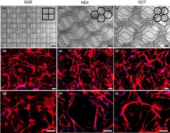
A PAM system was used to fabricate the scaffolds layer-by-layer from PTyC or PLLA. To investigate the effect of curvature on stem cell morphology, spreading, and differentiation, we focused on square (SQR), hexagonal (HEX), and octagonal (OCT) patterns [Figs. 1(a)–1(c)]. First, we characterize the mechanical properties of the scaffolds (Supplementary Fig. S1). The elastic modulus of the PTyC films (107 ± 14 MPa) was approximately two times larger than that of the PLLA films (46 ± 2 MPa). When scaffold architectures are considered, the moduli of SQR (PTyC = 50 ± 14 and PLLA = 25 ± 3 MPa) and HEX (PTyC = 43 ± 7 and PLLA = 21 ± 3 MPa) scaffolds were significantly higher than the OCT (PTyC = 11 ± 7 and PLLA = 18 ± 8 MPa) scaffolds. Here, we would like to note that the modulus values are well above the corresponding modulus values for fat tissue (~3 kPa) and pre-calcified bone (~30–40 kPa),[Reference Engler, Sen, Sweeney and Discher1, Reference Guvendiren and Burdick14] thus not a determining factor in our studies. Our results show that the architecture of the scaffold controls the overall stiffness of the scaffold. However, the overall scaffold stiffness values for different designs are not necessarily important for stem cell differentiation as cells initially reside on individual struts, and the stiffness they feel is the stiffness of the polymer rather than stiffness of the overall scaffold.
(a–c) Phase contrast images of the 3D scaffolds printed from PTyC using square (SQR), hexagon (HEX), and octagon (OCT) architecture. (d–i) Fluorescent images of hMSCs cultured on these scaffolds for 14 days (F-actin in red and cell nuclei in blue). Scale bars for the top two rows are 200 µm and for the bottom row are 100 µm. (Merged images of the phase contrast and fluorescence channels are given in the Supplementary Fig. S2.)

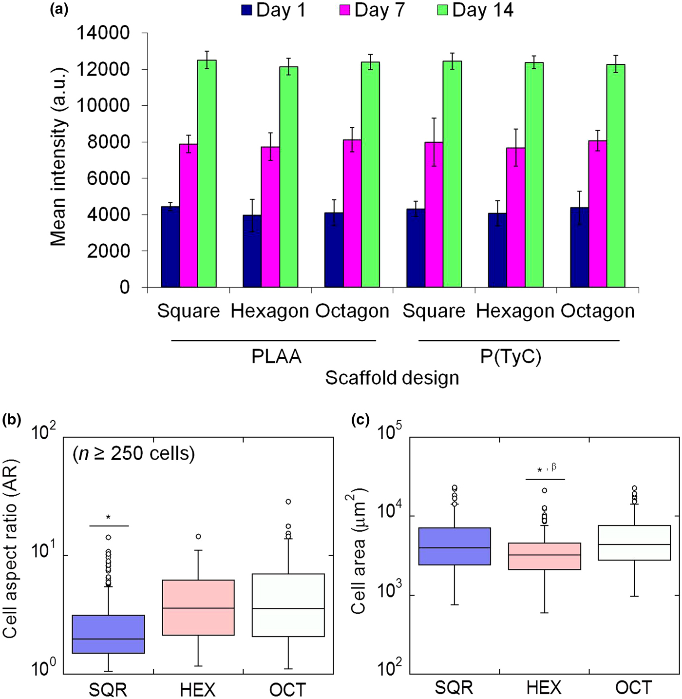
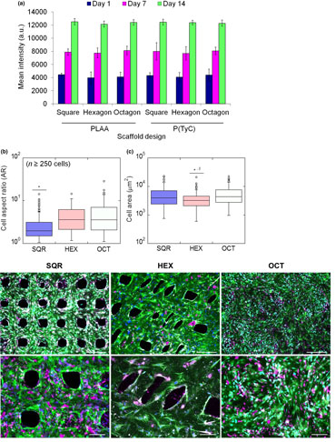
Our initial studies indicated poor cell adhesion and a lack of uniform cell distribution on our scaffolds even after 14-day culture for both PTyC and PLLA scaffolds. Thus, the scaffolds were incubated in a fibronectin solution, which enhanced stem cell adhesion and ensured uniform distribution of cells [Figs. 1(d)–1(i) and Supplementary Fig. S2]. Alamar Blue assay results indicated an increase in metabolic activity with culture time for both PLLA and PTyC scaffolds [Fig. 2(a)]. Stem cells attached and remained viable over time independent of the scaffold architecture and the polymer system. When the scaffolds with identical designs are compared, we did not observe any significant difference in cell morphology and spreading between PLLA and PTyC scaffolds (Supplementary Fig. S3). However, we observed significant differences in cell AR and cell area between scaffold designs [Figs. 2(b) and 2(c)]. We focused on PTyC scaffolds for the remainder of the study as we did not observe any difference between PTyC and PLLA.
(a) Alamar Blue assay results plotted as mean intensity versus scaffold design. Cell aspect ratio (AR) (b) and cell area (c) for hMSCs cultured on PTyC scaffolds with square (SQR), hexagonal (HEX), and octagonal (OCT) architecture plotted using Box-and-Whiskers diagram. For cell AR in (b) *P < 0.001 for SQR versus OCT and HEX. For mean cell area in (c), *P < 0.001 for HEX versus OCT, and βP < 0.01 for HEX versus SQR (n = 250 cells).
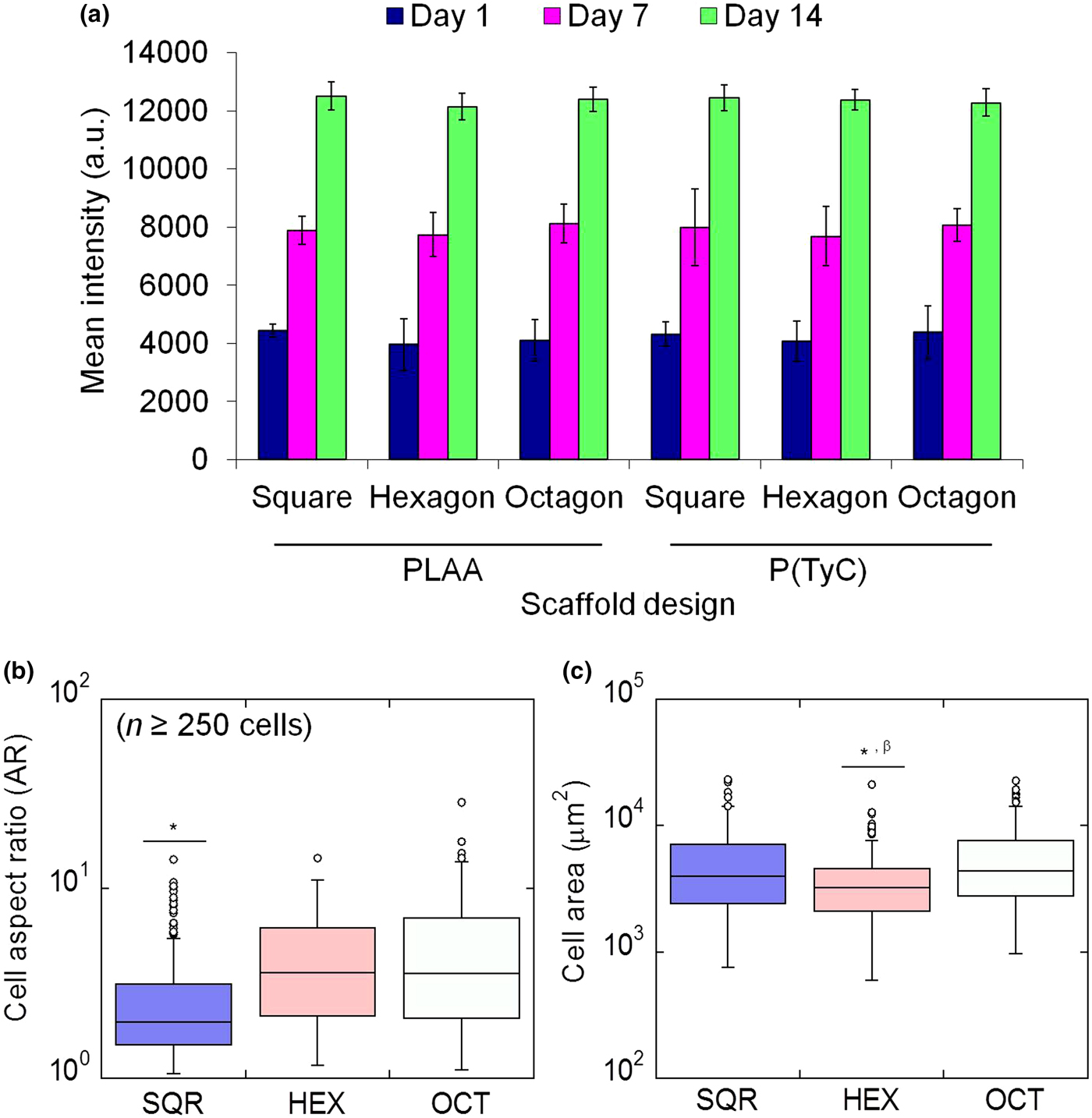
hMSCs were found to attach and spread on the scaffolds by aligning themselves along the printed struts [Figs. 1(g)–1(i)]. Stem cells were highly elongated with significantly higher AR for OCT (AR ~ 5.0) and HEX (AR ~ 4.4) as compared with SQR (AR ~ 2.5) scaffolds [Fig. 2(b)]. When cell spreading (defined by mean cell area) was considered [Fig. 2(c)], stem cells on SQR (mean cell area of 5100 µm2) and OCT (mean cell area of 5700 µm2) scaffolds had significantly higher cell spreading than that of HEX (mean cell area of 3900 µm2) scaffolds.
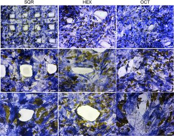
We examined whether these significant changes in cell morphology correlated with differentiation state. Here we would like to note that our study focuses on micro-scale surface patterns and curvature, which requires the use of induction media to evaluate the stem cell fate decision as shown in the literature. In contrast, nano-scale surface patterns are known to induce osteogenic differentiation of stem cells even in the absence of induction media.[Reference Dobbenga, Fratila-Apachitei and Zadpoor40] For this purpose, hMSCs were seeded on 3D printed PTyC scaffolds with SQR, HEX, or OCT patterns. Stem cells were allowed to attach and spread for 1 day in growth media. The media were then replaced with 1:1 adipogenic/osteogenic mixed induction media, and the cells were incubated for 14 days. The cells were stained for ALP as a marker of osteogenesis (Fig. 3). Note that the adipogenic cells appeared yellow after ALP staining due to oil droplets. Qualitatively, the majority of the cells stained positive for ALP.
Bright field images of hMSCs cultured on scaffolds in mixed osteogenic/adipogenic induction media for 14 days. [blue, Alkaline phosphatase indicating osteogenic (BONE) cells; yellow, Lipid droplets for adipogenic (FAT) cells.]. Scale bars are 200, 100, 50 µm, from top to bottom row, respectively.

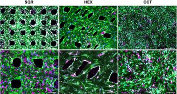
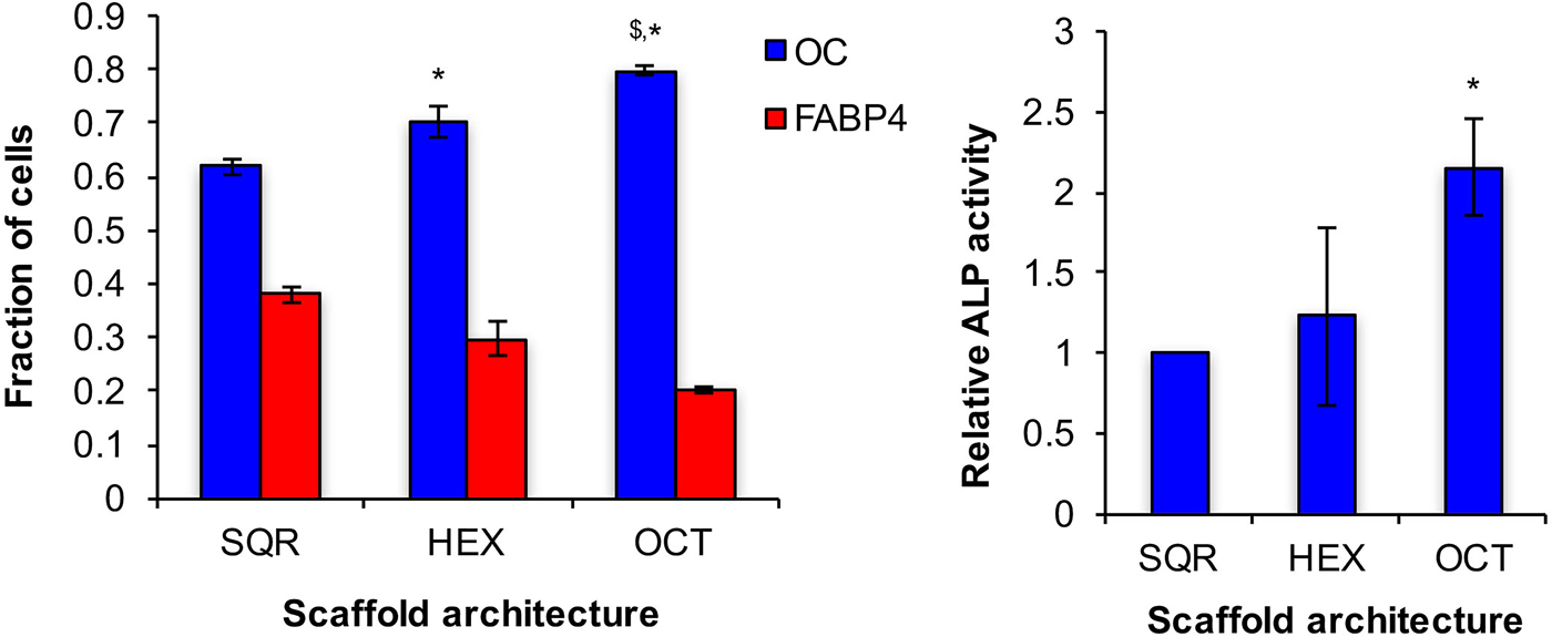
Another sample group was dual-immunostained for OC as a marker of osteogenesis and FABP4 as a marker of adipogenesis (Fig. 4). Due to the selection of secondary antibodies, OC-positive cells appeared green, whereas FABP4-positive cells appeared red under fluorescent microscope. When all cells were quantified, osteogenic differentiation was found to be upregulated (Fig. 5(a)) such that 62 ± 2% of the cells stained positive for OC for SQR scaffolds, followed by 70% ± 4 for HEX scaffolds, and 80 ± 1% for OCT scaffolds (we did not observe any dual-stained cells). ALP activity was also measured as a quantitative measure of osteogenic differentiation. Our analysis showed that the ALP activity followed a similar trend (Fig. 5(b)). The ALP activity of the cells on OCT scaffolds (relative to SQR scaffolds) was significantly higher than that of HEX (1.3 times) and SQR (2.2 times).
Fluorescent images of the dual-immunostained hMSCs cultured on scaffolds in mixed osteogenic/adipogenic induction media for 14 days [green, Osteocalcin indicating osteogenic (BONE) cells; magenta, FABP4 showing lipid droplets in adipogenic (FAT) cells]. Scale bars are 500 and 200 µm, for top and bottom rows, respectively.

(a) Quantitative analysis of immunostaining results showing fraction of cells stained positive for OC (bone) and FABP4 (fat). (b) ALP activity of hMSCs for scaffolds plotted relative to SQR (where relative ALP activity for SQR is set to 1). Relative ALP activity denotes mU ALP/μg DNA (normalized to SQR).

Conclusion
In summary, we used 3D printed scaffolds to investigate the effects of scaffold architecture on stem cell differentiation. The scaffolds were printed using PLLA or PTyC. We did not observe any significant differences between scaffolds fabricated from PLLA or PTyC in terms of stem cell attachment, morphology, and viability. However, we observed significant differences in stem cell spreading and AR as well as lineage commitment dependent on scaffold architecture. When stem cell morphology is considered, the following relationships were obtained: OCT > HEX ≫ SQR, for cell AR, and OCT ≥ SQR ≫ HEX, for mean cell area. Cells displayed the largest AR and cell area on OCT scaffolds compared those grown on other scaffolds. We believe that this is due to the highly curved surfaces displayed by the OCT scaffolds. Cells were elongated on HEX scaffolds, yet had the lowest mean cell area. On SQR scaffolds, cells were bulkier with low AR, but had high cell area. We observed significantly higher ALP activity and OC staining for hMSCs cultured on OCT scaffolds after 14 days of culture in the mixed induction media. Our results suggest that OCT architecture (with abundance of curved surfaces) is more efficient to drive stem cells into osteogenic lineage. Our study demonstrated the importance of scaffold architecture, which may be an important factor in the design of bone scaffolds.
Supplementary material
The supplementary material for this article can be found at https://doi.org/10.1557/mrc.2017.73.
Acknowledgments
Authors would like to thank Tyler Hoffman for his assistance on cell culture assays. This study was supported by RESBIO, the “Resource for polymeric biomaterials”, funded by the National Institute of Health, National Institute of Biomedical Imaging and Bioengineering Award Number P41EB001046, and by the National Science Foundation under Grant No. 1714882.