Middle East Respiratory Syndrome coronavirus (MERS-CoV) can cause a fatal respiratory disease, the human-to-human transmission and subclinical infections of which have been recognized in previous studies.Reference Assiri, McGeer and Perl 1 , Reference Drosten, Meyer and Muller 2 From May to July 2015, the Republic of Korea experienced the largest outbreak of MERS outside the Arabian Peninsula, and 92 of the 186 laboratory-confirmed cases of the entire outbreak were associated with our hospital, a 1,950-bed tertiary care center. 3 Initially, the largest unprotected exposure event occurred in the emergency department (ED); it generated 82 MERS-CoV infections including 8 cases among healthcare personnel (HCP).Reference Cho, Kang and Ha 4 , Reference Park, Ko and Peck 5 To prevent secondary transmissions from MERS-CoV–infected HCP, we implemented a thorough screening and monitoring strategy. Every symptomatic HCP was isolated, and his or her sputum was tested by real-time reverse transcriptase polymerase chain reaction (rRT-PCR). In addition, all the HCP exposed or assigned to MERS patients were quarantined for 14 days, and their sputum was tested by rRT-PCR as a screening test before returning to general patient care. Additional MERS-CoV infections in 7 assigned HCP were detected at the early course of the disease and were isolated immediately. Although no further transmission occurred from HCP, some rRT-PCR–negative HCP experienced fever and myalgia, which provoked suspicion of subclinical infection of MERS-CoV. To evaluate the appropriateness of our screening and monitoring strategy of detecting unrecognized MERS-CoV infections, we performed a serologic investigation of HCP after the end of outbreak.
METHODS
Study Population, Serum Sampling, and Data Collection
HCP who were closely exposed to or managed MERS patients during the outbreak were screened. HCP were classified into 2 groups according to the awareness of MERS-CoV infection and consequent application of personal protective equipment (PPE): HCP exposed to MERS patients without application of PPE due to lack of awareness of MERS-CoV infection of the patients (ie, exposed HCP); and HCP assigned to MERS patient care during the outbreak, all of whom used PPE (ie, assigned HCP). HCP whose MERS-CoV infections were confirmed by rRT-PCR were excluded from the study.
Sera of HCP who agreed to participate in the study with written informed consent were prospectively collected. Sampling of HCP sera was performed at 6 to 26 weeks post exposure. In total, 41 sera from the healthy population preserved in the biobank of Gangnam Severance Hospital were used as controls. In addition to administering to questionnaires to participating HCP, we retrospectively collected clinical data from electronic medical records and internal epidemiologic investigations during the outbreak. The institutional review board of our hospital approved the present study.
Definitions
MERS-like symptoms included fever, myalgia, cough, sputum, dyspnea, rhinorrhea, sore throat, nausea, vomiting, and diarrhea.Reference Assiri, McGeer and Perl 1 An HCP who experienced any symptom was considered symptomatic. Fever was defined as a single tympanic temperature measurement of ≥38.0°C or a temperature of ≥37.5°C sustained over a 1-hour period. We used the term ‘clinical MERS-CoV infection’ to represent MERS-CoV infection confirmed by rRT-PCR regardless of symptom presence, as the infectivity of an asymptomatic case remains uncertain. 6 In addition, ‘subclinical MERS-CoV infection’ represents MERS-CoV infection that was not detected by rRT-PCR screening but was confirmed by serologic tests. MERS patients who transmitted MERS-CoV to others were considered highly infective patients.
Laboratory Procedures
We primarily used commercial anti-MERS-CoV enzyme-linked immunosorbent assay (ELISA) IgG kits (EUROIMMUN, Lübeck, Germany) to detect antibody response.Reference Drosten, Meyer and Muller 2 , Reference Muller, Meyer and Corman 7 Sera were tested according to manufacturer instructions with 1:100 dilutions. We additionally used immunofluorescence assay (IFA) and plaque reduction neutralization test (PRNT) to substantiate ELISA results, which are known to be more specific than ELISA.Reference Drosten, Meyer and Muller 2 , Reference Muller, Meyer and Corman 7 Samples with an ELISA optical density (OD) ratio ≥0.2 were selected for these confirmatory tests, which is approximately 3-fold of the median value of control samples (0.06) in the present study. Although the manufacturer instructions suggested higher cutoff OD ratios of borderline (0.8–1.1) and positive (≥1.1) to minimize false positivity, we used low cutoff values for IFA and PRNT confirmation to avoid missing weak antibody responses of subclinical infections. This stepwise approach using a low ELISA cutoff was also used in previous serologic investigations of MERS.Reference Drosten, Meyer and Muller 2 , Reference Muller, Meyer and Corman 7 , Reference Breakwell, Pringle and Chea 8 We used commercial anti-MERS-CoV IFA IgM kits and IgG (EUROIMMUN) with 1:10 and 1:100 dilutions, respectively. We conducted PRNT as previously described and defined a 1:20 dilution as the lowest possible diagnostically significant titer.Reference Drosten, Meyer and Muller 2 , Reference Buchholz, Muller and Nitsche 9
Statistical Analysis
For comparison of ELISA OD ratios, Student t tests or Mann-Whitney U tests were used for 2-group comparisons, and 1-way analysis of variance (ANOVA) or Kruskal-Wallis tests were used for 3-group comparisons. Linear regression models were used for comparisons between age and ELISA OD ratios. All P values were 2-tailed, and those <.05 were considered statistically significant. SPSS Statistics version 20.0 for Windows (IBM, Armonk, NY) was used for all statistical analyses.
RESULTS
HCP in the Hospital-Associated Outbreak
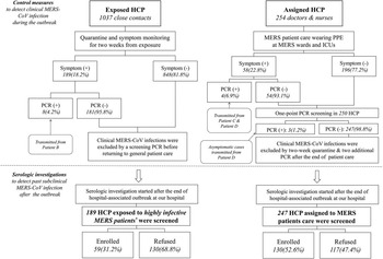
An overall description of the hospital-associated MERS outbreak was previously reported.Reference Park, Ko and Peck 5 HCP exposed and assigned to MERS patients during the outbreak are depicted in Figure 1 (detailed description is presented in the Supplementary Materials). During the outbreak, a total of 1,037 HCP were closely exposed to MERS patients without protection, 8 of whom were infected with MERS-CoV. Among 1,029 rRT-PCR–negative HCP, 189 HCP who were exposed to highly infective MERS patients were screened, and 59 of these HCP (31.2%) consented to this study for serologic investigation.
HCP exposed and assigned to MERS patients during the MERS outbreak of our hospital. HCP were classified into 2 groups according to the awareness of MERS-CoV infection and consequent application of PPE: (1) HCP exposed to MERS patients without application of PPE due to unawareness of MERS-CoV infection of the patients (exposed HCP) and (2) HCP assigned to MERS patient care during the outbreak, all of whom were applied with PPE (assigned HCP). Seven assigned HCP were infected with MERS-CoV while managing MERS patients despite application of PPE. Symptom monitoring, quarantine, and screening by sputum MERS-CoV rRT-PCR were performed during the outbreak, and serologic investigation was conducted to detect subclinical MERS-CoV infections. *Highly infective MERS patients were defined as those who additionally transmitted MERS-CoV to others (patients A, B, C, and D in this article, described in the Supplementary Materials in detail). HCP, healthcare personnel; MERS-CoV, Middle East Respiratory Syndrome coronavirus; rRT-PCR, real-time reverse transcriptase polymerase chain reaction; PPE, personal protective equipment.

In managing MERS patients, HCP were grouped in cohorts such that those assigned to MERS patient care would not care for patients in the general population. During the outbreak, 254 HCP were assigned to MERS patient care, 7 of whom were infected with MERS-CoV despite PPE application. After the outbreak, 247 rRT-PCR-negative HCP were screened, 130 of whom (52.6%) consented to the study.
Antibody Response of HCP Exposed to Highly Infective MERS Patients
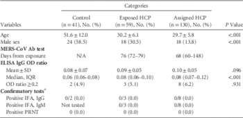
Characteristics and serologic test results of enrolled HCP exposed to 4 highly infective MERS patients are presented in Table 1 and Supplementary Table 2. The mean age was 30.2 years and 69.5% were female. Most of them did not have any underlying comorbidities (91.5%). A quarter of the HCP experienced MERS-like symptoms within 2 weeks of the exposure, among which fever or myalgia were most common. Sera sampling was performed at a median of 76 days after the exposure (range, 65–79 days). The median ELISA OD ratio was 0.08 (range, 0.05–0.26). In total, 3 sera (5.1%) with ELISA OD ratio ≥0.2 (0.206, 0.234, and 0.262, respectively) underwent confirmatory tests; all of these were negative both for IFA and PRNT.
Demographics and MERS-CoV Antibody Test Results of HCP and Controls

NOTE. MERS-CoV, Middle East Respiratory Syndrome coronavirus; HCP, healthcare personnel; N/A, not applicable; ELISA, enzyme-linked immunosorbent assay; OD, optical density; SD, standard deviation; IQR, interquartile range; IFA, immunofluorescence assay; PRNT, plaque reduction neutralization test.
a Samples with an ELISA OD ratio ≥0.2 were selected for confirmatory tests, which is approximately 3-fold of the median (0.06) value of control samples.
Antibody Response of HCP Assigned to MERS Patient Care
The mean age of 254 HCP assigned to MERS patient care was 29.7 years, and 86.2% were female (Table 1). Only 9.2% had underlying comorbidities. Approximately 20% of the HCP experienced MERS-like symptoms within 2 weeks. Sera sampling was performed at a median of 69 days after the exposure (range, 44–178 days). In total, 8 sera (6.2%) with ELISA OD ratio ≥0.2 (range, 0.22–0.26) underwent confirmatory tests, all of which were negative both for IFA and PRNT.
Comparison of MERS-CoV IgG ELISA Reactivity With Controls
ELISA OD ratios and their comparison according to 3 categories are presented in Table 1 and Supplementary Figure 1. The median ELISA OD ratio of controls was 0.06 (IQR, 0.06–0.08). The highest value of ELISA OD ratio (0.47) of the present study was observed in a single control (ie, serum of a 71-year-old female). Although all the sera with ELISA OD ratio ≥0.2 were confirmed to be negative, age, sex, and the medians of ELISA OD ratio were statistically different between the 3 categories. To clarify presence of any possible influence of age and sex to ELISA reactivity we performed additional statistical comparisons. There was no significant relationship between age and ELISA OD ratio for both controls and HCP (P=0.178 and P=0.416, respectively; Supplementary Figure 2) and between sex and ELISA OD ratio (P=0.601).
DISCUSSION
Serologic surveillance studies in MERS-CoV–exposed individuals played an important role in contact investigations.Reference Drosten, Meyer and Muller 2 , Reference Breakwell, Pringle and Chea 8 – Reference Hall, Tokars and Badreddine 10 In a household contact investigation in Saudi Arabia, sera sampled after day 13 post exposure were positive, while those of PCR-confirmed MERS patients taken within 2 weeks were all negative.Reference Drosten, Meyer and Muller 2 This implies that serologic surveillance of exposed individuals should use sera sampled at least 2 weeks after exposure, although exact seroconversion period should be further investigated. In the present investigation, we collected sera at 6–26 weeks after exposure to assure enough time for seroconversion.
Because adequate sampling interval for seroconversion is required for serologic surveillance, the rRT-PCR assay of respiratory samples should be emphasized in contact investigation conducted within 14 days from exposure. During the outbreak, we vigorously conducted screening of HCP exposed and assigned to MERS patients using rRT-PCR assays, which detected 15 MERS-CoV infected HCP at a very early stage of the disease. We primarily used sputum specimens for screening unlike previous studies used upper respiratory tract specimens,Reference Drosten, Meyer and Muller 2 , Reference Breakwell, Pringle and Chea 8 because we experienced much higher sensitivity of sputum specimen compared to nasopharyngeal swab even in patients without respiratory symptoms (Huh et al, in revision, September 2016). By testing sputum rRT-PCR before returning to general patient care, we excluded clinical MERS-CoV infections in HCP participating in the present serologic investigation.
During the MERS outbreak at our hospital, 1,037 HCP were closely exposed to MERS patients, and 8 of them were infected. Approximately 20%–25% of the exposed or assigned HCP experienced mild-to-moderate MERS-like symptoms, and some of them complained of prolonged symptoms despite consecutive negative rRT-PCR, provoking suspicion of subclinical infection undetected by rRT-PCR. We purposely applied a very low cutoff ELISA OD ratio of 0.2 for initial screening and subsequent IFA and PRNT confirmation. A low ELISA OD ratio cutoff was previously shown to detect subclinically infected individuals that may not experience a pronounced seroconversion.Reference Drosten, Meyer and Muller 2 However, in our study, none of the enrolled HCP was positive for anti-MERS-CoV antibody. Although not all exposed HCP were tested, the absence of serologic response implies that these symptoms were not directly related to MERS-CoV infections. In addition, it also suggests that 14-day monitoring and screening by sputum rRT-PCR could appropriately detect MERS-CoV infections during the outbreak.
Another important purpose of the present study was to evaluate the appropriateness of infection preventive measures for assigned HCP. In total, 7 assigned HCP were infected with MERS-CoV despite the application of standard PPE (ie, surgical gloves, surgical gown, eye shield, and N95 respirator). The appropriateness of standard PPE was questioned, and the application of PPE with hooded coverall and goggle was recommended thereafter. Also, the insufficient negative pressure (<2.5 Pa) of most of isolation rooms in MERS wards and intensive care units (ICUs) were assumed to be a cause of MERS-CoV transmission. However, in our serologic investigation, none of the tested sera of assigned HCP showed a positive result for anti-MERS-CoV antibody, which implies that the systemic problem of infection preventive measures is less likely to be a primary cause of transmission. Instead, possible transmission mechanisms of assigned HCP may include incidental contamination of unprotected body surfaces from excessive droplet generation or breakage of N95 respirator fit without recognition; most of these HCP had very close physical contact with severely coughing MERS patient including sputum induction, portable X-ray shooting, and patient transportation just before intubation.Reference Park, Ko and Peck 5 We think proper application of PPE should be extremely emphasized in caring for patients with severe respiratory symptoms.
Because the present study fully relied on the consent of participating HCP, we could not collect sera from all of the screened HCP. However, considering the psychological trauma of this fatal respiratory disease in the unexpected outbreak of MERS, a participation rate of 51.1% of the target population indicates that the HCP are quite dedicated. By testing 189 HCP, including highly exposed individuals, our cohort was at least twice as large as cohorts in previous serologic investigations of HCP.Reference Breakwell, Pringle and Chea 8 – Reference Hall, Tokars and Badreddine 10
In conclusion, the serologic investigation of 189 HCP exposed and assigned to MERS patients during the 2015 Korean MERS outbreak revealed that none of the tested sera were positive for MERS-CoV antibody. This absence of seroconversion implies that our screening strategy of 14-day symptom monitoring followed by sputum rRT-PCR appropriately detected MERS-CoV infections.
ACKNOWLEDGMENTS
We are extremely grateful for the participation of HCP in this investigation. We also greatly appreciate the HCP and staff members at Samsung Medical Center and all other hospitals who worked together to overcome the MERS outbreak. Finally, we thank Jinseob Kim for statistical advice and the graphical work, and Mingu Kang for IFA testing.
Financial support: This study was supported by the Samsung Biomedical Research Institute (SBRI grant no. SMX1161321). C.D. reports funding by EU grants Antigone (grant no. 278976) and Prepare (grant no. 602525).
Potential conflicts of interest: There are no potential conflicts of interest relevant to this article to report.
SUPPLEMENTARY MATERIAL
To view supplementary material for this article, please visit https://doi.org/10.1017/ice.2016.251.