Significant outcomes
-
Cisplatin significantly caused memory impairment.
-
Learning and memory impairment induced by cisplatin was accompanied with an oxidative stress status in the hippocampus, which was presented by an increased level of lipid peroxidation but decreased level of antioxidant activity.
-
Vitamin E attenuated learning and memory impairments induced by cisplatin.
-
Learning and memory-improving effect of vitamin E was accompanied with anti-oxidant effects, which was showed by a decrease in lipid peroxidation but an increase in antioxidant activity.
Limitations
-
Cortical levels of MDA, thiol, catalase, glutathione, and SOD are needed to be measured.
-
Precise evaluation at the molecular level is suggested to be done.
-
Measurements of lipid peroxidation and antioxidation in the other organs including kidney are suggested to be measured.
-
Evaluation of the function of other organs including kidney is suggested to be measured.
Chemotherapy is widely used for cancer treatment. Cisplatin is an eminent chemotherapeutic agent in the treatment of various cancers, including neuroblastoma, hepatoblastoma, germ cell tumours, and primary central nervous system neoplasms (Nakagawa Sponne et al. 2003., Reference Nakagawa, Fujita, Kubo, Tokiyoshi, Yamada, Kanayama, Hagiwara, Nakanomyo and Hiraoka1996; Radhi et al., Reference Radhi, Fulbright, Ginn and Guest2015). Patients administered with cisplatin often recover without life-threatening conditions; however, their treatment is still associated with significant challenges. Recent studies have denoted the highly toxic and dose-limiting side effects of cisplatin, such as nephrotoxicity, hepatotoxicity, and neurotoxicity (Song et al., Reference Song, Chen, Liao, Ou and Tsai2010; Sen et al., Reference Sen, De, Devanna and Chakraborty2013; Waseem & Parvez, Reference Waseem and Suhel2013). Chemo brain or cognitive impairment is a common side effect of chemotherapy drugs, which is frequently reported by patients treated for tumours that are outside the central nervous system (Ahles & Saykin, Reference Ahles and Saykin2007; O’Farrell, MacKenzie, & Collins, Reference O’Farrell, MacKenzie and Collins2013). Furthermore, animal studies have documented that cisplatin could decrease neurogenesis in the dentate gyrus in the hippocampus of adult rats (Hinduja et al., Reference Hinduja, Kraus, Manohar and Salvi2015), as well as in the patients undergoing chemotherapy who report impaired cognition and memory (Kaasa et al., Reference Kaasa, Olsnes, Thorud and Høst1988; Troy et al., Reference Troy, McFarland, Littman-Power, Kelly, Walpole, Wyld and Thomson2000). Neuropsychological studies have shown decreases in processing speed, memory, executive functioning, and attention. Indeed, neuroimaging techniques have shown structural alterations in white and grey matter. Moreover, there are specific regional changes in brain activity in patients suffering from chemotherapy-induced cognitive impairments (Ahles & Saykin, Reference Ahles and Saykin2007; Simó et al., Reference Simó, Rifà-Ros, Rodriguez-Fornells and Bruna2013). Cisplatin penetrates into the brain where it inhibits neuronal stem cell proliferation (Bost et al., Reference Bost, Sahra, Marchand-Brustel and Tanti2012).
Oxidative stress and depletion of antioxidant enzymes (Chen et al., Reference Chen, Jungsuwadee, Vore, Allan Butterfield and St Clair2007) are the mechanisms that may be involved in cisplatin-induced neurotoxicity (da Silva Machado et al., Reference da Silva Machado, Mendonça, de Paula Venancio, Bianchi and Antunes2013). Therefore, it seems that the reduction of the oxidative damage of proteins, polyunsaturated lipids, and DNA/RNA could be effective in this regard, limiting the side effects of this drug. According to the literature, the substances that could reduce oxidative stress have significant effects on memory improvement as well (Oz et al., Reference Oz, Atalik, Yerlikaya and Demir2015; Baghcheghi et al., Reference Baghcheghi, Beheshti, Shafei, Salmani, Sadeghnia, Soukhtanloo, Anaeigoudari and Hosseini2018).
Vitamin E is a hydrophobic, fat-soluble compound, which is found abundantly in numerous food sources, such as corn oil, peanuts, fruits, and vegetables (Chun et al., Reference Chun, Lee, Ye, Exler and Eitenmiller2006; Thomas & Gebhardt, Reference Thomas and Gebhardt2006). Previous findings have indicated that vitamin E could protect cells against oxidative stress and damage through the reduction of lipid peroxidation (Green et al., Reference Green, Hodges, Chipman, O’Donovan and Graham2008; Schneider, Reference Schneider2009). Moreover, some studies have denoted the protective effects of vitamin E against cisplatin-induced nephrotoxicity (Ajith et al., Reference Ajith, Usha and Nivitha2007, Reference Ajith, Abhishek, Roshny and Sudheesh2009). A study has shown that this vitamin was involved in memory enhancement through the reduction of free radicals in memory of juvenile hypothyroid rats (Baghcheghi et al., Reference Baghcheghi, Beheshti, Shafei, Salmani, Sadeghnia, Soukhtanloo, Anaeigoudari and Hosseini2018). Although there are a lot of studies about antioxidant effects of vitamin E in other organs, neuroprotective effects of vitamin E are less investigated. Indeed, there are no studies to investigate the effect of this vitamin on the cisplatin-induced cognitive deficits in rats.
Materials and methods
Experimental animals
In total, 40 male Wistar albino rats aged 8 weeks (weight: 240 ± 10 g) were provided by the animal centre of Sabzevar University of Medical Sciences, Iran. The animals were housed in Plexiglas cages (four per cage) at room temperature (23–25°C) within a 12-h light/dark cycle. Food and water were available to the animals’ ad libitum. This study was approved by the Ethics Committee for Animal Experiments at the Sabzevar University of Medical Sciences and all experimental works were done in accordance with the National Institute of Health Guide for the Care and Use of Laboratory Animals (Council, 2010).
Experimental protocol
The animals were randomly divided into five groups of eight, including group one (control; n = 8) receiving 0.9% saline once daily for 1 week intraperitoneally (i.p.), group two (cisplatin; n = 8) receiving cisplatin (2 mg/kg/7 day; i.p.) (John et al., Reference John, Lomeli and Bota2017), group three receiving cisplatin and vitamin E (n = 8; 2 and 200 mg/kg/7 day; i.p.) (Kiasalari et al., Reference Kiasalari, Khalili, Shafiee and Roghani2016), group four (n = 8) receiving vitamin E before cisplatin administration and pre-treatment with vitamin E (200 mg/kg/14 day; i.p.), followed by cisplatin (2 mg/kg/7 day; i.p.), and group five receiving vitamin E (n = 8; 200 mg/kg/7 day; i.p.).
In the present study, vitamin E was provided by Zahravi Pharma Company (Tehran, Iran). The behavioural assay [Morris water maze (MWM)] was performed at the same time of the day during the experimental procedures. The animals were euthanised under ketamine and xylazine anaesthesia, and their hippocampi were collected 1 week after the injection of cisplatin for biochemical measurements.
Behavioural studies
MWM was conducted to examine the effect of cisplatin on memory impairment and the potential of vitamin E for the improvement of spatial learning and memory.
The MWM apparatus was composed of a circular pool (diameter: 136 cm, height: 60 cm, depth: 30 cm). The temperature of water was maintained at 23–25°C. A circular platform (diameter: 10 cm, height: 28 cm) was placed at 2 cm below the surface of water at the centre of the north-western quadrant. Visible cues were present at various locations around the room. In each trial, the animals were released randomly in the pool at one of the four quadrants (northern, eastern, southern, and western). In this process, the rats were trained on learning the location of the hidden platform based on the mentioned visual cues. The experiments were repeated with four trials for 5 days. The animals were allowed to remain on the platform for 20 s before the next trial. The mean spent time and travelled distance was recorded using a camera hung above the centre of the pool which was connected to the computer containing Radiab software (was made by Nomiri, Mashhad, Iran). It is notable that the researchers were blinded to the treatments, on day 6 of the experiments, a probe test was performed in order to evaluate the ability of the animals to recall the location of the platform. To this end, the escape platform was removed, and the rats were placed inside the pool for 90 s. The spent time, path travelled in the target quadrant, and number of the platform crossings were measured (north-western quadrant [Q1]) and compared between the groups (Vafaee et al., Reference Vafaee, Hosseini, Sadeghinia, Al-reza Hadjzadeh, Soukhtanloo and Rahimi2014).
Biochemical measurements
After each experiment, the animals were euthanised, and their hippocampal tissues were extracted for biochemical measurements. The concentration of malodialdehyde (MDA) was measured in the hippocampal tissue manually (Pourganji et al., Reference Pourganji, Hosseini, Soukhtanloo, Zabihi and Al-reza Hadjzadeh2014). In brief, 1000 μl of the samples were added to 2000 μl of a complex solution containing thiobarbituric acid, trichloroacetic acid, and hydrochloric acid. Afterwards, the mixture was incubated in a hot water bath at the temperature of 100°C for 10 min. Finally, the solution was centrifuged (1000× g) for 10 min, and the absorbance was read at 535 nm.
The concentration of thiol was also measured manually. In brief, 50 μl of the tissue homogenates were added to 1000 μl of ethylenediaminetetraacetic acid-Tris buffer. Following that, 20 μl of 2, 2′-dinitro-5, 5′-dithiodibenzoic acid solution (10 mM) was mixed with the solution, and the mixture was incubated at room temperature for 15 min. Finally, the yellow complex showed the peak absorbance at 412 nm. The enzyme activity of superoxide dismutase (SOD) was measured based on the protocol of Madesh and Balasubramanian (Reference Madesh and Balasubramanian1998), and the absorbance of the supernatant was measured at 570 nm.
Statistical analysis
The obtained data were expressed as the mean and standard error of the mean (SEM). Data analysis was performed in SPSS version 19 (SPSS Inc., Chicago, IL, USA). The difference in the escape latency during 5 days of MWM was assessed using repeated-measures analysis of variance (ANOVA), followed by least significant difference (LSD) post-hoc comparison test. In addition, the data on swimming speed, the percentage of the time and distance in the target quadrant in the probe trial, and biochemical findings were compared using one-way ANOVA, followed by LSD post-hoc comparison test. In all the statistical analyses, p-value of less than 0.05 was considered significant.
Results
Behavioural findings
MWM test
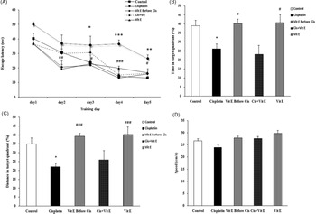
In the present study, the mean latency time gradually shortened during the behavioural test in all the experimental groups (Fig. 1(A)). The animals treated with cisplatin took longer to access the platform compared to the control group (p < 0.05 and p < 0.001, respectively). In addition, the mean latency was significantly shorter in the vitamin E group compared to the cisplatin group (p < 0.05 and p < 0.001, respectively) (Fig. 1(A)). According to the findings, vitamin E supplementation significantly decreased the mean latency on all the training days (p < 0.05, p < 0.001) (Fig. 1(A)).
Comparison of the time latency (A), percentage of time spent (B), percentage of travelled distance (C), and swim speed (D) in the target quadrant in MWM test between groups. The data are expressed as the mean ± SEM. ***p < 0.001, **p < 0.01, and *p < 0.05 compared to control group, ###p < 0.001, ##p < 0.01, and #p < 0.05 compared to cisplatin group (n = 8).

In the probe trial test, the percentage of the spent time and travelled distance in the target quadrant was calculated in order to evaluate the improvement of spatial learning and memory. The animals treated with cisplatin had a lower percentage of the spent time and travelled distance compared to the control group (p < 0.05) (Fig. 1(B) and (C)). However, the animals receiving vitamin E and vitamin E before cisplatin administration had higher percentages of the spent time and travelled distance in the target quadrant compared to the cisplatin group (p < 0.001 and p < 0.05, respectively) (Fig. 1(B) and (C)). However, no significant differences were observed in the spent time and travelled distance in the target quadrant between the animals receiving vitamin E plus cisplatin and those receiving vitamin E before cisplatin administration. Moreover, no significant differences were denoted between the control group, animals receiving vitamin E, those administered with vitamin E before cisplatin treatment, and the group receiving vitamin E plus cisplatin in terms of the mean swimming speed (Fig. 1(D)).
Biochemical findings
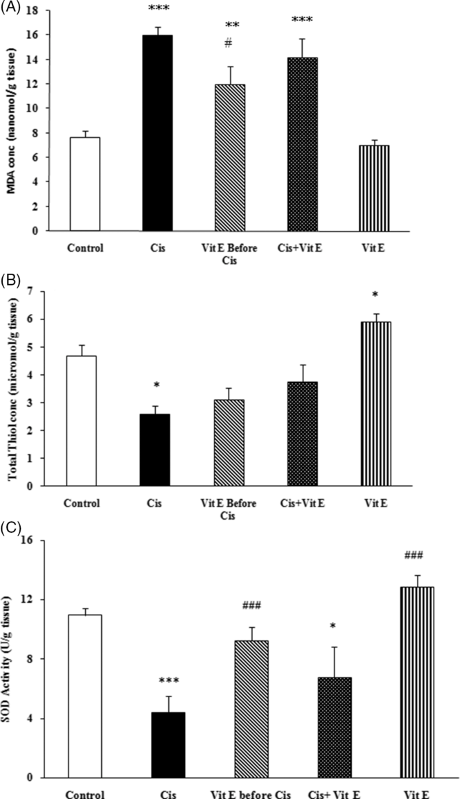
The hippocampal concentration of MDA significantly increased in the animals receiving cisplatin, vitamin E plus cisplatin, and vitamin E before cisplatin administration compared to the control group (p < 0.001, p < 0.01) (Fig. 2(A)). On the other hand, the injection of vitamin E decreased MDA concentration in the hippocampal tissues of the animals receiving vitamin E compared to the control group (p < 0.01) (Fig. 2(A)). Furthermore, pre-treatment with vitamin E 1 week before cisplatin administration decreased MDA concentration in the hippocampal tissues of the animals receiving vitamin E before cisplatin administration compared to the group treated with cisplatin (p < 0.001 and p < 0.01, respectively) (Fig. 2(A)). However, no significant difference was observed between the animals receiving vitamin E plus cisplatin and those receiving vitamin E before cisplatin administration in this regard.
Effect of vitamin E and cisplatin on hippocampus MDA level (A), thiol level (B), and SOD level (C). Data are expressed as mean ± SEM. ***p < 0.001, **p < 0.01, and *p < 0.05 compared to control group, ###p < 0.001, ##p < 0.01, and #p < 0.05 compared to cisplatin group (n = 8).

According to the obtained results, the concentration of thiol in the hippocampal tissues was lower in the animals treated with cisplatin compared to the control group (p < 0.05) (Fig. 2(B)). However, no significant difference was observed between the animals receiving vitamin E and the control group in this regard. On the other hand, the treatment of the animals with vitamin E during or before cisplatin injection increased the concentration of thiol in the hippocampus, while the difference was not considered significant. According to the findings, the hippocampal concentration of thiol was significantly higher in the animals receiving vitamin E compared to those treated with cisplatin, vitamin E plus cisplatin, and vitamin E before cisplatin administration (p < 0.05) (Fig. 2(B)).
As depicted in Fig. 2, the activity of SOD was significantly lower in the animals treated with cisplatin compared to the control group (p < 0.001) (Fig. 2(C)). On the other hand, treatment with vitamin E significantly increased the SOD activity in the animals receiving vitamin E and vitamin E before cisplatin administration (p < 0.001) (Fig 2(C)). It is also notable that no significant difference was observed between the pre-treatment and treatment with vitamin E in the mentioned groups (Fig. 2(C)).
Discussion
Neurotoxicity is considered to be a major side effect of anticancer chemotherapeutic agents, such as cisplatin. Anticancer drugs could cause damage to various cell lines in the peripheral and central nervous system (Mendonça et al., Reference Mendonça, da Silva Machado, Teixeira, de Freitas, de Lourdes Pires Bianchi and Antunes2013; Mejia, Reference Mejia2020). According to the results of the present study, there was a significant difference between the animals treated with cisplatin and control group in terms of the spatial memory. The escape latency time during the training days was significantly higher in the cisplatin group compared to the control group. In fact, the percentage of the spent time and travelled distance in the target quadrant on the final trial test day was lower in the groups injected with cisplatin. This finding is consistent with the results of the previous studies in this regard, which denoted the deficiency of learning and memory following cisplatin treatment (Oz et al., Reference Oz, Atalik, Yerlikaya and Demir2015).
Previous findings have confirmed the adverse effects of cisplatin on the rate of neurogenesis and proliferation of the hippocampus (Dietrich et al., Reference Dietrich, Han, Yang, Mayer-Pröschel and Noble2006; Manohar et al., Reference Manohar, Jamesdaniel and Salvi2014). Brain injuries in the cerebellar cortex following cisplatin treatment have also been reported in animal models (Hassan et al., Reference Hassan, Chibber, Khan and Naseem2013). Furthermore, previous studies have indicated the degeneration of the Purkinje cell line in the cerebellar cortex in cisplatin-treated subjects (Abou-Elghait et al., Reference Abou-Elghait, El-Gamal, Abdel-Sameea and Mohamed2010). Hippocampus is the vital organ for long-term memory, which is the potential target for the effects of chemotropic drugs; these agents commonly cause changes in memory and learning (Snyder et al., Reference Snyder, Hong, McDonald and Wojtowicz2005). In the medical literature, there are a few studies focusing on the cognitive impairment caused by cisplatin. However, severe cognitive malformation has been reported in the mice and rats treated with cisplatin (Troy et al., Reference Troy, McFarland, Littman-Power, Kelly, Walpole, Wyld and Thomson2000; Dietrich et al., Reference Dietrich, Han, Yang, Mayer-Pröschel and Noble2006).
According to the results of the present study, the concentration of MDA as a marker of oxidative stress was significantly higher in the cisplatin group compared to the control group, while the SOD activity in the hippocampus was observed to be lower. In addition, pre-treatment with vitamin E with and without cisplatin significantly changed the obtained results regarding the recovery of impaired memory. Cisplatin induces oxidative tissue damage, thereby leading to the production of free radicals and lipid peroxidation in various organs, such as the kidneys, hepatic tissues, and cochlea (Jilanchi et al., Reference Jilanchi, Nematbakhsh, Bahadorani, Talebi, Eshraghi-Jazi, Mansouri and Ashrafi2013; Waseem & Parvez, Reference Waseem and Suhel2013; Sakat et al., Reference Sakat, Kilic, Akdemir, Yildirim, Eser and Kiziltunc2018). In addition, various central and peripheral nervous system manifestations have been associated with the reduction in the plasma concentration of vitamin E in cancer patients following cisplatin therapy (Weijl et al., Reference Weijl, Wipkink-Bakker, Lentjes, Berger, Cleton and Osanto1998; Bove et al., Reference Bove, Picardo, Maresca, Jandolo and Pace2001; Halliwell & Gutteridge, Reference Halliwell and Gutteridge2015).
Recent studies have well established the protective effects of Vitamin E (Jilanchi et al., Reference Jilanchi, Nematbakhsh, Bahadorani, Talebi, Eshraghi-Jazi, Mansouri and Ashrafi2013). Vitamin E is a free radical scavenger against stress oxidative (Jain & Palmer, Reference Jain and Palmer1997). Due to its special shape, it is the only antioxidant that can be located in the cell membrane of the body and protect the cell membrane. Due to its fat solubility, it uses the same methods that cholesterol molecules use to move in the body. Another very important role of this antioxidant is when it is transferred in the body, which, along with other fat and cholesterol molecules, prevents them from oxidising in the arteries. This wonderful antioxidant prevents the ageing of the immune system and it keeps brain cells young and works better than any other prescription drug for delaying Alzheimer’s (Tucker & Townsend, Reference Tucker and Townsend2005; Catalgol & Ozer, Reference Catalgol and Nesrin2012; Nicolson & Ash, Reference Nicolson and Ash2014;). The elimination of reactive oxygen species (ROS) is a possible mechanism for the protective effects of vitamin E against cisplatin-induced peripheral nerve damage (Park et al., Reference Park, Choi, Bang, Huh and Kim2000). Several studies have indicated that the produced ROS could induce cell death and brain damage (Jang & Surh, Reference Jang and Young-Joon2002; Sponne et al., Reference Sponne, Fifre, Drouet, Klein, Koziel, Pinçon-Raymond, Olivier, Chambaz and Pillot2003). Moreover, electrophysiological findings have confirmed that vitamin E protects peripheral nerves against the toxic effect of cisplatin-based chemotropic therapy (Argyriou et al., Reference Argyriou, Chroni, Koutras, Iconomou, Papapetropoulos, Polychronopoulos and Kalofonos2006).
According to the findings of the current research, vitamin E increased the concentration of thiol in the hippocampal tissues, while it could not properly fix the antioxidant activity in co-administration with cisplatin to reach the levels of the control group. Therefore, it could be concluded that using antioxidants before chemotherapy is more effective compared to co-treatment. This could be due to the increased levels of antioxidants before the release of radicals in the tissues, which inhibit oxidative stress and tissue damage. Further biochemical studies are suggested in order to confirm these findings.
In conclusion, cisplatin may affect the brain, which is at least partially indirect. It is possible that cisplatin affects the kidneys or liver and then the brain is affected secondarily, which needs further investigation in future studies. Vitamin E could protect the male rats against cisplatin-induced neurotoxicity. The beneficial effects of vitamin E on cisplatin-induced memory impairment may attribute to its potential to decrease brain oxidative stress or because of improving oxidative status in other organs, which in turn improves brain function and memory. Furthermore, pre-treatment with vitamin E for several days prior to cisplatin administration may be a better protocol for benefitting from the protective effects of vitamin E on cisplatin-induced neurotoxicity.
Supplementary material
To view supplementary material for this article, please visit https://doi.org/10.1017/neu.2020.34
Acknowledgements
The authors would like to thank the Vice President of Research at Sabzevar University of Medical Sciences for the financial support.
Author contributions
All authors made substantive contributions to the study. M. Kafami designed the study and contributed to the statistical analyses. MH, AA, PH, FB, NM, and MGH collected data and contributed to the interpretations of the data. MH contributed to the statistical analyses. All authors have read and given final approval for the latest version of the manuscript.
Financial support
This research project was financially supported by the Vice Chancellor of Research at Sabzevar University of Medical Sciences, Iran.
Conflict of interest
The authors declared no conflicts of interest relevant to this article.
Ethical approval
The animal procedures were carried out in accordance with the standards of the Guide for the Care and Use of Laboratory Animals, and the study protocol was approved by the Committee on Animal Research at Sabzevar University of Medical Sciences (document number: IR.MEDSAB.REC.1397.084).