Introduction
There is convincing evidence that host nutritional factors can be decisive for the establishment and development of parasitic flatworms (Frazer et al., Reference Frazer, Reddy, Fried and Sherma1997; Oliveira et al., Reference Oliveira, Barbosa, Gomes, Machado-Silva, Barros, Neves and Coutinho2003; Bansal et al., Reference Bansal, Bhatti and Sehgal2005). For example, a vitamin B-deficient diet and a protein-free diet had a detrimental effect on the growth of Echinostoma caproni in ICR mice (Rosario & Fried, Reference Rosario and Fried1999; Simpkins & Fried, Reference Simpkins and Fried2001), and E. trivolvis developed poorly in avian hosts lacking fat-soluble vitamins (Simpkins & Fried, Reference Simpkins and Fried1999). Significantly greater amounts of phosphatidylcholine and phosphatidylethanolamine were found in worms from mice maintained on a high-fat diet as compared with worms from those on a standard diet, suggesting that the host diet influences the lipid content of adults of E. caproni (Frazer et al., Reference Frazer, Reddy, Fried and Sherma1997).
Lipids from the human blood-dwelling parasite Schistosoma mansoni have a number of interesting features (Wuhrer et al., Reference Wuhrer, Dennis, Doenhoff and Geyer2000) and important changes in lipid composition occur during schistosome growth and maturation (Rogers & McLaren, Reference Rogers and McLaren1987). The ability of schistosomula to convert cholesterol into its metabolites shows that it is an acquired property, observed in adults only and is specifically active during pairing. Because schistosomes are unable to synthesize acids or sterols de novo, they acquire these compounds from their hosts (Rumjanek & Simpson, Reference Rumjanek and Simpson1980). Males incorporate about three times more cholesterol than females (Haseeb et al., Reference Haseeb, Eveland and Fried1985). Earlier studies regarding the metabolism of cholesterol uptake by paired and unpaired worms of S. mansoni during pairing showed that labelled worms lost up to 65% of their cholesterol (Popiel & Basch, Reference Popiel and Basch1986). This suggests that normal cholesterol transfer in worm pairs is bi-directional and that it is facilitated by physical contact between juxtaposed membranes. In addition, cholesterol exchange in schistosome worm pairs may be partly or wholly a consequence of normal tegumental turnover of the molecule (Bansal et al., Reference Bansal, Bhatti and Sehgal2005).
Schistosomes use serum lipoproteins to circumvent deficits in their metabolic pathways and/or deflect immune recognition. Because parasite surface-bound host low-density lipoprotein (LDL) may provide the schistosome with cholesterol and other lipids as well as aiding immune avoidance, the understanding of this process may provide fundamental insights into lipid metabolism and host defence of schistosomes (Bica et al., Reference Bica, Hamer and Stadecker2000; Brindley, Reference Brindley2005; Braschi et al., Reference Braschi, Curwen, Ashton, Verjovski-Almeida and Wilson2006). Recent studies suggest that cholesterol plays a predominant role in sequestration of the surface membrane antigens of the S. mansoni lung-stage schistosomula (El Ridi et al., Reference El Ridi, Tallima, Mohamed and Montash2004; Tallima & El Ridi, Reference Tallima and El Ridi2005). Observations on S. mansoni-infected conventional laboratory mice indicate that patent schistosome infections could counteract the effects of an atherogenic diet by modulating host lipid metabolism and inducing a reduction in blood total cholesterol concentrations (Doenhoff et al., Reference Doenhoff, Stanley, Griffiths and Jackson2002).
Taking into account the few data about the effects of cholesterol in S. mansoni worms in vivo, the morphological characteristics of adult worms recovered from albino mice fed on a cholesterol-rich diet were investigated.
Material and methods
Host–parasite model
Thirty female Swiss Webster mice (3 weeks old) were obtained from the Laboratory mice Breeding Center (Oswaldo Cruz Foundation, Rio de Janeiro, Brazil) and were conventionally housed in a temperature- (21 ± 1°C) and humidity-controlled (60 ± 10%) room, exposed to a 12 h light and dark cycle (artificial lights) and to an air exhaustion cycle. This study has been authorized by the CEUA Fiocruz No. P0105-02. A S. mansoni–mouse model was used throughout this study.
Experimental diet
In the experimental group, mice were fed on a high-fat chow containing lard, egg yolks, wheat flour, cornstarch, casein, and vitamins and minerals mixture (47% carbohydrate, 24% protein and 29% lipid) (5.7 kcal (g body wt)− 1 day− 1) (Aguila et al., Reference Aguila, Loureiro, Pinheiro Ada and Mandarim-de-Lacerda2002) for over a period of 5 months. The ingredients were purchased from commercial sources and were prepared weekly in our laboratory and stored at +4°C. Mice of the control group were fed on a commercial standard chow (Nuvilab, Paraná, Brazil) containing 12% fat, 28% protein and 60% carbohydrate (4.6 kcal/g body wt/day). Water and food were freely available.
Experimental groups
After feeding on their respective diets, mice were subcutaneously infected with c. 50 S. mansoni cercariae per mouse (BH strain, Brazil), recovered by shedding from laboratory-reared Biomphalaria glabrata snails. The S. mansoni life cycle was maintained at the Malacology Department (Oswaldo Cruz Institute, Rio de Janeiro, Brazil), using B. glabrata snails and mice as intermediate and definitive hosts, respectively, and procedures for experimental infections have been described previously (Martinez et al., Reference Martinez, Neves, de Oliveira, Machado-Silva and Rey2003). Mice were then separated into two groups: infected mice fed on standard chow (ISC, n = 15) and infected mice fed on a high-fat chow (IHFC, n = 15).
Biochemical analysis
Blood samples were obtained by puncture of the retro-orbital sinus 1 day before experimental infection and at day 63 after infection. Plasma was collected from food-deprived (overnight) mice for plasma lipid measurements. Plasma samples were obtained after blood was centrifuged at 120 × g for 15 min and stored at − 80°C until the lipids were analysed. Total cholesterol (TC) was determined by the cholesterol esterase/cholesterol oxidase/peroxidase method, triglycerides (TG) were measured using the glycerol phosphate oxidase/peroxidase method. The low-density and very-low-density lipoproteins (LDL-c and VLDL-c) and chylomicron fractions were abundantly precipitated by the addition of phosphotungstic acid in the presence of magnesium ions. After centrifugation, the cholesterol concentration in the high-density lipoprotein (HDL-c) fraction, which remained in suspension, was determined (Aguila et al., Reference Aguila, Rodrigues-Apfel and Mandarim-de-Lacerda1998). Friedewald's formula (Friedewald et al., 1972) was used to determine the LDL cholesterol: LDL-c = TC–(HDL-c–VLDL-c), where VLDL-c = TG/5.
Recovery and processing of worms
Infected mice were euthanized at day 63 after infection by hypovolaemic shock and the liver, small and large intestines (from the junction between stomach and duodenum until the middle part of the rectum in the pelvic cavity) were removed. Worms were recovered from the portal and mesenteric veins and washed in saline. Schistosomes were processed for conventional bright-field microscopy as follows: specimens were fixed in AFA (alcohol, formalin and glacial acetic acid) at room temperature, stained with 2.5% hydrochloric carmine, dehydrated in an alcoholic series (70°Gl, 90°GL and absolute), clarified in methyl salicylate with Canada balsam (1: 2), and preserved as whole-mounts on glass slides (Neves et al., Reference Neves, Pereira, de Oliveira, Gomes and Machado-Silva1998).
Morphological analysis
The whole-mounts (n = 91, IHFC; n = 72, ISC) were examined microscopically (Olympus BX50 Miami, Florida, USA) to assess morphology by bright-field microscopy (Neves et al., Reference Neves, Pereira, de Oliveira, Gomes and Machado-Silva1998). The same whole-mounts (n = 10, IHFC; n = 10, ISC) were also used for further analysis by laser confocal microscopy (LSM 510–ZETA, Zeiss, Laser confocal Microscopy, Germany) (Neves et al., Reference Neves, Costa-Silva, Martinez, Branquinho, de Oliveira, Lenzi, Gomes and Machado-Silva2004). In both analyses, the reproductive organs were focused: testicular lobes, supernumerary testes, seminal vesicle, ovary, oocytes, seminal receptacle, vitelline glands, ootype and uterus (Neves et al., Reference Neves, de Lamare Biolchini, Machado-Silva, Carvalho, Branquinho, Lenzi, Hulstijn and Gomes2005).
Data analysis
The statistical program SPSS 9.0 for Windows (SPSS Inc., Chicago, Illinois, USA) was used to facilitate calculations. Variations in plasma lipid concentrations were compared using Student's t-test. To evaluate properly the accuracy of measurements, χ2 was calculated and tested for significance (two-tailed). Measurements with P values ≤ 0.05 were considered significantly different.
Results
Biochemical findings
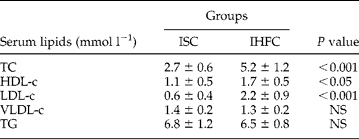
Table 1 shows the serum lipids in the two groups of mice 1 day before experimental infection. Usually, the infected mice fed on high-fat chow showed higher levels of serum lipoproteins than the infected mice fed on standard chow, except for VLDL-c and TG. Comparing IHFC mice with ISC mice, major differences were observed for TC (increased by 90%) and LDL-c (increased by 250%), while HDL-c showed an increase of about 50%.
Serum lipoproteins (mean±SD) found in the infected mice fed standard chow (ISC group) and in the infected mice fed high-fat chow (IHFC group).
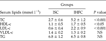
HDL-c, high-density lipoprotein cholesterol; LDL-c, low-density lipoprotein cholesterol; NS, not significant; TC, total cholesterol; TG, triglycerides; VLDL-c, very-low-density lipoprotein cholesterol.
TC levels were reduced (2.3 ± 0.5, P = 0.1602) in mice fed on standard chow (ISC group) and were significantly reduced (3.8 ± 0.7, P = 0.0002) in IHFC mice at the end of the experiment (day 63 after infection) when compared with the levels measured 1 day before experimental infection.
Morphological findings
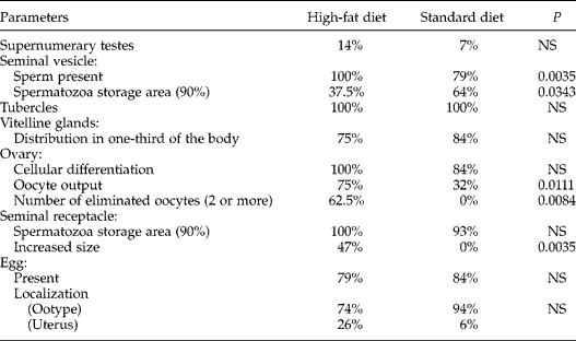
Light microscopy revealed some differences in the composition of the reproductive system between worms collected from each group of mice (table 2). Adult worms recovered from IHFC mice showed greater morphological differentiation regarding supernumerary testes, seminal vesicle and seminal receptacle than ISC mice. In the IHFC group, the ovary showed much higher oocyte-laying rates than in control females. The seminal vesicle of flukes retrieved from IHFC mice group showed a smaller amount of sperm. The dorsal surface of male worms presented tubercles, a common feature (100%) seen in worms recovered from mice of the two groups. About 80% of the females produced immature eggs in the ootype (74% and 94%) or uterus (26% and 6%), in IHFC and ISC control groups, respectively.
Light microscopy observations in the composition of the reproductive system between male and female adult worms from the high-fat diet and standard diet groups of mice.
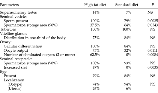
NS, not significant.
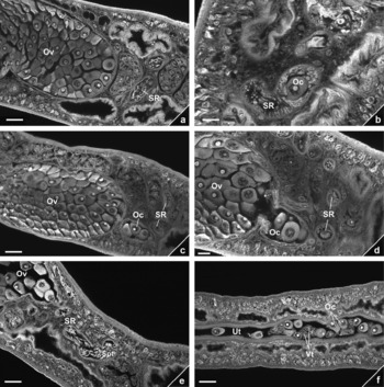
In addition, laser confocal microscopy also showed morphological differences between worms from mice of both groups. The reproductive system of females was distinguished by the presence of ovary, seminal receptacle, oviduct, oocytes and uterus (fig. 1). No modification in the ovary was observed. The ovaries had an elongated shape filled with oocytes (fig. 1a–c). At the posterior end, a protruding bulge, which enclosed oocytes leaving the ovary, was observed in females collected from mice on the high-fat diet (fig. 1d). The seminal receptacle closely posterior to the ovary, presented an increased size (fig. 1e) when compared to that observed in female controls (fig. 1b). In experimental female worms oocytes and vitelline cells were inside the uterus (fig. 1f). Both ootype and vitelline glands did not show any morphological alteration in worms of both groups.
Reproductive organs of female adult Schistosoma mansoni from mice fed on a standard diet (a, b) or a high-fat chow (c, d, e, f) and observed with confocal laser scanning microscopy. Ov, ovary; Oc, oocyte; SR, seminal receptacle; Spt, spermatozoa; Ut, uterus; Vt, vitelline cell. (a) Ovary with mature oocytes. (b) Seminal receptacle filled with spermatozoa and an oocyte. (c) Ovary with clumps of remaining oocytes. (d) Oocytes leaving the ovary. (e) Seminal receptacle lying closely posterior to the ovary presenting an increased size and filled with spermatozoa. (f) Oocytes and vitelline cells lying inside the uterus. Scale bars = 20 μm (a, c, e, f) and 10 μm (b, d).

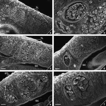
The male reproductive system was distinguished by the presence of testicular lobes, gynaecophoric canal and seminal vesicle (fig. 2). The seminal vesicle of control males presented an abundant amount of sperm, in contrast with those recovered from mice under a high-fat diet (fig. 2a, b, d). However, the number and morphological features of testicular lobes were no different between worms from mice of both groups (fig. 2a, c). Specimens recovered from mice under the high-fat diet showed more supernumerary testes, although the numbers varied (fig. 2e and f).
Reproductive organs of male adult Schistosoma mansoni from mice fed on a standard diet (a, b) or a high-fat chow (c, d, e, f) and observed with confocal laser scanning microscopy. DC, deferent canal; GC, gynaecophoric canal; Spt, spermatozoa; TL, normal testicular lobes; SV, seminal vesicle; STL, supernumerary testicular lobe. (a) Normal testicular lobes. (b) Detail of the reproductive organs: seminal vesicle and deferent canal leading to the gynaecophoric canal. (c) Normal testicular lobes. (d) Seminal vesicle with scarce spermatozoa. (e and f) Supernumerary testicular lobes showing large cells resembling mature oocytes. Scale bars = 20 μm (a, c, d, e, f) and 10 μm (b).

Discussion
Four major points can be made regarding the present results. The first refers to the use of a diet-induced model that has shown to be successful in the study of dietary effects on cholesterol and lipoprotein metabolism (Khovidhunkit et al., Reference Khovidhunkit, Kim, Memon, Shigenaga, Moser, Feingold and Grunfeld2004; Fernandez & Volek, Reference Fernandez and Volek2006). Present findings showed that a high-fat diet caused dyslipidaemia in mice.
The second point corroborates previous studies on the effects of nutrition upon Schistosoma mansoni infection (Coutinho et al., Reference Coutinho, Barros, Barbosa, Oliveira, Silva, Araujo and Andrade2003; Oliveira et al., Reference Oliveira, Silva, Barbosa, Ribeiro-Dos-Santos, Coutinho, Andrade and Soares2004). Reductions in the levels of cholesterol in the hosts were observed during experimental infections with S. mansoni (el-Marzouki & Amin, Reference el-Marzouki and Amin1997; Lima et al., Reference Lima, Sena, Stewart, Owen and Dolphin1998; Ramos et al., Reference Ramos, de Vasconcelos, de Carvalho and Lima2004), and schistosome infections counteracted the effects of an atherogenic diet by modulating host lipid metabolism and inducing a reduction in total blood cholesterol concentrations (Doenhoff et al., Reference Doenhoff, Stanley, Griffiths and Jackson2002). These changes in lipid metabolism reinforce the opinion that schistosomes utilize host lipids for their metabolism (Rumjanek & Simpson, Reference Rumjanek and Simpson1980).
The third point is related to the understanding of how schistosomes utilize compounds from their hosts. The physiological relationship between schistosomes and their hosts is complex and details of several biochemical processes still remain to be determined (Neves et al., Reference Neves, Machado-Silva, Pelajo-Machado, Oliveira, Coutinho, Lenzi and Gomes2001). For example, it is not yet understood whether fatty acids can enter the parasite by crossing the tegumental membranes or whether they must be absorbed through the gut, since the fatty acids acquired by schistosomes from their hosts are bound to the surface of the parasite (Brindley, Reference Brindley2005; Braschi et al., Reference Braschi, Curwen, Ashton, Verjovski-Almeida and Wilson2006). Furthermore, cholesterol exchange in schistosome worm pairs may be partly or wholly consequences of the normal tegumental turnover of the molecule (Bansal et al., Reference Bansal, Bhatti and Sehgal2005).
The surface of the adult worms is of particular importance, playing a central role in the physiology of schistosomes (Racoosin et al., Reference Racoosin, Davies and Pearce1999). No previous studies have been concerned with a presumed correlation between a cholesterol-rich diet and morphological features of the tegument. The results presented here suggest that high-fat chow does not affect the external aspect of the tubercles, which is identical in control males. Previously, it was demonstrated that worms recovered from mice maintained under a protein-deficient diet showed grouped and flat tubercles, and also subtegumental vacuolated areas (Neves et al., Reference Neves, Machado-Silva, Pelajo-Machado, Oliveira, Coutinho, Lenzi and Gomes2001).
The content and distribution of cholesterol is regulated dynamically by complex mechanisms (Sato et al., Reference Sato, Ishii, Makino, Iwabuchi, Yamaji-Hasegawa, Senoh, Nagaoka, Sakuraba and Kobayashi2004). Although labelled lipid in newly transformed schistosomula has been shown to be distributed in droplets throughout the organism (Furlong et al., Reference Furlong, Thibault, Morbelli, Quinn and Rogers1995), no studies have been addressed to discuss how lipids are distributed in adult worms. Interestingly, the distribution of cholesterol was studied by following the distribution of its fluorescent analogue, dehydroergosterol, using the nematode Caenorhabditis elegans as a laboratory model. In this case, it was demonstrated that dehydroergosterol is accumulated in oocytes and spermatozoa. Males displayed exceptionally strong labelling of spermatids, which suggested a possible role for cholesterol in sperm development (Matyash et al., Reference Matyash, Geier, Henske, Mukherjee, Hirsh, Thiele, Grant, Maxfield and Kurzchalia2001). Data accumulated in recent years have demonstrated that cholesterol is not only a structural component of the membrane, making it less fluid, but is also actively involved in the modulation of cell signalling and reproduction (e.g. sperm and oocyte development and egg laying) (Choi et al., Reference Choi, Chitwood and Paik2003). This modulation is thought to occur via cholesterol- and glycosphingolipid-enriched microdomains within the plasma membrane, often referred to as glycolipid-enriched membranes (GEMs) or ‘rafts’ (Simons & Ikonen, Reference Simons and Ikonen1997).
The fourth point is related to the most interesting observation in females grown in the group of mice on a high-fat diet, showing oocytes leaving the ovary and within the uterus in association with vitelline cells. During oogenesis, a rapid accumulation of proteins and lipids occurs in the oocytes, hence enabling the eggs to mature in a relatively short time. In addition, the seminal vesicle of control males contained an abundant amount of sperm, different from males derived from mice under the high-fat diet. The present results suggest that rich-cholesterol environments are advantageous, especially for reproduction. The rate of faecal egg-laying and tissue egg burden in mice fed on the high-fat chow were higher compared with those observed in mice fed on the standard diet (Neves et al., Reference Neves, Alencar, Costa-Silva, Águila, Mandarim-de-Lacerda, Machado-Silva and Gomes2007).
Morphological studies by means of light and laser confocal microscopy, show polyorchidism in male schistosomes. The lobes of these supernumerary testes contain variable amounts of fully developed oocytes instead of a testicular mass (Hulstijn et al., Reference Hulstijn, Barros, Neves, Moura and Machado-Silva2003). A considerable weight of evidence has been accumulated to show that the process underlying the expression of supernumerary testes with oocytes is, at least in part, host induced (Hulstijn et al., Reference Hulstijn, Barros, Neves, Moura, Gomes and Machado-Silva2006). In this context it is relevant to mention that similar morphological features were observed here. The present findings suggest that the rich-cholesterol environment could be actively involved in the modulation of cell signalling and reproduction, because the lobes contained fully developed oocytes in variable amounts, different from control males.
Acknowledgements
We wish to thank Dr Henrique L. Lenzi and Pedro Paulo de Abreu Manso for use of facilities and help with the confocal laser-scanning microscope, Dr Lygia R. Corrêa for providing cercariae, Bruno Eschinazi Vieira for technical photography, all members from the Oswaldo Cruz Institute, and Mr Charles Wendell Cropper from Flash Idiomas & Traduções for English revision of the text. This work was supported by a PhD scholarship from CAPES, and by fellowships from CNPq. We are also grateful to the Oswaldo Cruz Foundation (FIOCRUZ) and Rio de Janeiro State University (UERJ) for financial support.