Introduction
Cochlear implantation is an established treatment for severe to profound sensorineural hearing loss. The complication rate is generally low, and is classified as either major or minor.Reference Bhatia, Gibbin, Nikolopoulos and O'Donoghue 1 Post-implant haematoma is considered a minor complication, but can have serious consequences if not properly managed. For example, mismanagement could result in an infected haematoma, with the risk of total implant extrusion.Reference Filipo, D'Elia, Covelli, Bertoli, De Seta and Manganaro 2
Quoted to have an incidence of 0.4–3.7 per cent, the vast majority of haematomas occur in the immediate and early post-implant period.Reference Filipo, D'Elia, Covelli, Bertoli, De Seta and Manganaro 2 We present and discuss a series of patients who developed haematoma several months or even years after the initial cochlear implant surgery.
Materials and methods
The case records of all post-cochlear implant patients who presented to the senior author with delayed-onset haematoma formation around the cochlear implant site from 1996 to 2016 were retrospectively studied.
Results
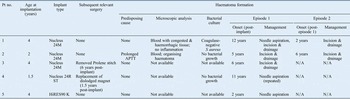
There were a total of five patients in our series (Table I). Each of them presented with a delayed-onset boggy swelling over the region of the implant receiver-stimulator. As two patients had recurrent episodes, a total of seven swellings were managed. The patients were all children who had undergone cochlear implant surgery aged 1.5–4 years (median, 2.5 years). The first haematoma episode occurred 2–12 years (median, 6 years) after cochlear implant surgery. Of the two patients who had recurrent episodes, one had recurrence at two years and the other at six years after the first episode. There was a median follow-up period of 3 years (range, 0.5–3 years) since the last episode had occurred.
Patient profile and management summary

Pt no. = patient number; APTT = activated partial thromboplastin time; N/A = not applicable
The development of swelling around the implant site was sudden and spontaneous, with patients eventually having difficulty in securing the magnetic coil over the receiver-stimulator.
None of the patients presented with a history of preceding trauma, such as a fall or a knock to the implanted area, as a predisposing cause. There was no history suggestive of coagulopathy, such as prolonged bleeding after sustaining wounds, or easy bruising of the skin, although one patient was found to have prolonged activated partial thromboplastin time (43.7 seconds, compared to a normal upper limit of 36.1 seconds).
Of the five patients, two had a history of a previous surgery relating to the cochlear implant. One patient had surgery (9.5 years prior to the development of swelling) to replace a magnet that had been dislodged from its Silastic® pocket. Another had surgery (seven years prior to the development of swelling) to remove a Prolene stitch (used in the earlier years of cochlear implant surgery as an anchoring suture), which had been causing skin irritation.
All the patients had associated pain and tenderness. None had signs of acute inflammation of the overlying skin or skin breakdown. The fluid content in all the swellings was similar in appearance (dark red blood). Microscopic analysis of the fluid, which was available for two of the patients, confirmed haematoma (Table I).
Of the three bacterial culture tests conducted, only one yielded a positive result. In this case, the sample, which was obtained via needle aspiration in an uncooperative child, grew coagulase-negative Staphylococcus aureus. The swelling persisted, and she was treated by incision and drainage 2 days later. From the contents of the swelling obtained from the incision and drainage procedure, no signs of purulence were evident, no bacteria could be cultured and no inflammation was detected on histological analysis. These observations suggested that the initial positive bacterial culture result was the result of contamination during the needle aspiration procedure.
Of the seven haematoma episodes, two were managed by needle aspiration alone, five by incision and drainage alone, and one by both needle aspiration and incision and drainage procedures. All patients had a mastoid pressure bandage applied for about 24 hours after each procedure. For the three haematomas managed by needle aspiration, one was successfully managed after aspirating only once. The other two swellings resolved after repeat aspiration and incision and drainage respectively. Successful resolution of the swelling was achieved for all episodes treated by incision and drainage.
Of those haematomas that were incised and drained, an incision of about 1–2 cm in length was made over each of the swellings. No active bleeding was evident in any of the patients after the blood, together with any clots, had been evacuated. In each patient, the cochlear implant device had been left in situ and no obvious source of bleeding was encountered in the surgical field.
Discussion
Although delayed haematoma after cochlear implant surgery is not rare, the cause remains unclear. The literature on post-cochlear implant haematoma occurring as a late complication is surprisingly scarce. A Medline search revealed only one paper that dealt specifically with such a complication. In a retrospective study on 22 subjects with post-cochlear implant haematoma, 4 subjects presented with delayed-onset lesions years after the cochlear implant surgery.Reference Filipo, D'Elia, Covelli, Bertoli, De Seta and Manganaro 2 Each of them had a distinct preceding cause: three had a definite history of trauma to the implant site and one had coagulation disorder. In our series, however, only one patient had coagulation abnormalities and none had a definite history of trauma as a preceding cause. The existence of a seemingly spontaneous delayed-onset haematoma after cochlear implant surgery as a distinct clinical entity is highlighted because of its diagnostic significance.
In our series, there were initial concerns that the presenting swelling could be the result of abscess formation. Although bacterial culture was positive in one case, we eventually thought it to be a contaminant rather than a cause. It should be differentiated from an abscess, which usually has overt signs and symptoms of infection, and often has skin breakdown.Reference Low, Rangabashyam and Wang 3 In a swelling of this nature, another differential diagnosis which should be considered is that caused by cerebrospinal fluid (CSF) accumulation. Extradural CSF collection has been reported several months following paediatric cochlear implantation.Reference Horton, Friedmann and Roland 4 , Reference Filipo, Covelli, D'elia and Mancini 5
Some post-cochlear implant patients may develop small haematomas that are asymptomatic and spontaneously resolve without medical attention even being sought. The patients in the present series, however, were all symptomatic: the swellings were painful and interfered with the wearing of the external cochlear implant device. Although it was possible that some cases would have resolved spontaneously over time, parental distress often dictated active measures that would resolve the problem sooner rather than later.
-
• The literature on post-cochlear implantation haematoma occurring as a late complication is scarce
-
• Most affected patients did not have a history of definite trauma or other predisposing causes
-
• Differential diagnoses of an abscess or cerebrospinal fluid leakage should be considered
-
• Therapeutic options include observation for spontaneous resolution, needle aspiration, and incision and drainage
-
• Needle aspiration is diagnostic and therapeutic; however, it risks an uncomplicated haematoma converting to a septic one
Needle aspiration alone, with a pressure dressing, was successfully used to resolve an episode of haematoma in two patients, although one patient required a repeat aspiration procedure. Another patient required incision and drainage when the swelling persisted after an aspiration procedure. Needle aspiration could be easily carried out in the clinic. Furthermore, it can immediately differentiate a haematoma from other causes of swelling such as CSF or pus. However, needle aspiration cannot remove clots or stop active bleeding if present.Reference Rojas, Eliason and Fredrick 6 Furthermore, the risk of inadvertently introducing infection and possibly resulting in conversion to an abscess remains a concern (as illustrated by patient one). Nevertheless, based on our experience, we believe that needle aspiration in the clinic is a good first-line treatment option in patients who are able to tolerate the entire procedure performed under aseptic conditions and allow a mastoid pressure dressing to be effectively applied for the next 24 hours or so. When this is not possible, such as in a fretful young child, incision and drainage under general anaesthesia might be a better option.
During cochlear implant surgery, deep bony wells are often created to prevent device migration and to maintain a low device profile, but such a technique may result in exposure of the underlying dura, particularly in children. Filipo et al. hypothesised that delayed-onset CSF swelling was due to chronic direct contact of the receiver with the dura, creating a local area of weakness predisposing to leakage.Reference Filipo, Covelli, D'elia and Mancini 5 Horton et al. suggested that even a minor, unnoticed trauma to the receiver site could induce a small violation of the dura, given its proximity to the device.Reference Horton, Friedmann and Roland 4 The reason(s) why haematoma should develop years after cochlear implant surgery without apparent predisposing causes is speculative. As deep bony wells had been used in these children, the same causative mechanisms proposed for delayed CSF leakage may well apply, resulting in the breaching of venous sinus walls that might have been exposed during surgery. The types of implant used by the patients in this study (Table I) are likely a reflection of the frequency of their use by the senior author, rather than being specifically associated with this complication.
Conclusion
The majority of delayed-onset haematoma episodes occurred in patients without obvious predisposing factors such as coagulopathy and definite trauma. Therapeutic options include an observational approach for spontaneous resolution, needle aspiration, and incision and drainage. Needle aspiration can differentiate a haematoma from an abscess or CSF leakage, and it provides an effective immediate therapeutic solution. However, aseptic techniques are emphasised to minimise the chances of an uncomplicated haematoma converting into a septic one.