Introduction
Therapeutic embolisation was first used in the management of acute gastrointestinal bleeding in 1972, by Rosch et al.,Reference Rosch, Dotter and Brown1 and has since become established in the management of many clinical conditions. Kingsley and O'Connor provided a review on the subject of embolisation in otolaryngology in 1982, noting its application in the management of acute haemorrhage, vascular tumours and angiomatous malformations.Reference Kingsley and O'Connor2
The aim of this review is to provide an update on the use of embolisation in the management of otorhinolaryngological conditions (see Appendix 1 for our search strategy).
Epistaxis
Sokoloff et al. first used embolisation successfully for refractory epistaxis in 1974.Reference Sokoloff, Wickbom and McDonald3 Local protocols have been developed for epistaxis management and have achieved control in 95 per cent of patients.Reference Daudia, Jaiswal and Jones4 If sphenopalatine artery ligation fails or the patient is unfit for general anaesthesia, angiographic embolisation is indicated.
The embolic materials used include polyvinyl alcohol particles, microcoils and Gelfoam® torpedoes.Reference Fukutsuji, Nishiike, Aihara, Uno, Harada and Gyoten5 Success rates range between 71 and 100 per cent.Reference Tseng, Narducci, Willing and Sillers6 External carotid artery ligation should be avoided as it prevents later embolisation. Angiography normally demonstrates a supply from large collaterals from the greater palatine artery and infraorbital branches of the internal maxillary artery, and it is uncommon to find other responsible aberrant vessels.
Pre-operative embolisation
Pre-operative tumour embolisation is undertaken via an endovascular approach, although direct percutaneous injection of an embolic agent into the tumour may be performed. Indications for embolisation include control of surgically inaccessible feeding vessels, which reduces blood loss and damage to adjacent structures, and that also allows better observation of the surgical field, reducing the risk of surgical complications.Reference Gupta, Purkayastha, Bodhey, Kapilamoorthy and Kesavadas7
Juvenile nasopharyngeal angiofibroma
These tumours have the potential to cause life-threatening bleeding during surgery.Reference Yiotakis, Eleftheriadou, Davilis, Giotakis, Ferekidou and Korres8 Recurrence is related to incomplete excision. Angiography allows embolisation of the feeding arteries pre-operatively, reducing intra-operative blood loss by 60 per cent. Embolisation is performed 24–72 hours before resection. Most vessels arise from the external carotid artery via the internal maxillary artery and ascending pharyngeal arteries, although the internal carotid artery can also contribute to large tumours via the vidian and middle meningeal arteries.Reference Yiotakis, Eleftheriadou, Davilis, Giotakis, Ferekidou and Korres8
In the pre-embolisation era, the average blood loss was 1136 ml. Studies have shown that this is greatly reduced with the use of embolisation (to 547 mlReference Shenoy, Grover, Janardhan, Jayakumar, Hedge and Satish9 and 677 ml,Reference Li, Qian, Shan and Wang10 variously) enabling an almost bloodless operative field and enhancing tumour resection.Reference Shenoy, Grover, Janardhan, Jayakumar, Hedge and Satish9
However, when there is deep invasion of the sphenoid, embolisation may make complete excision more difficult, as it means that fingers of tissue that extend down the vidian canal or behind the pterygoid plates may remain undetected as they do not bleed.Reference El-Banhawy, Ragab and El-Sharnoby11, Reference Carrillo, Albores, Ramirez-Ortega, Aiello-Crocifoglio and Onate-Ocana12
Tumour haemorrhage
This occurs frequently in patients with ulcerative tumours, due to vessel erosion by malignancy. Management is initially conservative, with local packing, but transarterial embolisation has been used with success.Reference Chou, Lu, Lin, Hong, Chen and Hsu13 Surgical treatment can be difficult, because of factors such as fistulae and radiation-induced necrosis, and is associated with a high mortality and complication rate. Surgical ligation can leave the patient at risk of re-bleeding from a collateral circulation, changing the vascular anatomy and making endovascular management more difficult.Reference Kakizawa, Toyota, Naito and Ito14 Emergency ligation of the internal or common carotid artery results in a mortality rate of 9–100 per cent, with neurological complications in 16–100 per cent of cases.
Arterial embolisation offers an effective, safe, fast method for controlling bleeding from tumours.Reference Imai, Kajihara, Kamei, Komaki and Tamada15 Transarterial embolisation has been reported to have a success rate of 92 per cent,Reference Kakizawa, Toyota, Naito and Ito14 as well as producing an average post-haemorrhage survival of 26 days, compared with 8 days in patients receiving angiography alone.Reference Chou, Lu, Lin, Hong, Chen and Hsu13
The haemostatic effect of embolisation is thought to last longer than that of ligation, as the embolic materials can reach the peripheral vessels.Reference Kakizawa, Toyota, Naito and Ito14, Reference Bhansali, Wilner and Jacons16 Even if re-bleeding occurs, repeated embolisation is relatively easy. The recurrence rate of haemorrhage after embolisation is 0–33 per cent.Reference Kakizawa, Toyota, Naito and Ito14
The optimal timing and application of transarterial embolisation is difficult in patients who are haemodynamically unstable due to massive bleeding, and the mortality of the procedure is high in such patients. In those with minor bleeding, it can be hard to identify the source of the bleeding, and radiologists may hesitate to use transarterial embolisation if no definite source can be found.
Palliative embolisation
The use of palliative embolisation to control haemorrhage from terminal head and neck cancers has been reported. Most bleeding arises from the oropharynx; therefore, local packing is distressing and often impractical.
Ligation of the carotid artery is an alternative, although it has significant mortality and morbidity. Pharyngocutaneous fistulae or tumour may make external access to the carotid system difficult. Reports favour transarterial embolisation over open techniques due to its safety and efficacy, with low complication rates and satisfactory survival rates being reported.Reference Sesterhenn, Iwinska-Zelder and Dalchow17
Paraganglioma
The first successful report of carotid body tumour embolisation was in 1983 by Shick et al.Reference Schick, Hieshima and White18 Blood loss has been shown to be significantly reduced with the use of pre-operative embolisation. Direct puncture therapeutic embolisation can be considered in the palliative management of these tumours.Reference Van Den Berg19
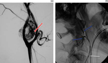
In cases of carotid body tumour resection, mean blood replacements of 2.1 and 4 units have been reported in cases in which embolisation was not performed, reduced to 200 ml with the use of pre-operative embolisation.Reference Schick, Hieshima and White18 This procedure can also reduce tumour size by as much as 25 per cent, an important consideration as cranial nerves are easier to identify and preserve and it is thus uncommon to have to clamp or resect the internal or external carotid artery.Reference Persky, Setton, Niimi, Hartman, Frank and Bernstein20 Pre-operative embolisation of tumours larger than 2 cm has been advocated, and the operative procedure should be performed within 48 hours of embolisation (Figure 1).Reference Schick, Hieshima and White18
(a) A lateral angiogram of the common carotid artery showing the typical appearance of a carotid body tumour splaying the carotids, supplied by the ascending pharyngeal artery (red arrow). (b) Lateral angiogram showing pre-operative embolisation with particles and coils (blue arrows); the catheter tip (white arrow) is seen in the common carotid.

Embolisation in cases of vagal paraganglioma has led to a reported reduction of mean blood loss from 990 to 425 ml, of operative time from 431 to 334 minutes, of hospital stay from 18.3 to 7.4 days, and of post-operative hypoglossal nerve paralysis incidence.Reference Miller, Boon, Atkins and Lowry21
Carotid blow-out syndrome
Rupture of the extracranial carotid arteries or their major branches is life-threatening.Reference Chang, Lirng, Luo, Wang, Wu and Guo22 Carotid blow-out syndrome is a pre-mortem event, and emergency surgical ligation may be inappropriate due to the poor condition of the patient.Reference Luo, Teng, Chang and Chang23 Surgical management is difficult, due to previously irradiated or infected fields. The mortality rate for emergency ligation of the internal or common carotid artery is 9–100 per cent, with major neurological morbidity rates of 9–84 per cent.Reference Roh, Suh, Kim, Lee, Choi and Choi24, Reference Bates and Shamsham25
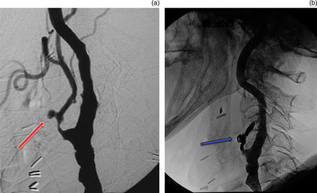
Recent developments in the use of covered stents have decreased morbidity rates to 0–8 per cent and mortality rates to 0 per cent, although the long-term sequelae have not been determined.Reference Roh, Suh, Kim, Lee, Choi and Choi24 When the internal, common or proximal external carotid artery trunk is involved, the best approach is occlusion with detachable balloons. Branches of the external carotid artery are best approached via superselective embolisation so the branches adjacent to the affected artery are preserved, reducing the risk of ischaemia involving the face and neck, and the risk of impairment of cranial nerve function (Figure 2).Reference Luo, Teng, Chang and Chang23
(a) A lateral angiogram of the common carotid artery in a patient with a laryngeal tumour treated with radiotherapy and neck dissection, who presented with neck haemorrhage from a pseudo-aneurysm (red arrow) of the external carotid artery. (b) Lateral angiogram showing treatment via occlusion of the external carotid artery with coils (blue arrow).

There is a 15–20 per cent incidence of delayed cerebral ischaemic complications. Re-bleeding and complication rates can be high, due to infected wounds and tumour progression.Reference Roh, Suh, Kim, Lee, Choi and Choi24 Intracranial complications can occur due to backflow of embolic material into the internal carotid artery or vertebral artery.Reference Luo, Teng, Chang and Chang23 However, immediate and long-term success has been reported in up to 100 per cent of cases, with a major complication rate of 7 per cent.Reference Luo, Teng, Chang, Chang and Guo26
It has been concluded that a combination of coil embolisation and stent graft isolation of the common and internal carotid artery can control acute bleeding from vascular tumour invasion and prevent further carotid bleeding episodes in patients with advanced tumours.Reference Bates and Shamsham25
Other uses
Chylothorax secondary to injury to the thoracic duct or one of its branches has been treated by percutaneous embolisation of the thoracic duct proximal to the transection, with subsequent normalisation of chylous output.Reference Patel, Lewandowski, Bove, Nemcek and Salem27
Trauma
Severe craniofacial injury may lead to significant oronasal bleeding. The presumed location and lateralisation of bleeding is often misleading; therefore, bilateral internal carotid artery and external carotid artery angiography is helpful.Reference Komiyama, Nishikawa, Kan, Shigemoto and Kaji28
In cases of ethmoid artery bleeding, embolisation would be via the ophthalmic artery, which carries an unacceptable risk of blindness due to occlusion of the retinal and posterior ciliary branches; surgical clipping is thus advocated.Reference Komiyama, Nishikawa, Kan, Shigemoto and Kaji28
Many asymptomatic patients have significant vascular injuries, and management of zone I or III injuries can require extensive surgery. There is currently a debate concerning the merits of mandatory neck exploration versus the use of angiography and embolisation, since delayed management of vascular and aerodigestive tract injuries is associated with a high morbidity rate. One study deemed interventional surgery to have been unnecessary in up to 63 per cent of patients.Reference Golueke, Goldstein and Scalafani29 In contrast, it has been demonstrated that patients with a normal initial angiogram have a less than 2 per cent incidence of missed injuries.Reference Germiller, Myers, Harris and Bradford30
Definitive control of haemorrhage can be achieved with the use of transarterial embolisation, especially in cases of zone III trauma, in which haemorrhage can be difficult to access operatively. However, Golueke demonstrated no difference between surgical exploration and selective management, in terms of length of stay, morbidity and mortality.Reference Golueke, Goldstein and Scalafani29
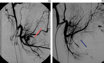
In cases of penetrating neck trauma, the liberal use of cervical angiography and transarterial embolisation has been advocated due to its safety and reliability.Reference Golueke, Goldstein and Scalafani29 Therapeutic embolisation has been shown to be effective in arterial neck trauma, and should be considered a viable method of management when open access is not deemed essential (Figure 3).Reference Sclafani, Panetta, Goldstein, Phillips and Hotson31
(a) A right lateral upper cervical angiogram of an 18-year-old patient, attacked with a broken glass bottle, who subsequently presented with an expanding submandibular mass, showing a pseudo-aneurysm (red arrow) in the region of the lingual artery. (b) The same right lateral upper cervical angiogram view as illustrated in Fig 3 (a) showing subsequent embolisation of the pseudo-aneurysm (blue arrow).

Hereditary haemorrhagic telangiectasia
The treatment of epistaxis in patients with this condition is initially conservative. There is no definitive cure; rather, therapeutic measures aim to reduce the severity and frequency of epistaxis, to improve quality of life.Reference Layton, Kallmes, Gray and Cloft32 Increased vascularity due to aberrant angiogenesis may make permanent treatment with embolisation difficult.Reference Layton, Kallmes, Gray and Cloft32 In one series utilising embolisation, success rates of 80 per cent were reported in patients with idiopathic epistaxis, compared with 20–25 per cent in those with hereditary haemorrhagic telangiectasia.Reference Roh, Suh, Kim, Lee, Choi and Choi24, Reference Roland, McRae and McCombe33
The role of embolisation in hereditary haemorrhagic telangiectasia has not been established, because of high levels of recurrence and technical difficulties with embolisation of the anterior ethmoids.Reference Layton, Kallmes, Gray and Cloft32
Arteriovenous malformation
Angiography and selective embolisation is usually performed prior to surgical excision of arteriovenous malformations, when this is indicated, to reduce the vascularity of the lesion and to define feeder vessels. This aids long-term cure, since incomplete excision leads to recurrence. Embolisation has been shown to decrease operative blood loss and lesion size and to decrease the extent of surgical excision.Reference Righi, Bade, Coleman and Allen34 Due to the potential for rapid expansion in the period between embolisation and resection, the time between procedures should be 24–48 hours.Reference Kohout, Hansen, Pribaz and Mulliken35
Treatment of external carotid artery arteriovenous malformations with embolisation alone is indicated if the lesion is deemed inoperable and surgical resection would increase morbidity, or the patient is unfit for surgery.Reference Righi, Bade, Coleman and Allen34
Recommended indications for treatment with embolisation and resection are based on the clinical staging according to the modified Schobinger classification, i.e. (1) quiescence, (2) expansion, (3) destruction and (4) decompensation.Reference Kohout, Hansen, Pribaz and Mulliken35 Early stage lesions (i.e. stages one and two) can be resected and reconstructed easily. Painful or rapidly enlarging lesions (stages two and three) warrant early intervention owing to the high probability of progression and the risk of serious haemorrhage.
Intervention for extensive stage one lesions remains controversial, for three reasons: (1) the risk of progression cannot be predicted, (2) resection of very extensive stage one lesions is more likely to be incomplete, and (3) the deformity produced by extensive resection and reconstruction may be worse than that caused by the original lesion.Reference Kohout, Hansen, Pribaz and Mulliken35, Reference Aslan, Yavuz, Cagici and Kizilkilic36 Successful use of embolisation to treat arteriovenous malformations has been reported in 60 per cent of cases.Reference Kohout, Hansen, Pribaz and Mulliken35
Carotid–cavernous fistula
Carotid–cavernous fistulae involve an abnormal arteriovenous anastomosis between the carotid artery and the cavernous sinus.Reference Bizri, Al-Ajam, Zaytoun and al-Kutoubi37 They can be spontaneous, in patients with collagen vascular disease, fibromuscular disease of the cerebral arteries, or rupture of intracavernous internal carotid artery aneurysms.Reference Bizri, Al-Ajam, Zaytoun and al-Kutoubi37, Reference Yu, Lei, Chen, He, Chen and Li38 Acquired cases occur as a result of trauma, transsphenoidal surgery, cavernous sinus meningioma embolisation and rhinocerebral mucormycosis.
This condition can present with epistaxis and, sometimes, signs of ophthalmic vein occlusion (i.e. dilated forehead veins, headache and raised intra-orbital pressure). Patients may also develop pulsatile proptosis and visual disturbances.Reference Yu, Lei, Chen, He, Chen and Li38, Reference Pothula, Reddy and Nixon39
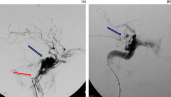
Reviews of carotid–cavernous fistula management have shown that angiography is the key to diagnosis and management. Endovascular embolisation has become an established treatment method. Stenting is also a feasible option due to its ability to maintain patency of the internal carotid artery (Figure 4).Reference Yu, Lei, Chen, He, Chen and Li38
(a) A lateral angiogram of the skull base in a 70-year-old man who presented with a history of recent head trauma without fracture, with subsequent proptosis and chemosis, showing a direct fistula between the cavernous sinus (blue arrow) and the internal carotid artery (red arrow). (b) A later sequence of the lateral angiogram of the skull base post embolisation showing the first coil placed in the cavernous sinus (blue arrow).

Tonsillar haemorrhage
Post-tonsillectomy haemorrhage due to a pseudo-aneurysm of the lingual or facial artery usually presents hours or days after the surgery.Reference Walshe, Ramos, Low, Thomas, McWilliams and Hone40 Angiography should be performed when an aneurysm or pseudo-aneurysm is suspected in the tonsillar fossa, and can be treated with embolisation.Reference Walshe, Ramos, Low, Thomas, McWilliams and Hone40
Tracheostomy-related haemorrhage
This is a recognised complication, and the majority can be managed conservatively. However, a tracheo-innominate artery fistula has a high mortality rate if untreated.Reference Johnson, Tabee, Fitz-James, Pass and Serres41, Reference Takasakia, Enatsua and Hasus42 Rates of 0.1–1 per cent have been reported after surgical tracheostomy, with a peak incidence at 7–14 days post-procedure.Reference Grant, Dempsey, Harrison and Jones43
If conservative measures fail, surgical management is required. However, technical limitations include difficulty in locating the bleeding vessels due to scarring from prior procedures. Thus, angiography with embolisation has been advocated as the preferred method of management, and has been used with success.Reference Johnson, Tabee, Fitz-James, Pass and Serres41,Reference Takasakia, Enatsua and Hasus42
Transsphenoidal surgery
Delayed epistaxis resulting from damage to the sphenopalatine branches of the external carotid artery is an infrequent but serious complication of transsphenoidal surgery, which can be managed by sphenopalatine artery ligation or embolisation.Reference Cockroft, Carew, Trost and Fraser44 False aneurysms of the sphenopalatine artery can occur after transsphenoidal surgery.Reference Nishioka, Ohno, Ikeda, Ohashi and Haraoka45
Significant vascular complications related to internal carotid artery injury during transsphenoidal surgery include carotid laceration, carotid–cavernous fistula, traumatic aneurysm, subarachnoid haemorrhage and cerebral infarction. Arterial injuries that manifest during or after transsphenoidal surgery are rare (1 per cent), but they are associated with significant morbidity (24 per cent) and mortality (14 per cent).Reference Cockroft, Carew, Trost and Fraser44, Reference Nishioka, Ohno, Ikeda, Ohashi and Haraoka45
Imaging of both the internal and external carotid circulations is important. Although internal carotid artery injuries are more common, negative findings on an internal carotid artery angiogram do not necessarily rule out a vascular injury.
Skull base tumours
When a significant portion of a skull base tumour is supplied directly by the internal carotid artery, particle embolisation from within the carotid artery may be performed.
For large, recurrent tumours, which may not be supplied directly by vessels typical of newly diagnosed lesions, direct percutaneous embolisation may help.
Pre-operative embolisation of a primary jugular foramen tumour should only be performed when it can reduce operative morbidity and blood loss.Reference Noonan and Choi46 In skull base surgery involving the region of the jugular foramen, the jugular bulb is opened. This can be associated with significant blood loss, prolonged operative time, and the risk of compression injury to cranial nerves IX, X and XI from overpacking. Embolisation of feeder vessels to the jugular bulb before surgery reduces the risk of these complications. There is a theoretical risk of injury to lower cranial nerves, but this is thought to be less than the risk incurred from surgical packing.Reference Carrier, Arriaga, Gorum, Dahlen and Johnson47
Functional endoscopic sinus surgery
Bleeding is a recognised complication of endoscopic sinus surgery.Reference Roland, McRae and McCombe33 Iatrogenic injury of the internal carotid artery is rare; however, if this happens then pressure to control bleeding, followed by angiography and embolisation, is life-saving. Pepper and colleagues' guidelines for internal carotid artery bleeding recommend the use of angiographic control (using tamponade balloons) to control bleeding, together with emergency coil embolisation.Reference Pepper, Wadwa, Tsai, Shibuya and Wong48
Complications
Tseng et al. have classified the complications of embolisation into major and minor events.Reference Tseng, Narducci, Willing and Sillers6 The commonest minor side effect is fever and localised pain due to tissue necrosis.Reference Kingsley and O'Connor2 Minor complications are transient, and include facial pain, headaches, mental confusion, paraesthesia, jaw pain, groin pain, numbness and facial oedema.
Major complications of embolisation include cerebrovascular accident (CVA), blindness, ophthalmoplegia, facial nerve palsy and soft tissue necrosis.Reference Tseng, Narducci, Willing and Sillers6 The most serious risk is inadvertent embolisation of the internal carotid artery, leading to a CVA. General complications, such as sensitivity to the embolic or contrast material, are rare.Reference Kingsley and O'Connor2
Barlow et al. stated that the complication rate for embolisation in epistaxis resembles that of arterial ligation, being 13–48 per cent.Reference Barlow, Deleyiannis and Pinczower49 A literature review of the success rates and complications related to embolisation for epistaxis found that, of 572 patients embolised for persistent epistaxis, five suffered significant long-term morbidity (a prevalence of <1 per cent) and two suffered minor long-term morbidity (i.e. facial scarring). The risk of CVA was less than 1 per cent.Reference Sadri, Midwinter, Ahmed and Parker50
Post-embolisation spasm of the accessory meningeal artery may cause ischaemia and hypoaesthesia of the third branch of the trigeminal nerve, whereas occlusion of the petrosal artery may cause ischaemia of the horizontal segment of the facial nerve, resulting in paralysis. Loss of vision can result from anastomosis between the internal maxillary artery and ophthalmic artery branches, although pre-operative angiography should demonstrate a choroidal ‘blush’ if this is the case.
Soft tissue necrosis is a rare complication, due to the extensive collateral blood supply of the head and neck. Unilateral necrosis of the mucosa overlying the hard palate has been reported in a patient following bilateral internal maxillary artery embolisation for epistaxis. The patient also received balloon tamponade and bilateral nasal packs for two days post-procedure, and it was suggested that packing should be removed as soon as possible after embolisation.Reference Guss, Cohen and Mirza51
Herdman suggested that if the stylomastoid artery is occluded, the facial nerve is not usually in danger, due to supply from the middle or accessory meningeal artery (present in 90 per cent of the population). In the remaining 10 per cent, as well as in cases of embolisation of the middle meningeal artery, facial nerve dysfunction should be expected. However, permanent embolic agents are more likely to cause a permanent palsy; if these agents are needed (for example when surgery is not an option), the provocation test (involving pre-embolic injection of lidocaine) may assess the risk of a lasting palsy.Reference Marangos and Schumacher52
The complications of embolisation for post-tonsillectomy haemorrhage include: vessel perforations with subsequent extravasation of embolic material; ischaemic injury to mucosal surfaces and cranial nerves; inadvertent involvement of the internal carotid artery; catheter-induced vasospasm; and post-procedure pain.Reference Opatowsky, Browne, McGuirt and Morris53
If hypovolaemic and vasoconstrictive drugs are used in the management of the trauma patient, there is the possibility of external carotid artery territory vasospasm. It is important to be aware that embolisation into these vessels may cause reflux of embolic material into the internal carotid artery, resulting in intracerebral complications.Reference Komiyama, Nishikawa, Kan, Shigemoto and Kaji28
Conclusion
Embolisation has a significant role to play in many aspects of otolaryngological practice. It is well established in the management of intractable epistaxis, being effective and having low complication rates.
Pre-operative embolisation of vascular tumours is available in most centres. The benefits extend beyond a reduction in blood loss; embolisation also improves visualisation of the surgical field, enabling greater ease of dissection, and reduces the duration of the operative procedure. Embolisation plays an increasingly important role in the palliation of advanced head and neck tumours, and in the management of the sequelae of these pathologies.
In cases of head and neck trauma, bilateral angiography with a view to embolisation should be considered as a diagnostic and therapeutic intervention, especially if surgical management is being considered.
Embolisation has proved an important adjunct in the management of haemangiomas, arteriovenous malformations and telangiectatic conditions, and also of post-operative complications.
In experienced units, embolisation is a safe, effective and important therapeutic adjunct in the management of a wide variety of otolaryngological conditions, and could have an important role to play in most areas of ENT practice.
Appendix 1. Search strategy
The search strategy used encompassed the Medline, Embase, PubMed and Cochrane databases. The search was performed in November 2008, with the strategy outlined below. All types of study were considered, and no language or publication date restrictions were applied. Results were discounted if not related to ENT (e.g. embolisation of uterine fibroids).
The number of articles retrieved is shown below
Embase
Otolaryngology plus embolisation: 0
Otolaryngology: 484 081
Embolisation: 20 286
Combined: 3002
Applicable: 107
Medline
Otolaryngology embolisation: 529
Applicable: 27
PubMed
Embolisation otolaryngology (plus related articles): 147
Cochrane
Library search for embolisation and otolaryngology: 0
A–Z search: 0
Search by topic ‘ENT disorders’: malignant disease = 0, non-malignant disease = 4