Introduction
The discovery of the non-encapsulated Trichinella zimbabwensis in crocodiles from commercial farms in Zimbabwe in 1995 and the descriptions thereof (Foggin et al., Reference Foggin, Vassilev and Widdowson1997; Mukaratirwa & Foggin, Reference Mukaratirwa and Foggin1999; Pozio et al., Reference Pozio, Foggin, Marucci, La Rosa, Sacchi, Corona, Rossi and Mukaratirwa2002) has led to several studies that include the phylogeny, ecological characteristics and geographic history of the species (Pozio & Murrell, Reference Pozio and Murrell2006), but also specifically the host range and extent of geographical occurrence in Africa. These studies have shown that T. zimbabwensis naturally infects crocodiles in Zimbabwe, Mozambique, Ethiopia and South Africa (Pozio et al., Reference Pozio, Foggin, Gelanew, Marucci, Hailu, Rossi and Gomez-Moralez2007; La Grange et al., Reference La Grange, Marucci and Pozio2009), as well as Nile monitor lizards (Varanus niloticus) in Zimbabwe (Pozio et al., Reference Pozio, Foggin, Gelanew, Marucci, Hailu, Rossi and Gomez-Moralez2007). South African surveillance studies reported a 38.5% prevalence of T. zimbabwensis among wild crocodiles from the Mpumalanga province and Kruger National Park (KNP) (La Grange et al., Reference La Grange, Marucci and Pozio2010). Despite experimental studies demonstrating the ability of T. zimbabwensis to infect mammalian hosts including rodents, pigs, carnivores and primates (Mukaratirwa & Foggin, Reference Mukaratirwa and Foggin1999; Pozio et al., Reference Pozio, Marucci, Casulli, Sacchi, Mukaratirwa, Foggin and La Rosa2004; Mukaratirwa et al., Reference Mukaratirwa, Dzoma, Matenga, Ruziwa, Sacchi and Pozio2008) there has only been a single report of a naturally infected mammal to date (La Grange et al., Reference La Grange, Marucci and Pozio2010) and the continued surveillance of other potential natural host species is vital in order to fully unravel the epidemiology of this parasite.
A recent survey in KNP to determine the extent of pansteatitis in crocodiles additionally provided an opportunity to establish the occurrence and distribution of T. zimbabwensis in wild Nile crocodiles representing the whole of KNP and to evaluate muscle predilection patterns in naturally infected crocodiles. Studies on different Trichinella species in various mammalian hosts have shown muscle predilection to be influenced by host and parasite species characteristics as well as infection intensity and age (Serrano et al., Reference Serrano, Pérez-Martín, Reina, Navarrete and Kapel1999; Kapel et al., Reference Kapel, Webster and Gamble2005). Host characteristics appear to be the most important determinant for predilection (Kapel et al., Reference Kapel, Webster and Gamble2005). Predilection patterns of T. zimbabwensis in red foxes (Vulpes vulpes) and other species revealed little deviation from that of other Trichinella species in the same host (Mukaratirwa & Foggin, Reference Mukaratirwa and Foggin1999; Hurníková et al., Reference Hurníková, Dubinský, Mukaratirwa, Foggin and Kapel2004) and similarly insignificant deviations were observed between T. spiralis, T. britovi, T. nativa and T. pseudospiralis infections in pigs, wild boars and horses (Kapel et al., Reference Kapel, Webster and Gamble2005). However, T. zimbabwensis has a proven propensity in nature towards infecting reptilian hosts and, more specifically, Nile crocodiles (Crocodylus niloticus) and Nile monitor lizards (V. niloticus) (Pozio et al., Reference Pozio, Foggin, Gelanew, Marucci, Hailu, Rossi and Gomez-Moralez2007; La Grange et al., Reference La Grange, Marucci and Pozio2009), whereas the encapsulated taxa reported in Africa, T. britovi, T. nelsoni and Trichinella T8, only infect mammalian hosts (Pozio et al., Reference Pozio, De Meneghi, Roelke-Parker and La Rosa1997, Reference Pozio, Pagani, Marucci, Zarlenga, Hoberg, De Meneghi, La Rosa and Rossi2005; Marucci et al., Reference Marucci, La Grange, La Rosa and Pozio2009). The importance of host characteristics, natural infection patterns of T. zimbabwensis and the physiological and anatomical differences between mammals and reptiles provides the incentive for studies aimed at elucidating predilection patterns of this species to reptilian hosts. The aim of this study was to determine the occurrence, distribution and predilection patterns of T. zimbabwensis muscle larvae in naturally infected Nile crocodiles from the KNP, South Africa.
Materials and methods
Source of crocodiles
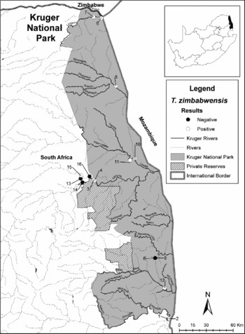
Following the deaths of more than 300 crocodiles attributed to pansteatitis in the Olifants and Letaba rivers near the western border of the KNP, South Africa, during 2008–2010, a survey of the disease in crocodiles from KNP perennial rivers was conducted in July 2010. A total of 12 Nile crocodiles from the five perennial rivers that originate outside the KNP, one large seasonal river (Shingwedzi River) and Shiloweni dam on an internal stream, were lethally sampled (fig. 1). Four crocodiles were also obtained in February 2011 from mine tailing dams in the Phalaborwa area, approximately 40 km upstream from where the crocodile deaths were detected (fig. 1). Larger crocodiles (>2.5 m in length) were targeted for the survey as they were deemed to have a greater chance of developing pansteatitis as a result of their propensity towards cannibalism and the increased amounts of food consumed compared to smaller or younger animals. This criterion also favoured the Trichinella surveillance as larger and older animals generally have a greater chance of becoming infected, for the same reasons. All animals were humanely slaughtered by means of firing a free projectile through the brain with a large-calibre rifle before full necropsies were conducted.
Map of Kruger National Park (KNP) showing major river systems and the localities where crocodiles (Crocodylus niloticus) were culled, and their status with regard to Trichinella zimbabwensis infection. 1 and 2, Ressano Garcia weir, Crocodile River; 3 and 4, Mamba weir, Olifants River; 5, Kanidood dam, Shingwedzi River; 6, Old Pafuri picnic spot, Luvuvhu River; 7, bridge, Luvuvhu River; 8 and 9, internal catchment, Shiloweni dam; 10, Engelhardt dam, Letaba River; 11, Hlanganini mouth, Letaba River; 12, Lower Sabie weir, Sabie River; 13, Selati dam, Phalaborwa; 14, Phalaborwa barrage; 15, Van Ryissen dam, Phalaborwa; 16, Tailings dam, Phalaborwa Mining Company.

Detection of larvae in muscles
During necropsy, muscle samples were collected from the head (base of tongue and caudal pterygoid), neck (sternomastoid), back (longissimus complex), internal intercostals, anterior legs (triceps brachii), posterior legs (external tibial flexor) and tail (longissimus caudalis and illoischiodalis). Additional samples were collected superficially from the lateral aspects of the tail base. These additional samples were collected bearing in mind that a biopsy sample would primarily consist of superficial musculature and the aim of this was to determine whether the superficial musculature could be deemed suitable for the detection of T. zimbabwensis. Fifty grams of muscle tissue were collected from each of the muscle groups. A total amount of 100 g of muscle tissue from the various muscle groups for each animal was pooled together for digestion. The muscle tissue was artificially digested as described by Nöckler & Kapel (Reference Nöckler, Kapel, Dupouy-Camet and Murrell2007) to determine the presence of Trichinella sp. first-stage larvae. After digestion, larvae from each of the positive animals were harvested, preserved in absolute ethyl alcohol and a proportion of larvae from each animal was referred to the International Trichinella Reference Centre in Rome, Italy, for species identification. From the remaining samples, 25 g from each of the muscle groups from positive animals were tested individually to determine intensity of infection in each individual muscle or group of muscles.
Data analysis
The occurrence of infection was calculated as the number of crocodiles positive for Trichinella sp. first-stage larvae in muscle divided by the total number of crocodiles tested and expressed as a percentage. Intensity of infection was quantified by the number of first-stage larvae in 1 g (lpg) of muscle, muscle group or tongue. Analysis of variance was used to determine the differences in the mean intensity of infection among the different muscles/muscle groups and the level of significance was set at P≤ 0.05.
Results and discussion
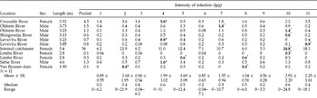
Of the 12 crocodiles originating from KNP, ten tested positive for Trichinella sp. muscle larvae, and only one of the four animals originating outside KNP, which were donated for the survey, tested positive (table 1). All the specimens of Trichinella sp. muscle larvae were sent to the International Trichinella Reference Centre for species identification and were confirmed as T. zimbabwensis. Larval burdens in pooled samples from each crocodile ranged from 0.1 lpg of muscle to 36 lpg, with a mean larval burden of 4.3 lpg. The intensities of infection for individual muscles/group of muscles are shown in table 1.
Intensity of infection of Trichinella zimbabwensis larvae in muscles/muscle groups of naturally infected crocodiles (Crocodylus niloticus) from the Kruger National Park, South Africa.

1, Tongue; 2, pterygoid; 3, sternomastoid; 4, tricep; 5, intercostal; 6, longissimus complex; 7, external tibial flexor; 8, tail – longissimus caudalis; 9, tail – iloischiodalis; 10, tail – dorsal biopsy; 11, tail – ventral biopsy.
lpg, larvae per gram of muscle.
N= 11, depicts the number of muscles/muscle groups tested for each animal.
* Depicts the muscle/muscle group with the highest intensity of infection for each animal sampled.
The pterygoid muscle harboured the highest mean number of larvae (2.68 lpg) followed by the triceps muscle (1.99 lpg). An interesting observation was that the dorsal tail musculature appeared to harbour a higher mean number of larvae (1.04 lpg) than ventral muscles (0.56 lpg). Similarly, the superficial musculature of the dorsal tail on average harboured more larvae (2.92 lpg) than the deeper muscles (2.25 lpg) and this trend was consistently observed in all but three of the positive animals. The results from the analysis of variance revealed no significant differences in the intensity of infection among the muscles sampled (F= 0.45, P= 0.92). However, the results from the dorsal tail, which mimic biopsies in live animals, indicate the potential that biopsies have for surveillance of T. zimbabwensis in crocodiles, since in 10 of the 11 positive animals (91%), even with relatively low levels of infection, larvae were successfully detected with biopsy samples of only 10 g.
The prevalence of T. zimbabwensis in wild crocodiles tested previously from South Africa, particularly the Mpumalanga province, was reported to be 38.5% (La Grange et al., Reference La Grange, Marucci and Pozio2009). The prevalence within KNP was much higher at 80%, but these previous results from KNP represented only a small sample of five animals that all originated from the Olifants Gorge area (La Grange et al., Reference La Grange, Marucci and Pozio2009). At that time it was thought that the high prevalence could have resulted from a possible increase in the frequency of cannibalism following the large number of crocodile deaths attributed to pansteatitis. The results of the present survey show the occurrence among the tested animals to be as high as 83.3%. Results also show the high number of infected crocodiles not to be limited to any specific area or the result of unusual circumstances, but suggest a natural phenomenon within the general crocodile population of KNP. In contrast with the findings of experimental studies conducted on encapsulated Trichinella sp. in mammals (Serrano et al., Reference Serrano, Pérez-Martín, Reina, Navarrete and Kapel1999; Kapel et al., Reference Kapel, Webster and Gamble2005) and non-encapsulated Trichinella sp. in varans (Varanus exanthematicus) (Pozio et al., Reference Pozio, Marucci, Casulli, Sacchi, Mukaratirwa, Foggin and La Rosa2004), where the tongue was the most important predilection site, in this study the tongue did not harbour the highest number of lpg. In studies with non-encapsulated Trichinella sp. in caimans (Caiman crocodylus), however, the anterior legs harboured, on average, more larvae than the tongue, and the intercostal muscles and posterior legs were also shown to be important predilection sites (Pozio et al., Reference Pozio, Marucci, Casulli, Sacchi, Mukaratirwa, Foggin and La Rosa2004). The results from the present study support these findings. The lower larval burdens observed in the tongue musculature when compared to mammals may be explained in part by the fact that in crocodiles, the tongue is fixed to the floor of the oral cavity, making it less mobile than that of mammals and thus less dependent on a high vascular supply associated with higher cellular metabolic requirements of moveable musculature (Huchzermeyer, pers. comm., 2010). This phenomenon was also seen in experimental studies involving T. pseudospiralis in monkeys (Macaca fasciolaris) (Kociecka et al., Reference Kociecka, van Knapen, Ruitenberg, Kim, Ruitenberg and Teppema1980), and does not seem to support the aforementioned. Additionally, the results of the present study did not support the findings of experimental studies conducted with non-encapsulated species in mammals where the diaphragm appeared to be the most important predilection site (Kapel et al., Reference Kapel, Webster and Gamble2005; Mukaratirwa et al., Reference Mukaratirwa, Dzoma, Matenga, Ruziwa, Sacchi and Pozio2008). These inconsistencies do, however, support the important role of host characteristics in muscle predilection and the pattern of infection of the host in nature, as alluded to in previous studies (Serrano et al., Reference Serrano, Pérez-Martín, Reina, Navarrete and Kapel1999; Kapel et al., Reference Kapel, Webster and Gamble2005). It is also important to note that the results from the present study are based on natural infections where the initial infection doses are not known.
More structured surveys involving the primary prey items of crocodiles are needed to elucidate the existence of other natural hosts that may play a role in the epidemiology of the parasites. Additional research with experimental infection of animals of uniform size and age, where the initial level of infection is controlled, is also required to remove any bias in predilection that may result from differences in age, immunological status and initial infection levels. The results of this study confirm the importance of the KNP as a natural refuge for T. zimbabwensis circulating among its wildlife (La Grange et al., Reference La Grange, Marucci and Pozio2010). These results also support the importance of host characteristics in muscle predilection by the larvae and show that muscle predilection of non-encapsulated T. zimbabwensis in reptilian hosts follows distinctly different patterns from those observed in mammals.
Acknowledgements
The authors wish to thank the rangers and scientific services staff of KNP responsible for the capture, euthanasia and transport of crocodiles. Our sincere thanks for the assistance received from the state veterinarian, Nelspruit, with the collection of samples; Dr Huchzermeyer for his assistance with necropsies; the invaluable insight into crocodilian anatomy and physiology provided by Drs Huchzermeyer, Webb, Manolis and Richardson; to Mrs T. La Grange, staff and students of the School for Biological and Conservation Sciences of the University of KwaZulu-Natal for their assistance in testing of samples; the International Trichinella Reference Centre for species confirmation; and Mr F. Koegelenberg for his assistance in the drafting of fig. 1.