Introduction
Vascular rings are congenital vascular anomalies of the aortic arch complex which cause compression of the trachea and/or oesophagus. These lesions often manifest with airway compromise due to extrinsic tracheal compression, or, less commonly, with feeding problems secondary to oesophageal compression. Patients with airway symptoms usually present earlier, while those with oesophageal symptoms present later in childhood, often when solid food is introduced. Congenital vascular anomalies can be broadly categorised as anatomically complete (e.g. double aortic arch) or anatomically incomplete (e.g. other vascular rings).Reference Kaiser, Kron and Spray1–Reference Woods, Sharp and Holcomb3
A variety of investigations may lead to the diagnosis of a vascular ring. Barium swallow, computed tomography (CT) scanning, magnetic resonance imaging and magnetic resonance angiography can provide diagnostic information necessary for surgical planning.Reference Turner, Gavel and Coutts4
Bronchoscopy is a very important diagnostic tool in the case of infants and young children with stridor. In a child with a vascular ring, bronchoscopic examination may demonstrate extrinsic (often pulsatile) compression of the trachea.Reference Bonnard, Auber, Fourcade, Marchac, Emond and Révillon5 Bronchoscopy is also useful to check vocal fold mobility prior to any planned cardiothoracic surgery, and to exclude any associated airway anomaly.Reference O'Connor and Cooney6
Computed tomography technology has improved dramatically, and now constitutes the backbone of aortic disease imaging and evaluation. Image acquisition is accomplished using the multi-detector row CT technique, while new reconstruction algorithms are used for image processing. Together, these techniques provide clinicians with high-resolution images which can be visualised as complex, three-dimensional renditions.Reference Jones, Jonas and Healy7 The use of multi-detector row CT enables new imaging possibilities, but also requires changes in radiological viewing methods and data handling. The most commonly used two- and three-dimensional post-processing techniques are multiplanar reformation, shaded surface display, maximum intensity projection and volume rendering. Any one of these post-processing techniques may be used as the initial and only imaging modality for assessment of aortic vascular pathology and for surgical planning.Reference Turkvatan, Buyukbayraktar, Olçer and Cumhur8 Multi-detector row CT with three-dimensional volume rendering has enhanced the conventional roles of thoracic CT and challenged the supremacy of other imaging modalities.Reference Jones, Jonas and Healy7
Aim
This study aimed to assess and compare the roles of bronchoscopy and multi-detector row CT in the diagnosis of congenital vascular ring.
Patients and methods
This study was conducted prospectively in the otolaryngology and head and neck surgery department and the radiology department of the Faculty of Medicine, Alexandria University, Egypt. We included infants and children below the age of 16 years who presented with a congenital vascular ring between April 2009 and April 2010. Clinical and demographic data were recorded. Any previous imaging studies were also assessed, including plain chest X-ray and echocardiography.
All patients underwent rigid bronchoscopy under general anaesthesia, with spontaneous respiration, using a rigid Storz® ventilating bronchoscope (Karl Storz, GmbH & Co. KG, Tuttingen, Germany) of the appropriate diameter and length for the patient's age. Visualisation and photo documentation were performed using a rigid telescope passed through the lumen of the bronchoscope, attached to a digital camera (Nikon 3.4 P; Nikon, Nikon Digital Camera 3.4 mega pixel, Nikon Inc., USA). The lumen of the trachea was assessed for any extrinsic compression. Any associated laryngeal or tracheal anomalies were documented.
All CT scans were taken using a 16 multi-detector row CT system (Aquilion 16; Toshiba Medical Systems, Tustin, Ca, USA) using a 0.4 second gantry rotation speed, 120 kV tube voltage, 300 mA tube current, 1 mm × 16 slices, 1.5 helical pitch, 15 mm per rotation table speed, 18 cm field of view and 512 × 512 Matrix size. All scans were acquired after the initiation of antecubital intravenous (IV) injection of 100 ml of nonionic iodinated contrast medium (300–350 mg of iodine per ml) (Iohexol, Omnipaque 300–350; Nycomed Amersham, Princeton, New Hampshire, USA). For all imaging procedures, contrast material was injected via a 20-gauge IV catheter placed in an antecubital vein, at a rate of 4 ml/sec. The contrast material was administered using a double-headed power injector (Stellant Medrad; Warren dale, PA, USA). Synchronisation between the flow of contrast material and the acquisition of CT images was achieved by using a real-time bolus-tracking system (Sure Start; Toshiba Medical Systems, Tustin, Ca, USA) assessing the aortic arch region.
Image analysis and interpretation
Helical data were reconstructed in the axial plane as 1.0-mm sections at 50 per cent overlap before storage. Data were then transferred to a three-dimensional workstation (Vitrea 2; Vital Images, Minnetonka, Minnesota, USA), where the reconstructed axial helical sections were reformatted in multiplanar, oblique multiplanar, curved multiplanar, volume-rendering and maximum intensity projection formats.
Rendering techniques
Appropriate reconstruction of data on a three-dimensional workstation was essential to the interpretation of the CT examination. Rendering techniques included three-dimensional volume rendering, maximum intensity projection, curved multiplanar reformation multiplanar reconstructions and source data.
Results
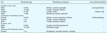
A total of 10 patients with congenital vascular ring were identified: six boys and four girls. Their age at presentation ranged from three months to four years, with a mean age of 15.9 months. Fifty per cent of cases presented within the first year of life (Table I).
Patient data

Mth = months; yr = years; R = right
Persistent cough was the most common presenting symptom (six of 10), followed by stridor (three of 10) and recurrent cyanotic episodes (three of 10). In two of the three patients presenting with stridor, multiple endotracheal intubations had previously been performed to maintain the airway. Other reported symptoms consisted of recurrent asthma (two patients), recurrent pneumonia (one patient), recurrent choking (one patient) and recurrent dysphagia (one patient) (Table I).
The types of congenital vascular ring detected in our patients comprised a double aortic arch in four cases (40 per cent), innominate artery compression in three cases (30 per cent), right aortic arch in two cases (20 per cent) and aberrant subclavian artery in one case (10 per cent) (Table I).
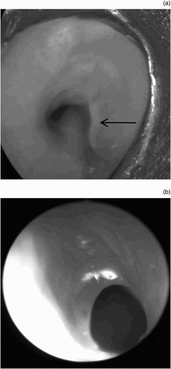
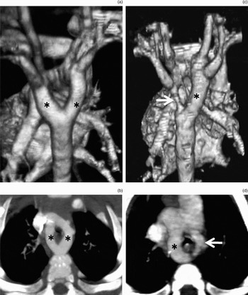
A double aortic arch was the most common anomaly encountered in our series. Bronchoscopy revealed significant, non-pulsatile compression of the lower segment of the trachea, arising posteriorly and laterally and mainly from the right side in all cases (Figure 1a). Two cases had associated anomalies: associated laryngomalacia in the first and a small glottic web in the second (Figure 1b). Radiologically, all four cases showed the ascending aorta running anterior to the trachea and then bifurcating into two arches which passed either side of the trachea, before reuniting posterior to the oesophagus to form the descending aorta. Typically, the descending aorta lay to the left of the spine. The right arch was the dominant limb, being more cephalad in position and larger in luminal diameter. There was a separate common carotid artery, subclavian artery and ductus arteriosus arising from each arch. Two types of double aortic arch were visualised: a type I double archReference Alkadhi, Wildermuth and Desbiolles9 in three cases (i.e. both arches are patent and the right ductus arteriosus regresses; Figure 2a and 2b); and a type II double archReference Alkadhi, Wildermuth and Desbiolles9 in one case (i.e. the right arch is patent while the left arch is intact but partially atretic; Figure 2c and 2d).
Laryngobronchoscopic view of double aortic arch. (a) Bronchoscopic view of 9-month-old female with double aortic arch, showing tracheal compression of the posterior and lateral walls mainly on the right side (arrow). (b) Laryngoscopic view of the same case, showing associated small glottic web.

Computed tomography (CT) scans of double aortic arch. (a) Volume-rendered posterior view and (b) axial CT image, both of type I double aortic arch, showing that both arches (asterisk) are patent. (c) Volume-rendered posterior view and (d) axial CT image, both of type II double aortic arch, showing a patent right arch (asterisk) but an atretic left arch (arrow).

Innominate artery compression was encountered in three patients, i.e. an anomalous innominate artery was found to be compressing the trachea. Bronchoscopy revealed compression of the anterior wall of the upper thoracic trachea (Figure 3a). This compression was pulsatile in one case. The compression was easily bypassed by the bronchoscope, and the distal trachea appeared normal. In one case, severe tracheomalacia was seen, with collapse of more than 50 per cent of the tracheal lumen during inspiration. Multi-detector row CT revealed an anomalous course of the innominate artery, with tracheal compression at the level of the artery (Figure 3b and 3c).
Views of anomalous innominate artery. (a) Bronchoscopic view of the trachea showing compression of the anterior wall (arrow) from left to right at the site of the innominate artery. (b) Axial computed tomography (CT) scan showing anomalous course of the innominate artery (arrow), compressing the anterior tracheal wall. (c) Volume-rendered CT image showing abnormal course of the innominate artery (arrow).

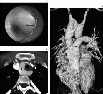
A right aortic arch was diagnosed in three cases. Bronchoscopy revealed compression of the lower segment of the trachea from the right side, obscuring the lumen of the right main bronchus (Figure 4a). Radiologically, the right aortic arch was visualised with mirror-image branching (Figure 4b and 4c). In such cases, the left innominate artery is the first vessel to arise from the aorta, and crosses anterior to the trachea; the right common carotid artery is the next vessel, followed by the right subclavian artery.
Views of right aortic arch. (a) Bronchoscopic view of the trachea revealing the classical appearance of the right aortic arch, with compression on the right anterolateral wall (arrow) obscuring the lumen of the right main bronchus. (b) Axial computed tomography (CT) scan showing the right aortic arch (asterisk). (c) Volume-rendered CT scan with right lateral view, showing right aortic arch (asterisk).

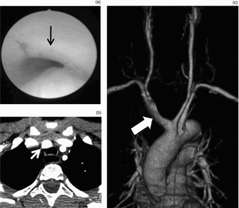
An aberrant subclavian artery was encountered in one case. Bronchoscopy of this case was irrelevant. Radiologically, the right subclavian artery was seen to arise just distal to the left subclavian artery as the last branch of the aortic arch (beyond the ligamentum arteriosum), and to run to the right posterior to the oesophagus (Figure 5a and 5b).
Views of aberrant right subclavian artery. (a) Axial computed tomography (CT) scan showing aberrant subclavian artery (asterisk) extending behind the trachea and oesophagus with a retroesophageal course. (b) Volume-rendered CT scan showing aberrant right subclavian artery (arrow) arising from the true lumen as the last artery of the aortic arch, and extending behind the trachea and oesophagus with a retroesophageal course.

Discussion
A vascular ring is defined as an anomaly of the great arteries (i.e. the aortic arch and its branches) which compresses the trachea or the oesophagus.Reference Kriklin, Kriklin and Barratt Boyes10 Such anomalies are very rare, and usually become symptomatic in infancy or early childhood.Reference Bakker, Berger, Wistenburg and Bogers11
We report 10 children with symptomatic vascular rings. Their mean age at presentation was 15.9 months, and 50 per cent presented within the first year of life. In contrast, Turner et al. Reference Turner, Gavel and Coutts4 reported 24 cases with a median age at presentation of 4.5 months. Of our patients, those with double aortic arch had the earliest presenting age (mean age, 9.5 months), while those with anomalous innominate artery had the latest presenting age (mean age, 25.3 months). Tuo et al. Reference Tuo, Volpe and Bava12 stated that the postnatal age at which double aortic arch is identified may vary, although it is diagnosed in early infancy in most patients.
Vascular rings rarely present in adults. However, Andreas et al. Reference Andreas, Reinhold, Micheal and Beate13 reported a vascular ring causing tracheal compression in a 65-year-old woman.
Common presenting symptoms of vascular ring include stridor, cyanosis and wheezing, and a characteristic high-pitched, ‘brassy’ cough. Additional findings include a history of asthma, recurrent pneumonia or dysphagia.Reference Turner, Gavel and Coutts4 In our series, cough was the most common presenting symptom (60 per cent of cases), followed by stridor (30 per cent). Turner et al. Reference Turner, Gavel and Coutts4 found stridor to be the most common presenting symptom in their series (14 out of 24 cases). We believe that cough may be under-appreciated as a presenting symptom of vascular ring, by both parents and physicians, and that this may lead to delay in diagnosis. However, the distressing symptom of stridor may provide more motivation for early investigation and thus early diagnosis.
The most common types of great vessel anomaly, in decreasing order of frequency, are: double aortic arch, right-sided aortic arch, innominate artery compression, aberrant right subclavian artery, pulmonary artery sling and aberrant left subclavian artery.Reference Woods, Sharp and Holcomb3 In our series, double aortic arch was the most common anomaly (40 per cent), followed by innominate artery compression (30 per cent), right-sided aortic arch (20 per cent) and aberrant right subclavian artery (10 per cent). Ruzmetov et al. Reference Ruzmetov, Vijay, Rodefeld, Turrentine and Brown14 stated that, in most surgical series of patients undergoing vascular ring repair, 45–65 per cent had a double aortic arch.
The diagnosis of a vascular ring requires a heightened index of clinical suspicion. Once the diagnosis has been made, it is important to obtain different imaging studies to confirm and further delineate the anatomical arrangement.Reference Backer, Ilbawi, Idriss and DeLeon15 The current study aimed to assess and compare the roles of bronchoscopy and multi-detector row CT in the diagnosis of congenital vascular ring.
All our patients underwent bronchoscopy using a rigid bronchoscope, under general anaesthesia. In our opinion, the patient's airway can be better controlled using a rigid ventilating bronchoscope. However, Cakir et al. Reference Cakir, Ersu, Uyan, Oktem and Karadag16 performed flexible bronchoscopy for 113 children presenting with persistent chest wheeze (two of whom proved to have a vascular ring), and declared this procedure safe for children. In our series, bronchoscopy was able to visualise an external, pulsating mass compressing the trachea in nine out of 10 cases. In one case (with aberrant subclavian artery), no bronchoscopic signs were noted except for mild mucosal congestion secondary to repeated aspiration. In three of our 10 cases, bronchoscopy detected associated anomalies: laryngomalacia in a case of double aortic arch; a glottic web in a patient with a double aortic arch; and tracheomalacia in a case of anomalous innominate artery. O'Connor and CooneyReference O'Connor and Cooney6 have stated that, in cases of congenital vascular ring, bronchoscopy is also useful to document normal vocal fold movement prior to cardiothoracic surgery, and to detect coexisting airway pathology.
The role of CT in the diagnosis of thoracic aortic disease has been debated for at least the last two decades.Reference Lawler and Fishman17 Thoracic CT has been used to diagnose or exclude aortic lesions since the mid- to late 1980s, and has continuously challenged the more well established use of thoracic angiography as the reference standard for diagnosis.Reference Sinclair18 Several trends have resulted in increased preference for CT over angiography, including: a greater general reliance on CT in the assessment of the patient with multiple aortic lesions; the steady improvement in the technological sophistication of CT, first with the introduction of helical scanning and later with multi-detector row CT; and the development of advanced CT workstations which allow rapid generation of tailored, multiplanar, three-dimensional volume rendering and endovascular presentations of aortic anatomy, improving diagnostic accuracy.
• Vascular rings are congenital vascular anomalies of the aortic arch complex which cause compression of the trachea and/or oesophagus
• A variety of investigations may lead to the diagnosis of vascular ring, including bronchoscopy and computed tomography
• This study assessed the relative value of bronchoscopy and multi-detector row computed tomography (CT) in managing congenital vascular rings
• Both investigations are essential for the diagnosis of congenital vascular ring
• Multi-detector row CT provides more information about the nature of the anomaly, and facilitates better communication between clinicians, compared with conventional CT
The development of CT technology has created the capacity for fast acquisition of high quality data sets, and the potential to view them in a practical, powerful, three-dimensional, real-time presentation.Reference Gotway and Dawn19 In many cases, although axial images may be adequate for diagnosis, volume-rendered, three-dimensional images can provide more information about the nature of the disease and facilitate better communication between clinicians.Reference Alkadhi, Wildermuth and Desbiolles9 In our series, multi-detector row CT accurately established the diagnosis in 10 out of 10 cases (100 per cent). However, this imaging modality could not detect any associated airway anomalies.
We encountered four cases of double aortic arch. This anomaly is created by persistence of the embryological aortic arch system. The ascending aorta runs anterior to the trachea and bifurcates into two arches which pass either side of the trachea before joining posterior to the oesophagus to form the descending aorta.Reference Lee, Siegel, Hildebolt, Gutierrez, Bhalla and Fallah20 Two types of double aortic arch have been denoted. In type I, the most common form, both arches are patent and the right ductus arteriosus regresses in most cases; this type was seen in three of our patients.Reference Alkadhi, Wildermuth and Desbiolles9, Reference Mirvis21 In type II, the right arch is patent and the left arch is intact but has an atretic portion; this type was seen in one of our patients. Both types involve a complete vascular ring.Reference Alkadhi, Wildermuth and Desbiolles9, Reference Mirvis21 Although rarely found in conjunction with intracardiac congenital heart disease, the double aortic arch forms a tight vascular ring which almost always requires surgical intervention.Reference Mirvis21
Three cases of innominate artery compression were encountered. All presented late (mean presenting age, 25.6 months) compared with other vascular ring types. One case was associated with tracheomalacia. An anomalous course of the innominate artery is a rare form of incomplete vascular ring. Most patients have mild symptoms, but some cases may have severe stridor, cyanosis and apnoea.Reference Yusuf, Ersin and Esce22 Several techniques have been described for the correction of this pathology; suspension of the innominate artery to the sternum is the most widely accepted treatment.Reference Adler, Isaacson and Balsara23
A right aortic arch was encountered in two patients, including one with a right aortic arch with mirror-image branching (i.e. type I right aortic arch) and one with a right aortic arch with aberrant left subclavian artery (i.e. type II right aortic arch). Mirror-image branching of a right aortic arch is less common, and results from interruption of the embryonic left arch between the left subclavian artery and the descending aorta, usually dorsal to the left ductus. It is almost always (98 per cent) associated with congenital heart disease, most commonly tetralogy of Fallot (in about 75 per cent of affected patients).Reference Alkadhi, Wildermuth and Desbiolles9
This study included one case of aberrant right subclavian artery. The right subclavian artery arises just distal to the left subclavian artery as the last branch of the aortic arch (beyond the ligamentum arteriosum) and runs to the right posterior to the oesophagus, thus producing a posterior indentation from the lower left to the upper right of the oesophagus on a barium oesophagram. Affected patients are typically asymptomatic, but the condition may cause dysphagia (‘dysphagia lusoria’) and recurrent aspiration.Reference Mirvis21 In our case, bronchoscopy was performed to investigate the cause of recurrent apnoea; however, findings were normal apart from mucosal congestion probably due to repeated aspiration. Multi-detector row CT was able to visualise accurately the course of the aberrant vessel, explaining the patient's symptoms.
Conclusion
Congenital vascular rings are rare anomalies of the aorta and great vessels which result in compression of the trachea and/or oesophagus. They usually manifest in infancy and early childhood. Diagnosis requires a high index of clinical suspicion. Bronchoscopy and radiological evaluation are essential for diagnosis and pre-operative evaluation. Bronchoscopy is superior in assessing the endoluminal airway and the vocal fold mobility, and in diagnosing associated airway lesions. Computed tomography can precisely delineate the exact anatomy of the compressing vessel, enabling optimal surgical intervention. Multi-detector row computed tomography can provide more information about the nature of the anomaly, and facilitate better communication between clinicians, compared with conventional CT scanning.