I. INTRODUCTION
Interest in thermosensitive microgels has persisted over several decades, and a great deal of research has been devoted to these systems. Most of the thermosensitive microgels are based on the monomer N-isopropylacrylamide (NIPAM), of which the polymer (PNIPAM) behaves slightly different compared with traditional hard-sphere colloids, for example, poly(methyl methacrylate), polystyrene, or silica nanoparticles. Reference Karg, Pastoriza-Santos, Liz-Marzan and Hellweg1 These spherical microgels display a strong thermoresponsivity, where below a lower critical solution temperature (LCST) they are highly swollen in water, but after heating they shrink rapidly to a collapsed polymer globule. This distinctive property brings a great advantage to the polymer for several applications. Reference Zhang2
However, the mechanical strength in bulk microgels is quite weak, which limits its applications. Reference Hellweg3 Recently, the core/shell structural microgels have attracted rather more attention, because they combined characteristics of thermosensitive polymer and an inorganic material to produce new fascinating composite materials with higher mechanical strength. Reference Dai, Ma and Cheng4 These microgels have a great potential to be used in some areas, such as smart carrier drug delivery, Reference Liu, Guo and Yang5 nanoreactors, Reference Lu, Proch and Schrinner6,Reference Wu, Kaiser, Drechsler, Ballauff and Lu7 and immobilization of enzymes. Reference Lu and Ballauff8 According to the previous works, core/shell thermosensitive microgels exist in essentially three architectures. One is based on a thermosensitive polymer coating on an inorganic core material (silica, metal, or oxidic nanoparticles). Reference Deng, Yang, Wang and Fu9–Reference Shamim, Hong, Hidajat and Uddin12 The second architecture is purely polymer and obtained by coating one polymer particle core with a different crosslinked polymer shell. Reference Facundo, Soria and Rosales13–Reference Koniger, Plack, Kohler, Siebenburger and Ballauff15 In this case, one polymer component might be inserted with respect to external stimuli like polystyrene. The third architecture adopts anisometric, star-shape, or totally irregular cores, whose systems usually require at least one more preparation step compared with the first two core/shell structures. Reference Lee, Lin, Wang and Chiu16 Most of the above-mentioned microgels have showed more advantages than “simple” microgels in practical applications. Reference Macková, Horák, Petrovský and Kovářová17
Carbon microspheres (CMSs), a new kind of carbon material, have drawn extensive attention from researchers in various fields, Reference Li, Shen, Li and Ye18 as they have excellent performances and attractive prospects, such as large surface area, good chemical, mechanical, and thermal stabilities. Reference Yang, Song, Li, Liu and Xu19 Yang and co-workers Reference Yang, Liu, Guo, Li, Liu and Xu20,Reference Liu, Zhao, Yang, Liu and Xu21 fabricated novel molecularly imprinted polymer on the surfaces of CMSs and obtained a better adsorption property toward dibenzothiophene compared with non-imprinted polymers on the surfaces of CMSs, showing its potential for deep desulfurization, which exemplified that the use of CMSs as carrier particles is reasonable and indeed effective. However, the core/shell thermosensitive microgels with a carbon core have been little reported.
In this work, the thermosensitive microgel as a shell onto a CMS core is reported, as shown in Scheme 1. The synthesis consisted of three steps. First, the oxidized-carbon microspheres were prepared by mixed-acid oxidizing the CMSs. Second, a silane coupling agent, 3-(trimethoxysilyl)propyl methacrylate (MPS), was used to modify the surfaces of the oxidized-CMSs, which generated a hydrophobic surface on the oxidized-CMSs for afterward reaction in the precipitation polymerization allowing for chemical coupling between the PNIPAM network and the inorganic material. At last, the thermosensitive monomer was grafted in the presence of a crosslinking agent, an initiator, and the functionalized CMSs. The obtained microspheres were named as CMS/PNIPAM here, which describes a core/shell microgel with CMS as core and PNIPAM as shell.
Preparation of CMS/PNIPAM.

II. MATERIALS AND METHODS
A. Materials
CMSs with the size of about 350 nm were synthesized by chemical vapor deposition with C2H2 as carbon source and Ar as carrier gas. Reference Yang, Liu, Guo and Xu22 Pure microgel PNIPAM was also prepared according to the previous work. Reference Zhang2 H2SO4 and HNO3 were obtained from Taiyuan Fertilizer Factory Chemical Reagent Factory, China. NIPAM, acetosalicylic acid (ASA), N,N′-methylenebisacrylamide (MBA), and phosphate buffered saline (PBS, pH = 7.4) were purchased from Aladdin. Ammonium persulfate (APS), acetic acid, and 3-(trimethoxysilyl)propyl methacrylate (MPS) were purchased from Tianjin Dongli Chemical Reagent Factory, China. Distilled water was used in all experiments.
B. Synthesis of oxidized-CMSs
The acid-oxidation of CMSs was used to improve the surface activity of parent CMSs, as the oxygen-containing groups were introduced onto the surfaces of CMSs. CMSs (0.5 g) were dispersed in mixed acid (120 mL, 96 wt.% H2SO4 and 65 wt.% HNO3 in volume ratio 3:1) under ultrasonication for 60 min at 70 °C. Then the mixture was transferred into 2 L distilled water and kept overnight at room temperature. Afterward, the products were filtered, and the filter cake was rinsed with distilled water until the pH value approached neutrality, then dried at 40 °C under vacuum to a constant weight.
C. Synthesis of silanized-CMSs
To realize the encapsulation of such oxidized-CMSs template cores via precipitation polymerization, a silane coupling agent, MPS, was used to modify the surfaces of the oxidized-CMSs, Reference Yang, Song, Li, Liu and Xu19 which leads to the formation of terminal C=C bonds on the surfaces of the cores. Oxidized-CMSs (0.3 g) were added to 60 mL of mixed solvent of ethanol and water (v/v = 3:1), followed by the addition of 10 mL of MPS and the adjustment of pH of reaction system to 5.0 by acetic acid. Then the mixture was refluxed in N2 atmosphere at 65 °C for 2 h under magnetic stirring. At last, the samples were washed with ethanol repeatedly to remove excess MPS, and dried, resulting in the formation of silanized-CMSs.
D. Synthesis of CMS/PNIPAM microspheres
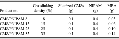
The NIPAM monomer was polymerized with a crosslinker MBA by seed precipitation with silanized-CMSs as seeds to prepare the crosslinked CMS/PNIPAM core/shell microspheres. As a typical reaction, a solution of 0.4 g of NIPAM and 0.06 g of MBA in 25 mL of distilled water was prepared in a three-neck flask equipped with a reflux condenser and a magnetic stirrer. The solution was heated to 40 °C, and then 0.1 g of silanized-CMSs was added. The mixture was heated to 70 °C and kept under N2 atmosphere to remove O2. For the polymerization, a solution of 1 mg of initiator APS dissolved in 1 mL of distilled water was rapidly added. The polymerization proceeded for 12 h at 70 °C, and then cooled to room temperature. The prepared microspheres were cleaned by centrifugation and redispersion in distilled water several times until the UV spectra measurement cannot detect the unreacted monomer NIPAM, as it has an adsorption at 222 nm. A series of polymerization was carried out according to the recipe mentioned in Table I. As is known, the monomer concentration plays an important role in a typical polymerization reaction, Reference Deng, Yang, Wang and Fu9 however, during the preparation of CMS/PNIPAM, the amount of NIPAM was excessive compared with that of CMSs and the thickness of polymer layer could be adjusted by the amount of crosslinker according to the experimental results. Therefore, the crosslinking degree, which is defined as the percentage of the dosage of crosslinking agent accounting for the amount of the monomer, was discussed as the main factor in this paper.
The recipe of the preparation of CMS/PNIPAM microspheres.

E. Release behavior of CMS/PNIPAM
First, 1 g/L ASA solution was prepared by adding 100 mg of ASA into 100 mL of PBS/ethanol mixing solution with a volume ratio of 95:5. Then 20 mg of PNIPAM, silanized-CMS, or CMS/PNIPAM was dispersed into 20 mL of drug solution and kept stirring at 25 °C for 24 h until they were fully swelled and saturated with ASA. The drug-loading microspheres were centrifugated, washed with distilled water, and dried. The soaking liquid was collected and analyzed by gas chromatography (GC). The residual amount of ASA was determined from the calibration plot. The encapsulation efficiency (η) of ASA was calculated as: η = (m 0 − m 1)/m 0, Reference Zhang, Zhang and Fan23 where m 0 and m 1 are the weight of the initial and residual ASA, respectively.
The controlled release behavior was investigated at 25 °C and 37 °C, separately. Drug-loading microspheres (20 mg) were dispersed into 40 mL of PBS/ethanol mixing solution with a volume ratio of 95:5 and the mixture was stirred by a magnetic stirrer with a rotational speed of 60 rpm. At definite time intervals, aliquots (4 mL) of the solutions were taken out and analyzed by GC to determine the releasing rate of the drug. Furthermore, 4.0 mL of fresh buffer solution was added after each sampling to keep the total volume of the solution constant. The cumulative release was expressed as the total percentage of released drug molecules.
F. Characterization and measurements
The morphology of all CMSs was characterized by field emission scanning electron microscopy (FESEM, JSM-6700F, operated at 10 kV, JEOL, Tokyo, Japan) with x-ray energy dispersive spectroscopy (EDS). Transmission electron microscopy (TEM) measurements were carried out on a JEM 2100 electron microscope operating at an acceleration voltage of 60 kV (JEOL, Tokyo, Japan). Fourier transformation infrared spectroscopy (FTIR, BRUKER TENSOR 27, Bremen, Germany) was used to prove the introduction of various functional groups onto the surfaces of CMSs. Thermogravimetry (TG) analysis was carried out on a Netzsch TG 209 F3 instrument from 40 to 900 °C in N2 atmosphere with a heating rate of 10 °C/min (NETZSCH-Geraetebau GmbH, Germany). Drug loading and release properties were determined by gas chromatography (GC; HXSP GC-950, FID detector temperature 300 °C, gasification temperature 300 °C, column temperature 180 °C, Haixin Shanghai, China). Average hydrodynamic diameter of the microspheres was measured using dynamic light scattering (DLS) of a Zetasizer Nano-ZS90 (Malvern Instruments, Worcestershire, United Kingdom). To measure the size at different temperatures, the suspension of microspheres was equilibrated at a particular temperature for 6 min before measurements and was measured four times with each run. The wettability of the samples was determined by monitoring water contact angle with a homemade setup and a microscope equipped with a charge-coupled device camera.
III. RESULTS AND DISCUSSION
A. Modification of CMSs
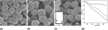
FESEM images presented in Fig. 1 clearly show different surfaces morphology of parent CMSs, oxidized-CMSs, and silanized-CMSs. It can be seen from Fig. 1(b) that oxidized-CMSs were coarsened after being oxidized, whereas parent CMSs [Fig. 1(a)] exhibited regular spherical shapes and smooth surface. The contact angle measurement result demonstrates that the CMSs are hydrophobic, whereas oxidized-CMSs are superhydrophilic, as shown in Fig. S1. This can be explained by the fact that the mixed acid not only eroded the CMSs surface, but also provided a large number of polar functional groups as active sites. After silanization, the surfaces of silanized-CMSs [Fig. 1(c)] became rather smooth, while the covering layer was suggested to be grafted MPS, as indicated in the EDS measurement in lower-left corner of Fig. 1(c). Figure 1(d) gives the TG curves of as-synthesized CMSs. The original CMSs had only 4.9% (wt.%) weight loss at 900 °C in N2 atmosphere, which was mainly caused by the loss of the water molecules adsorbed by CMSs in the air. Oxidized-CMSs began degradation at lower temperature, owing to the decomposition of oxygen-containing functional groups on the surfaces of the CMSs, and ultimately gave a weight loss of 16.8%. As shown in Fig. 1(d, iii), silanized-CMSs had a weight loss of about 21.6%, 4.8% more than oxidized-CMSs, attributed to the weight loss of MPS.
FESEM images of (a) as-synthesized CMSs, (b) oxidized-CMSs, (c) silanized-CMSs, and (d) their TG curves ((i) CMSs, (ii)oxidized-CMSs, (iii) silanized-CMSs).

B. Synthesis of CMS/PNIPAM
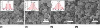
Figure 2 shows FESEM images of CMS/PNIPAM with different crosslinking densities. It can be observed that the microsphere sizes of the CMS/PNIPAM initially increased, and subsequently decreased slightly with the increase of the crosslinking degree from 8% to 25% [Figs. 2(a)–2(c)]. When the crosslinking degree was up to 35%, the self-polymerization took place [Fig. 2(d)]. The polymer microgels not only coated around CMSs, but also aggregated and existed independently.
FESEM images of CMS/PNIPAM with different crosslinking degree: (a) CMS/PNIPAM-8, (b) CMS/PNIPAM-15, (c) CMS/PNIPAM-25, and (d) CMS/PNIPAM-35.

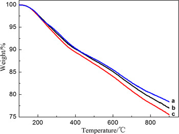
The amount of PNIPAM bound on the surfaces of the prepared microspheres was estimated from percentage of weight loss according to TG curve represented in Fig. 3. Compared with the weight loss of the silanized-CMSs shown in Fig. 1(d, iii), that of CMS/PNIPAM-8 [Fig. 3(a)] almost had no increase, which demonstrates that the concentration of crosslinker was insufficient and no polymer layer was grafted onto the CMSs. The weight losses of CMS/PNIPAM-15 [Fig. 3(b)] and CMS/PNIPAM-25 [Fig. 3(c)] were higher than that of silanized-CMSs. It clearly originated from the increased polymer's decomposition, even pyrolyzation. The weight percentage of grafted polymer layer can be calculated by subtracting the weight loss of the silanized-CMSs from that of CMS/PNIPAM-15 and CMS/PNIPAM-25. Results showed that it increased from 1.40% to 2.87% when the initial amount of MBA was raised from 0.06 to 0.10 g.
TG curves of (a) CMS/PNIPAM-8, (b) CMS/PNIPAM-15, and (c) CMS/PNIPAM-25.

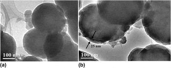
To confirm the core/shell structure, TEM measurements were carried out on the product silanized-CMSs and CMS/PNIPAM-15, as shown in Fig. 4. Compared with silanized-CMSs, CMS/PNIPAM-15 has core/shell structures with about 25 nm of the thermosensitive polymer layer onto the carrier CMSs surface. There also exist some protuberances around the CMS/PNIPAM-15 surface, which may be produced by the uneven polymerization.
TEM images of (a) silanized-CMSs and (b) CMS/PNIPAM-15.

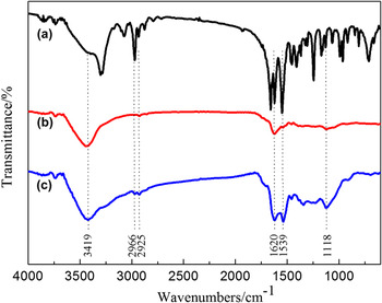
Figure 5 gives the FTIR spectra of pure NIPAM (a), the silanized-CMSs (b), and the CMS/PNIPAM-15 microspheres (c). As the FTIR spectra of the oxidized-CMSs do not have obvious difference from those of silanized-CMSs, it is not given in the figure. It can be clearly seen that the FTIR spectra of CMS/PNIPAM-15 microspheres possess both characteristic bands of pure NIPAM and silanized-CMSs. The wide band for CMS/PNIPAM-15 [Fig. 5(c)] at about 3419 cm−1 indicates that the –OH groups remained on the surfaces of microspheres, while the stretching vibration of N–H groups of PNIPAM was overlapped at the same band. Bands at 2966 cm−1 and 2853 cm−1 are attributed to the asymmetrical stretching vibration and the symmetrical stretching vibration of –CH2 – and –CH3 groups. The absorbance band at 1620 cm−1 is attributed to the stretching vibration of C=O, which is assigned to the C=O from the silanized-CMSs and the amide I band from PNIPAM. The typical absorbance band of the amide II band (the bending vibration of N–H and the stretching vibration of C–N) appeared at 1539 cm−1. The bands at 1118 cm−1 are ascribed to the skeleton stretching vibration of C–C from –CH(CH3)2. It is suggested that the thermosensitive polymer was crosslinked on the surfaces of silanized-CMSs.
FTIR spectra of (a) pure NIPAM, (b) silanized-CMSs, and (c) CMS/PNIPAM-15 microspheres.

C. Temperature effect on hydrodynamic diameter
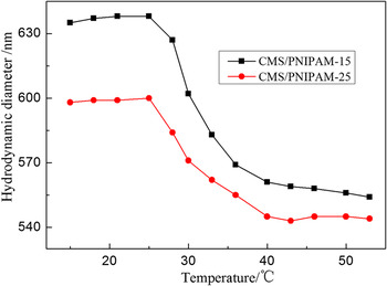
The hydrodynamic diameter of CMS/PNIPAM as a function of temperature is given in Fig. 6. The microspheres of CMS/PNIPAM-15 and CMS/PNIPAM-25 were found to show the decrease in hydrodynamic diameter upon heating with a variation of temperature in aqueous solution. As expected, this behavior comes from the shrinkage of the PNIPAM shell on the surfaces of CMSs. The LCST of CMS/PNIPAM-15 and CMS/PNIPAM-25 took place at around 30 °C, a shift away from that of lightly crosslinked pure PNIPAM microgel microspheres (35 °C Reference Zhang2 ) from the inorganic cores. Such a shift was also found by Deng Reference Deng, Yang, Wang and Fu9 and Nabzar Reference Nabzar, Duracher, Elaissari, Chauveteau and Pichot24 et al. when they studied thermosensitive microspheres with a magnetic or polystyrene core and PNIPAM shell. Meanwhile, there was few difference between the CMS/PNIPAM-15 and CMS/PNIPAM-25, as the hydrogel volume phase transition is determined by the composition of the hydrogel network structure of the hydrophobic and hydrophilic groups. It is also mentionable that PNIPAM hydrogel chains collapse above its LCST, eventually being dehydrated by expelling water molecule outside of the hydrogel shell, which results in shrinkage. Taking CMS/PNIPAM-15 for example, the hydrodynamic diameter shrinked from 635 to 554 nm, as the temperature increased from 18 to 53 °C. At temperature below LCST, hydrogel bonds between hydrophilic groups of polymer chains are dominant. These bonds become weaker and hydrophobic interactions between polymer chains become stronger when temperature is elevated over the LCST. The phenomenon is reversible and hydrogels return in its swollen state when temperature is lowered below the LCST. Compared with that of CMS/PNIPAM-15, the hydrodynamic diameter of the CMS/PNIPAM-25 at 18 °C was 37 nm less.
Hydrodynamic diameters of CMS/PNIPAM-15 and CMS/PNIPAM-25 as a function of temperature.

It is noted that hydrodynamic diameter of CMS/PNIPAM obtained by DLS was about 200 nm higher than the diameter measured by SEM. It can be illustrated by the fact that the hydrodynamic diameter was measured in the aqueous solution, so the hydrogels were abundantly swelled and there also existed a hydrated layer around the surfaces of CMS/PNIPAM.
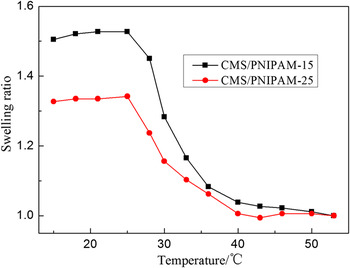
The swelling ratio (SR) is an important indicator to evaluate hydrogel properties. In this paper, it was calculated by the following equation: SR = V/V C, Reference Rahman, Chehimi, Fessi and Elaissari11 where V and V C are the volume of microspheres at a given temperature and collapsed state (in this case volume at 53 °C), respectively. Figure 7 shows the SRs of thermosensitive microspheres as a function of temperature. It can be seen that the SRs for CMS/PNIPAM decreased sharply in a certain range, showing typical thermosensitivity. The SR of CMS/PNIPAM-25 was lower. The most probable reason can be illustrated in two aspects. On one hand, the higher the crosslinking degree was, the compacter the hydrogels networks became. Then the water molecules were isolated to reduce their chance of transferring outside. On the other hand, it probably as Hoffman Reference Xue, Hoffman and Yager25 proposed: when the temperature was above LCST, the hydrogel’s layer began to shrink to the equilibrated water content from the surface to inside of the hydrogel. Therefore, it can initially form a dense skin on its surface to inhibit the hydrogel’s deswelling further.
SRs for CMS/PNIPAM-15 and CMS/PNIPAM-25.

D. Release behavior of the microspheres
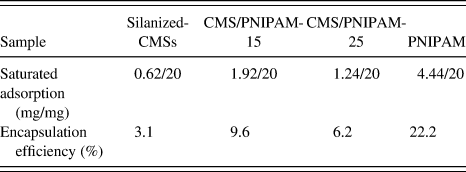
The potential application of the functional microspheres for the controlled release of drugs was also investigated with ASA as a model hydrophobic drug. According to GC analysis, the saturated adsorption and encapsulation efficiency of silanized-CMSs, CMS/PNIPAM-15, CMS/PNIPAM-25, and PNIPAM were calculated and listed in Table II. As the two indexes are only dependent on the component of polymer shell, it is deserved that the pure PNIPAM had the highest encapsulation efficiency. However, all the drug-loading amounts were relatively low, and this can be attributed to two reasons. First, on the basis of TG analysis, the amount of grafted polymer layer on the surfaces of CMSs was low. Second, PNIPAM chains are hydrophilic in the water at 25 °C, whereas ASA is hydrophobic. The adsorption was only induced by the concentration difference and physical adsorption, and such forces of these interactions are weak. It also can be seen from Table II that the encapsulation efficiency increased gradually from silanized-CMSs, CMS/PNIPAM-25, and CMS/PNIPAM-15, which can be related with the fact that the thickness of polymer shell increased.
Saturated adsorption and encapsulation efficiency of the microspheres.

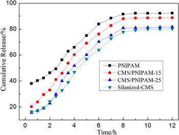
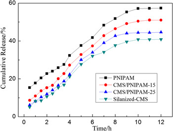
The release behaviors of the drug-loading microspheres with different polymer shells were investigated under the temperatures below (25 °C) and above (37 °C) their LCST of about 30 °C. The time dependence of the cumulative release of ASA is presented in Fig. 8 and Fig. 9, respectively. It can be found that the cumulative release amount of silanized-CMSs, CMS/PNIPAM-25, CMS/PNIPAM-15, and PNIPAM was up to 80.26%, 81.68%, 88.74%, and 92.32% at 25 °C, and decreased to 40.54%, 44.60%, 51.00%, and 57.32% at 37 °C, accordingly, which illustrates that the cumulative release increased with increasing thickness of polymer shell and encapsulation efficiency of the microspheres. The CMS/PNIPAM-15 showed better release behavior than the CMS/PNIPAM-25. Moreover, the amount of cumulative release at 25 °C reached the maximum value within 8 h, whereas that at 37 °C reached the maximum value within about 11 h. It indicates that the functional microspheres exhibited the temperature-responsive controlled releasing property owing to the PNIPAM blocks therein. Reference Lu, Mei, Drechsler and Ballauff26 At a certain temperature, the release rate and release percentage of the microspheres are determined by the solubility of the drug, drug diffusion coefficient, and the microgel's swelling rate. Although the swell of the microgels can control drug release behavior, the drug's hydrophilicity or hydrophobicity also affects microgel's swelling behavior. Reference Vera, Bravo, Medina and Claverie27
The time-dependence of drug release from the microspheres at 25 °C.

The time-dependence of drug release from the microspheres at 37 °C.

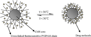
Figure 10 shows CMS/PNIPAM in a schematic fashion: the core consists of CMSs whereas the network consists of PNIPAM chains. The PNIPAM shell undergoes a volume transition at around 30 °C in which most of the water is expelled. The composite microspheres are suspended in water at a low temperature (T < 30 °C), which swells the thermosensitive network attached to the surfaces of the core microspheres, and drug molecules are easily absorbed or released in such a network by the internal and external concentration difference. At higher temperatures (T > 30 °C), the network shrinks and the microgel microspheres become hydrophobic, and the marked shrinking of the network slows down the diffusion of the drug. Moreover, as the ASA is a hydrophobic model drug, the hydrophobic interaction between PNIPAM and ASA increases at 37 °C, resulting in a lower cumulative release and release rate.
Schematic showing the drug release effect of CMS/PNIPAM.

IV. CONCLUSIONS
The novel thermosensitive core/shell microgels were prepared by precipitation polymerization with the CMS as core to enhance its mechanical strength. After acid oxidation of CMSs, oxidized-CMSs were initially functionalized by silane agent MPS, and were further subjected to crosslinking with NIPAM using MBA and APS as crosslinker and initiator accordingly. Results show that the amount of PNIPAM bound on the surfaces of the prepared microspheres and the shell's thickness could be adjusted by dosage of the crosslinker. When the initial weight ratio of silanized-CMSs to NIPAM was 1:4, the thickest polymer shell (25 nm) was obtained under the conditions of 15% crosslinking degree, reaction for 12 h at 70 °C. DLS results indicate that the hybrid microspheres have apparent thermosensitivity with a LCST at around 30 °C. Besides, the controlled release behavior was evaluated by a model drug ASA, and it was improved by increasing the thickness of thermosensitive polymer shell or decreasing the solution temperature from 37 to 25 °C. The CMS/PNIPAM-15 with a 25-nm-thick polymer shell showed higher release efficiency at 25 °C.
Supplementary Material
To view supplementary material for this article, please visit http://dx.doi.org/jmr.2014.92.
ACKNOWLEDGMENTS
The authors acknowledge financial support from National Natural Science Foundation of China (21176169), Shanxi Provincial Key Innovative Research Team in Science and Technology (2012041011), International Science & Technology Cooperation Program of China (2012DFR50460), Research Project supported by Shanxi Scholarship Council of China (2012-038), and Program for Changjiang Scholar and Innovative Research Team in University (IRT0972).