

The pattern of arterial blood supply to the thalamus is arranged such that perfusion territories of the posterolateral choroidal artery, the postero- medial choroidal artery, the inferotemporal arteries, the paramedial thalamic artery and the tubero-thalamic artery can be mapped (Barraquer-Bordas, Illa, Escartin, Ruscalleda, & Marti-Vilalta, Reference Barraquer-Bordas, Illa, Escartin, Ruscalleda and Marti-Vilalta1981; Chui & Howng, Reference Chui and Howng1989; Kumral, Kocaer, Ertubey, & Kumral, Reference Kumral, Kocaer, Ertubey and Kumral1995; Schmahmann, Reference Schmahmann2003; Song, Reference Song2011). The neuroanatomical description of the various thalamic nuclei and the thalamic fibre tracts, the thalamic peduncles, are equally well described (Schmahmann, Reference Schmahmann2003; Song, Reference Song2011). Despite the anatomical clarity, the clinical sequelae of thalamic stroke offer a complex picture of physical, neuropsychological and functional effects, which are difficult to separate out into syndrome groupings (Amici, Reference Amici2012; Fisher, Reference Fisher1959; Kumral et al., Reference Kumral, Kocaer, Ertubey and Kumral1995).
Thalamic haemorrhages are generally classified by their topographic location, with a conventionally used differentiation into anterior, medial, posterolateral and dorsal areas. However, the definition of location and the topographic boundaries are not identified consistently in the literature (Alastruey, Parker, Peiro, Byrd, & Sherwin, Reference Alastruey, Parker, Peiro, Byrd and Sherwin2007; Chung et al., Reference Chung, Caplan, Han, Pessin, Lee and Kim1996; Schmahmann, Reference Schmahmann2003) and so this convention is not without problems. Nevertheless, it has been proposed that outcomes and deficits can be associated with these different lesion locations, together with the additional general rubric that ‘the larger the lesion the greater the deficit’ (especially if the lesion is greater than 4 ml in volume) (Kumral et al., Reference Kumral, Kocaer, Ertubey and Kumral1995; Shah, Kalita, Misra, Mandal, & Srivastava, Reference Shah, Kalita, Misra, Mandal and Srivastava2005; Song, Reference Song2011). Anterior thalamic injury has been associated with cognitive and behavioural disturbances, including those of memory and language, acalculia, visual memory and cognition (Amici, Reference Amici2012; Barraquer-Bordas et al., Reference Barraquer-Bordas, Illa, Escartin, Ruscalleda and Marti-Vilalta1981; Chui & Howng, Reference Chui and Howng1989; Schmahmann, Reference Schmahmann2003), in addition to deficits that have been identified with the neocortical frontal dysexecutive syndrome by some researchers (Chui & Howng, Reference Chui and Howng1989; Fisher, Reference Fisher1959; Schmahmann, Reference Schmahmann2003; Summers, Reference Summers2002). Medial thalamic injury is associated in the longer term with severe memory loss and disorientation, together with motor dysfunction and some personality changes (Amici, Reference Amici2012; Kumral et al., Reference Kumral, Kocaer, Ertubey and Kumral1995; Schmahmann, Reference Schmahmann2003). Posterolateral thalamic injury can lead to the effects of ‘sensorimotor stroke’ with associated sensory loss (hemineglect), motor incapacity and cognitive deficits such as perceptual (‘Pusher syndrome’), language and memory problems (Amici, Reference Amici2012; Karnath, Johannsen, Broetz, & Kueker, Reference Karnath, Johannsen, Broetz and Kueker2005). Lastly, dorsal thalamic stroke has been associated with mild transient hemiparesis but also with longer-term language and memory deficits (Amici, Reference Amici2012; Kumral et al, Reference Kumral, Kocaer, Ertubey and Kumral1995; Schmahmann, Reference Schmahmann2003).
A variety of risk factors are associated with thalamic stroke. These include pre-existing physical conditions such as cardiovascular disease and hypertension, as well as environmental, or ‘lifestyle’, factors such as hypercholesterolaemia, smoking and alcohol consumption. Of particular interest here is the risk associated with congenital anatomical variables of the basilar artery diameter (Deng et al., Reference Deng, Cheng, Zhang, Zhou, Feng and Feng2012; Pico, Labreuche, Gourfinkel-An, & Amarenco, Reference Pico, Labreuche, Gourfinkel-An and Amarenco2006) and the structural variations in the circle of Willis (Alastruey et al., Reference Alastruey, Parker, Peiro, Byrd and Sherwin2007; El-Barhoun, Gledhill, & Pitman, Reference El-Barhoun, Gledhill and Pitman2009; Li et al., Reference Li, Li, Lv, Li, Luo and Xie2011). The mean diameter of a normal basilar artery is approximately 3.2 mm (Deng et al., Reference Deng, Cheng, Zhang, Zhou, Feng and Feng2012; Pico et al., Reference Pico, Labreuche, Gourfinkel-An and Amarenco2006; Smoker, Price, Keyes, Corbett, & Gentry, Reference Smoker, Price, Keyes, Corbett and Gentry1986). Higher mortality is associated with larger basilar artery diameters (Pico et al., Reference Pico, Labreuche, Gourfinkel-An and Amarenco2006). Anatomical variances in the circle of Willis are also associated with increased risk of thalamic stroke (De Silva, Silva, Amaratunga, Gunasekera, & Jayesekera, Reference De Silva, Silva, Amaratunga, Gunasekera and Jayesekera2011; El-Barhoun et al., Reference El-Barhoun, Gledhill and Pitman2009; Kapoor, Singh, & Dewan, Reference Kapoor, Singh and Dewan2008), although there is some lack of consensus in the literature as to the incidence of normal and abnormal patterns of the circle of Willis. Reported prevalence rates of abnormality range from 5 to over 70 per cent (De Silva, Silva, Gunasekera, & Jayesekera, Reference De Silva, Silva, Gunasekera and Jayesekera1998; Kapoor et al., Reference Kapoor, Singh and Dewan2008; Tanaka et al., Reference Tanaka, Fujita, Enoki, Matsumoto, Watanabe, Murase and Nakamura2006). Studies have identified over 20 variants in the circle of Willis, including variations in the posterior communicating artery (PCOM) (De Silva et al., Reference De Silva, Silva, Amaratunga, Gunasekera and Jayesekera2011), from which the thalamic perfusing arteries arise. The absence of the PCOM and hypoplasticity of the PCOM are both variants. Absence of the PCOM has been reported in about 1 per cent of the population (Kapoor et al., Reference Kapoor, Singh and Dewan2008), although other studies have found absence rates as high as 30 per cent (Li et al., Reference Li, Li, Lv, Li, Luo and Xie2011). Hypoplasticity of the PCOM is reported at rates of around 13 per cent, but without consistency in reported incidence rates. The evidence suggests that people with an incomplete circle of Willis may be at higher risk for stroke (Alastruey et al., Reference Alastruey, Parker, Peiro, Byrd and Sherwin2007).
Here we report 10 cases of patients who sustained thalamic strokes and who were examined for risk factors and post-stroke changes in behaviour and cognitive function.
Method
Participants
The participant sample comprises a subsample of all cases of thalamic stroke referred to the John Hunter Hospital Stroke Service for the period November 2009 to November 2011. The overall population of thalamic stroke patients (N = 23) was identified by examination of the Stroke Service Register by the attending neurologists. The neurologists then coordinated with the patients’ primary-care physicians in order to determine suitability for participation, which was determined by the following criteria: vital status known; no recurrence of further stroke subsequent to the index thalamic event; residing at home; ambulatory and living independently or with minimal care support. Potential participants were then informed of the research and voluntary consent to participate was sought. Of the 23 patients identified, 6 were deemed not suitable for testing; 5 either declined to participate or could not be contacted, and 2 did not complete testing. The resulting participant sample comprised 10 patients having sustained thalamic strokes. Patients were aged between 66 and 83 years (M = 75.67, SD = 5.7). Four of the participants were female and six were male.
Instruments
The Audio Recorded Cognitive Screen (ARCS) is a cognitive test that is administered to participants by tape recorder or compact disc (CD). Participants write their responses in a booklet for later scoring (Schofield, Lechner-Scott, Loughland, & Moore, Reference Schofield, Lechner-Scott, Loughland, Moore and Gauthier2013; Schofield, Lee, Lyall, Zunong, & Kwan, Reference Schofield, Lee, Lyall, Zunong and Kwan2008; Schofield et al., Reference Schofield, Lee, Lewin, Lyall, Moyle, Attia and McEvoy2010). Tests within the ARCS cover the key cognitive domains of memory, verbal fluency, visuospatial functioning, language, attention/executive function and speed of writing (Schofield et al., Reference Schofield, Lee, Lewin, Lyall, Moyle, Attia and McEvoy2010, Reference Schofield, Lechner-Scott, Loughland, Moore and Gauthier2013). The ARCS includes a 12-word list-learning task that assesses verbal episodic memory with both immediate and delayed recall. There are three verbal fluency tests: category fluency, letter fluency and action fluency. A clock-drawing task assesses visuospatial functioning and language is assessed through picture-naming tasks. Executive function is assessed with an attention test. In this test participants are presented with a series of lower-case letters and they must write the corresponding upper case version for as many letters as possible within a 30 s time limit. In the second part of the task the letters are randomly circled or not, with participants being required to write lower-case letters next to circled, and upper-case letters next to non-circled stimulus letters during the 30 s time limit. The final part of the ARCS is a speed of writing task, in which participants are required to write a given word repeatedly as many times as possible in 30 s.
The ARCS possesses a good psychometric profile when compared with other neuropsychological test batteries (Schofield et al., Reference Schofield, Lechner-Scott, Loughland, Moore and Gauthier2013) and has been found to be sensitive to mild cognitive impairment in clinical samples (Lechner-Scott et al., Reference Lechner-Scott, Kerr, Spencer, Agland, Lydon and Schofield2010; Loughland et al., Reference Loughland, Allen, Gianacas, Schofield, Lewin, Hunter and Carr2010; Schofield et al., Reference Schofield, Lee, Lyall, Zunong and Kwan2008). The test takes approximately 30 min to complete. It was designed to measure functional abilities in the elderly and normative data for healthy elderly groups are available for comparison (Schofield et al., Reference Schofield, Lee, Lyall, Zunong and Kwan2008). The ARCS provides individual performance profiles in order to compare test results against age-adjusted norms, with a mean of 100 and standard deviation of 15. Here we use a criterion of 1 standard deviation below the age-adjusted mean to define a performance deficit
Procedure
All patient diagnoses of thalamic stroke were confirmed by two independent neurologists on clinico-radiological criteria: sudden focal neurological deficit of vascular aetiology and magnetic resonance (MR) brain imaging of thalamic stroke (either infarction or haemorrhage) consistent with the acute clinical presentation. Participants attended one testing session, which took 60–90 min to complete. The first part of the appointment consisted of an interview in which details of the participant's medical history, and pre- and post-stroke functioning were elicited. Five participants attended this appointment with a partner or care-giver, and input from this support person was encouraged during the interview. In the second part of the session participants completed the ARCS. Partners/care-givers were not present during ARCS testing. One participant was unable to complete any of the ARCS.
With the participants’ consent, their clinical records were also accessed in order to assess MRI images of the participants’ stroke lesions. These images were used to determine the location and size of the stroke lesion and any anatomical variations in the anatomy of the circle of Willis. The volume of the stroke lesion was estimated using the area measures from the MRI slices, multiplied by the thickness of the slices. Measurements of the basilar artery diameter at the level of the mid-pons, were also taken from the ‘time of flight’ MR angiographic source image scans. Clinical records were also screened in order to determine the presence or absence of other risk factors, namely, coronary artery disease, peripheral vascular disease, diabetes, smoking (current or previous), hyperlipidaemia (hypercholesterolaemia), atrial fibrillation, prior stroke or other brain injury.
Results
The 10 patients involved in this study were all aged over 66 years (mean age 76) and living in their own homes. One of the 10 participants required a high level of care from their spouse, and one received regular community assistance for showering and shopping. The remaining participants all reported living relatively independently. The mean time between stroke and neuropsychological testing was 17.6 months.
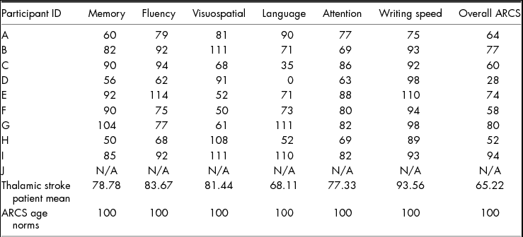
The results from the ARCS cognitive function tests (scaled scores for each cognitive domain) are presented in Table 1. ARCS scores are presented with the mean normed scores from an aged healthy population (bottom row). As can be seen from Table 1, patient J failed to perform any of the tasks and was removed from further analyses of behavioural data. Of the nine patients with measurable performances, the individual profiles show mixed levels of cognitive function. Patients A, D and H showed memory deficit; patients D and H performed poorly on the fluency tests; patients C, E, F and G all performed poorly on the visuospatial task; patients C, D and H showed language deficits; patients B, D and H exhibited attention deficits; and patient A was particularly slow in the speed of writing task.
Scaled ARCS Domain and Overall Scores for each Participant

N/A, not available.
Overall, the participants exhibited a range of cognitive deficits and, as a group, their performance shows clear impairment when compared with age-matched healthy control ability scores taken from the published ARCS standardised scores. There were statistically significant differences between the thalamic stroke patients group and the normed healthy elderly for memory, fluency, language, attention and for overall ARCS scores (domain comparisons: memory, t(8) = −3.396, p = .009; fluency, t(8) = −3.082, p = .015; language, t(8) = −2.691, p = .027; attention, t(8) = 7.959, p = .000; and overall ARCS scaled score, t(8) = 5.476, p = .001).
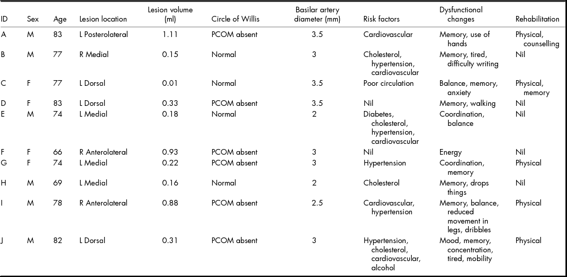
Measurements of the basilar artery diameter (in mm), hemisphere (right or left) of lesion, abnormality of circle of Willis (normal or abnormal), volume of lesion (in ml), lesion location (left versus right + anterior, medial, dorsal or postero- lateral) and pre-stroke risk factors, together with age and gender of participant, are all shown in Table 2. The mean basilar artery measurement taken from this sample (M = 2.9 mm) was compared with the average diameter taken from published data (Pico et al., Reference Pico, Labreuche, Gourfinkel-An and Amarenco2006; Smoker et al., Reference Smoker, Price, Keyes, Corbett and Gentry1986) (M = 3.17 mm) with no statistical difference being found (t(9) = 1.504, p = .167). The lesion volumes reported here ranged from 0.01 ml to 1.11 ml (M = 0.43 ml) – all less than 4 ml (Kumral et al., Reference Kumral, Kocaer, Ertubey and Kumral1995) and so all were considered to be of small size. Taking the overall ARCS score as a measure of overall cognitive functioning, the three patients with the largest lesions (patients A, F and I) had the highest plus two mid-range ARCS overall scores. Taken overall there was no statistical difference shown in ARCS scores when patients were grouped according to lesion volume, based on a median split of volumes.
Participant Data Summary
However, lesion volume was associated with abnormalities of the circle of Willis, such that the lesion volume for participants with a circle of Willis abnormality (M = 0.63 ml) was greater than that for those patients with a normal circle of Willis (M = 0.125 ml) (z = 2.558, p = .011). Non-parametric tests were used to compare ARCS domain and overall scores for participants with or without circle of Willis variations and no statistically significant differences were found. Likewise, no statistically significant differences were found when ARCS domain scores were used to compare the hemisphere (left or right) of the thalamic lesion location. The small sample size prevented statistical comparison of the sub-areas (1, posterolateral; 2, anterior; 3, dorsal and 4, medial lesions) of the thalamic lesion location.
Discussion
Despite showing clear impairment, the test outcomes for this group provide very little in terms of interpretable patterns of deficit. Inspection of the best and worst overall performance shows that patient I, with the best overall performance, was assessed with absence of the PCOM, a lesion volume of 0.88 ml (towards the upper end for the group) and a basilar artery diameter of 2.5 mm, whereas patient D, with the worst overall performance, also had an absent PCOM, a lesion volume of 0.33 ml and a basilar artery of 3.5 mm diameter. Some elements of a pattern of deficit might be suggested. For example, of the three participants with left dorsal lesions, one could not perform any of the tasks and the other two performed very poorly on the language subscale. Indeed, one of the two tested (patient D) performed poorest overall and the other (patient C) had the fourth poorest overall ARCS score, yet with the smallest lesion.
Generally, the patients with dorsal lesions showed deficit on all but the speed of writing task. Patient A, with a left posterolateral lesion, showed most impairment on the speed of writing task, in line with a ‘sensorimotor stroke’, but also showed impaired memory function. Indeed, he was below average on all but the language scale. The two participants with right anterior thalamic lesions (patients F and I) showed a very mixed pattern of responding, with one (patient F), showing deficit in visuospatial functioning, whereas the other (patient I) achieved the best ARCS overall score even with a lesion that was the third largest in volume. The four patients with right (patient B) and left (patients E, G and H) medial lesions also showed a very mixed pattern, but with a particular deficit in memory and language in one participant (patient H) and a deficit in visuospatial function in two (patients E and G). However, speculation about a pattern of deficit is hardly warranted since these data provide little in terms of any identification of patterns of cognitive deficit following thalamic stroke, either in terms of the size or the site of the lesion. And yet the performance scores demonstrate that, compared with appropriately matched normed scores, the patients are functionally very disabled.
The results of this study are clearly affected by the low statistical power offered by a small and, in terms of their thalamic lesion types, quite hetero- geneous sample. The only inference that can be drawn concerns the general cognitive dysfunction shown by these patients post stroke. Inspection of their rehabilitation programme shows that their physical needs have tended to receive intervention whereas their cognitive deficits have not. Table 2 illustrates that problems with their coordination, balance and mobility have received appropriate physiotherapy in their rehabilitation programme. The availability of rehabilitation for their cognitive dysfunctions is significant by its absence. Yet these same cognitive deficits are the cause of major functional difficulties for these patients. Further research aimed at disentangling the complex nature of relationship between various thalamic strokes and functional outcomes needs to be matched by efforts to provide cognitive rehabilitation programmes that will help patients to adapt and overcome these consequent difficulties.
Acknowledgments
This research was made possible through the patience and assistance of administrative and medical staff of the Hunter New England Area Health system, as well as the developers of the ARCS. It was made a reality by the participants, who freely gave their time and energy in the hopes of helping future stroke sufferers.
Sources of Funding
Funding was provided by the Faculties of Science & IT and of Medicine & Health Sciences, University of Newcastle, NSW, Australia.
Conflicts of Interest
There are no conflicts of interest to disclose.
Ethical Standards
Ethical approval for the research was granted by the Hunter New England Human Research Ethics Committee (HNEHREC reference number: 10/09/15/4.02).