Introduction
Most mammals are dichromats, acquiring color vision by contrasting signals originating from two classes of cone photoreceptors that are maximally sensitive to short (S) or long (L) wavelengths. Old World primates such as humans, great apes, and macaque monkeys, in addition have acquired a third cone type that is maximally sensitive to medium (M) wavelengths and thus enjoy trichromatic color vision. Most New World primates such as marmoset, capuchin, and squirrel monkeys show a sex-linked polymorphism of color vision whereby all males possess only two cone types (S plus M or L) and thus have dichromatic (“red-green color-blind”) vision. Females may have dichromatic or trichromatic color vision. For reviews on this topic, see articles by Jacobs and colleagues (e.g., Jacobs, Reference Jacobs2007, Reference Jacobs2008; Jacobs & Nathans, Reference Jacobs and Nathans2009). Here we review morphological and functional similarities and differences in the processing of S-cone signals in the inner plexiform layer of primate and nonprimate retinas. Consistent with the idea that the S-/L-cone system in evolutionary terms is considered a phylogenetically ancient color system (Mollon, Reference Mollon1989), the anatomical components of the blue-ON pathway are identical in Old World and New World primates; and S-cone selective (blue cone) ON bipolar cells have been identified anatomically and electrophysiologically in the retinas of nonprimate mammals. The blue-OFF pathway is less well understood, and there are discrepancies between Old World and New World primates with respect to the presence of an S-cone contacting blue-OFF midget bipolar cell. The morphological and physiological evidence for the involvement of a sign-inverting amacrine cell (recently found in a nonprimate mammal) to create the blue-OFF response and its role in blue signal processing is discussed.
Old World and New World primates
S-cones in primates
The S-cones in primates usually make up between 5% and 10% of all cones (Fig. 1A) and are found across the retina (reviewed by Ahnelt & Kolb, Reference Ahnelt and Kolb2000; Calkins, Reference Calkins2001). The peak density of S-cones is near the fovea, but they may be absent from the center of the fovea (Marc & Sperling, Reference Marc and Sperling1977; Wikler & Rakic, Reference Wikler and Rakic1990; Curcio et al., Reference Curcio, Allen, Sloan, Lerea, Hurley, Klock and Milam1991; Bumsted & Hendrickson, Reference Bumsted and Hendrickson1999; Martin & Grünert, Reference Martin and Grünert1999; Martin et al., Reference Martin, Grünert, Chan and Bumsted2000). There are some exceptions in nocturnal primate species, that is, the owl monkey Aotus (a New World monkey) and the lemur Galago (bush baby) lack S-cones (Jacobs, Reference Jacobs2008); and in the tarsier, S-cones are present but have a peak density in the far periphery (Hendrickson et al., Reference Hendrickson, Djajadi, Nakamura, Possin and Sajuthi2000).
The blue-ON/yellow-OFF pathway in primate retina. (A) Micrograph of the short wavelength sensitive (S-)cone mosaic in marmoset retina. S-cones were labeled with antibodies against the S-cone opsin kindly provided by J. Nathans. (B) Golgi-impregnated blue cone bipolar (BB) cell in macaque retina. Image kindly provided by H. Wässle. (C) Cholecystokinin (CCK) immunoreactive blue cone bipolar cells in macaque retina. (D) Double-labeled vertical section through macaque retina showing an S-opsin positive cone contacting a CCK immunoreactive blue cone bipolar cell. (E) Analysis of connections between S-cones and blue cone bipolar cells in marmoset retina. The dendrites of blue cone bipolar cells converge onto the S-cone pedicles (shown in blue). Modified from Luo et al. (Reference Luo, Ghosh, Martin and Grünert1999). (F) Schematic diagram showing the components of the blue-ON/yellow-OFF pathway. M, medium wavelength sensitive cone; L, long wavelength sensitive cone. DB, diffuse bipolar cell; and SBS, small bistratified ganglion cell. Modified from Martin (Reference Martin1998). (G) Vertical section through a Neurobiotin-injected small bistratified ganglion cell that was processed for CCK immunoreactivity. CCK immunoreactive blue cone bipolar axons costratify with the inner dendrites of SBS cells (marmoset retina). The arrows point to the inner and outer dendritic tier of the SBS cell. Modified from Ghosh et al. (Reference Ghosh, Martin and Grünert1997). (H) Whole mount view of the inner tier of dendrites of a small bistratified cell in marmoset retina that was injected with Neurobiotin and visualized with Streptavidin coupled to Texas Red. Modified from Ghosh et al. (Reference Ghosh, Martin and Grünert1997). Scale bar shown in E = 25 µm, valid for A–G. Scale bar in H = 25 µm.

The blue-ON/yellow-OFF pathway
In the S-cone pathway, the signals from S-cones are compared to the signals from M/L-cones (reviewed in Calkins, Reference Calkins2001). The anatomical components of the blue-ON pathway (Fig. 1) comprise S-cones, blue cone bipolar cells (Mariani, Reference Mariani1984), and small bistratified ganglion cells (Rodieck, Reference Rodieck, Valberg and Lee1991; Dacey, Reference Dacey1993). Please note that in the following, we will refer to short wavelength sensitive cones as “S-cones” because this term correctly describes their physiological properties. For historical reasons and for simplicity, we will refer to the ON bipolar cells that receive selective input from S-cones as “blue cone bipolar cells” (see also Boycott & Wässle, Reference Boycott and Wässle1991; Wässle et al., Reference Wässle, Grünert, Martin and Boycott1994; Joo et al., Reference Joo, Peterson, Haun and Dacey2011). This bipolar type has also been named “S-cone bipolar cell” (Calkins et al., Reference Calkins, Tsukamoto and Sterling1998), “S-bipolar cell” (Calkins, Reference Calkins2000, Reference Calkins2001), “S-cone ON bipolar cell” (Klug et al., Reference Klug, Herr, Ngo, Sterling and Schein2003; Schein et al., Reference Schein, Sterling, Ngo, Huang and Herr2004), or “inner S-cone bipolar cell” (Herr et al., Reference Herr, Klug, Sterling and Schein2003).
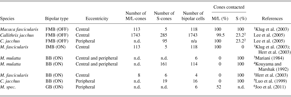
The blue-ON/yellow-OFF pathway was first described in Old World primates, but all components of this pathway have also been found in New World monkeys. A bipolar type that contacted a subpopulation of cones (Fig. 1B and 1C) was first described in Golgi preparations of macaque retina (Mariani, Reference Mariani1984). This bipolar type was assumed to be specific for S-cones and thus named blue cone bipolar cell. Subsequent studies in the retinas of macaque (Kouyama & Marshak, Reference Kouyama and Marshak1992) and marmoset (Ghosh et al., Reference Ghosh, Martin and Grünert1997; Luo et al., Reference Luo, Ghosh, Martin and Grünert1999) used double labeling and demonstrated this selectivity directly (Fig. 1D and 1E; Table 1). Quantitative analysis showed that in both macaque and marmoset, individual S-cones contact between two and four blue cone bipolar cells (Kouyama & Marshak, Reference Kouyama and Marshak1992; Wässle et al., Reference Wässle, Grünert, Martin and Boycott1994; Luo et al., Reference Luo, Ghosh, Martin and Grünert1999; Schein et al., Reference Schein, Sterling, Ngo, Huang and Herr2004).
Cone contacts of midget bipolar, blue cone bipolar, and giant bipolar cells in primate retina
1 The results are based on electron microscopic reconstruction.
2 Sparse connections between immunolabeled S-cones and CD15 immunoreactive bipolar dendrites may derive from FMB and/or DB6 cells.
3 The bipolar cells were identified in Golgi-impregnated retinas.
4 The bipolar cells were identified by CCK-immunohistochemistry and the S-cones by histochemistry.
5 The bipolar cells and the S-cones were identified immunohistochemically.
6 The bipolar cells were identified in Golgi-impregnated retina.
FMB, flat midget bipolar; IMB, invaginating midget bipolar; BB, blue cone bipolar; GB, giant bipolar; n.d. not determined.
Blue cone bipolar cells are assumed to give ON responses to light because their dendrites make invaginating synapses at cone pedicles (Mariani, Reference Mariani1984; Kouyama & Marshak, Reference Kouyama and Marshak1992) and express mGluR6 receptors (Vardi et al., Reference Vardi, Duvoisin, Wu and Sterling2000), and because their axons terminate in the ON sublamina (Mariani, Reference Mariani1984). Direct electrophysiological evidence showing that blue cone bipolar cells are ON cells so far has only been provided in nonprimate mammals (see below).
A ganglion cell type, named small bistratified cell, displaying blue-ON/yellow-OFF responses was characterized in macaque retina by Dacey and Lee (Reference Dacey and Lee1994). Small bistratified ganglion cells resemble the “shrub” cells found in Golgi-impregnated retinas in a number of Old World primate species (Polyak, Reference Polyak1941). Cells with similar morphology have been found in the retinas of New World monkeys (Fig. 1H), that is, in marmosets and capuchin monkeys (Ghosh et al., Reference Ghosh, Martin and Grünert1997; Silveira et al., Reference Silveira, Lee, Yamada, Kremers, Hunt, Martin and Gomes1999; Szmajda et al., Reference Szmajda, Grünert and Martin2008; Percival et al., Reference Percival, Jusuf, Martin and Grünert2009), but small bistratified cells appear to be absent in primates that lack S-cones (Yamada et al., Reference Yamada, Marshak, Silveira and Casagrande1998, Reference Yamada, Silveira, Perry and Franco2001). The inner dendritic tier of small bistratified cells is located close to the ganglion cell layer, and its outer dendritic tier is located in the OFF sublamina of the inner plexiform layer (Fig. 1F). In peripheral retina of macaque, the small bistratified cells make up about 6–10% of the ganglion cell population (Dacey, Reference Dacey1993); small bistratified cells have also been detected in central retina but their proportion in the fovea has not yet been determined (Calkins et al., Reference Calkins, Tsukamoto and Sterling1998). Anatomical evidence for a synaptic connectivity between blue cone bipolar cells and the inner dendrites of small bistratified ganglion cells (Fig. 1G) came from electron microscopic reconstructions in macaque retina (Calkins et al., Reference Calkins, Tsukamoto and Sterling1998) and from intracellular injection in combination with immunohistochemistry in marmoset (Ghosh et al., Reference Ghosh, Martin and Grünert1997). The yellow-OFF response of small bistratified cells presumably derives from diffuse OFF bipolar cells providing input to the outer dendrites (Dacey & Lee, Reference Dacey and Lee1994; Crook et al., Reference Crook, Davenport, Peterson, Packer, Detwiler and Dacey2009) but see also Field and Chichilnisky (Reference Field and Chichilnisky2007) and Packer et al. (Reference Packer, Verweij, Li, Schnapf and Dacey2010). Consistent with this hypothesis, anatomical studies in macaque (Calkins et al., Reference Calkins, Tsukamoto and Sterling1998) as well as in marmoset (Ghosh et al., Reference Ghosh, Martin and Grünert1997; Ghosh & Grünert, Reference Ghosh and Grünert1999; Percival et al., Reference Percival, Jusuf, Martin and Grünert2009) provided evidence for a synaptic connectivity between OFF bipolar cell types (DB2 and DB3) and the dendrites of small bistratified cells (Fig. 1F).
It should be noted here that electrophysiological recordings from a few examples of large bistratified cells suggested that these cells display similar blue-ON/yellow-OFF responses as the small bistratified cells (Dacey et al., Reference Dacey, Peterson, Robinson and Gamlin2003). Consistently, the inner dendrites of large bistratified cells costratify with the inner dendrites of small bistratified cells suggesting that both cell types receive their input via the blue cone bipolar cells. On the other hand, it has been suggested for the fovea that the postsynaptic ganglion cell profile at the blue cone bipolar synapse always derives from a small bistratified cell (Calkins et al., Reference Calkins, Tsukamoto and Sterling1998). The functional significance of a second type of blue-ON/yellow-OFF cells remains unclear. More studies are needed to determine the response characteristics and synaptic circuitry of large bistratified cells.
In both macaque (Calkins et al., Reference Calkins, Tsukamoto and Sterling1998) and marmoset (Ghosh & Grünert, Reference Ghosh and Grünert1999; Percival et al., Reference Percival, Jusuf, Martin and Grünert2009), the two dendritic tiers of small bistratified cells receive a significant proportion of synapses from amacrine cells (in addition to the bipolar input), but the amacrine cell type(s) providing this input is not yet known. Electrophysiological recordings from small bistratified cells in macaque retina demonstrated that their response characteristics did not change in the presence of GABA or glycine receptor antagonists suggesting that the yellow-OFF response is not generated by inhibition in the inner retina (Crook et al., Reference Crook, Davenport, Peterson, Packer, Detwiler and Dacey2009). Taken together, the functional significance of the amacrine input to small bistratified cells remains unclear.
Recently, two electrophysiological studies demonstrated that after dark adaptation small bistratified cells received input from rods (Crook et al., Reference Crook, Davenport, Peterson, Packer, Detwiler and Dacey2009; Field et al., Reference Field, Greschner, Gauthier, Rangel, Shlens, Sher, Marshak, Litke and Chichilnisky2009). This input is thought to be transmitted through the rod pathway, that is, via gap junctions between AII amacrine and blue cone bipolar cells (Field et al., Reference Field, Greschner, Gauthier, Rangel, Shlens, Sher, Marshak, Litke and Chichilnisky2009). The rod ON input to small bistratified cells is consistent with psychophysical data (Cao et al., Reference Cao, Pokorny, Smith and Zele2008). On the other hand, for the OFF sublamina, the data from Crook et al. (Reference Crook, Davenport, Peterson, Packer, Detwiler and Dacey2009) suggested that AII amacrine cells do not contact the OFF bipolar cell type(s) providing input to small bistratified ganglion cells.
In summary, anatomical and electrophysiological findings indicate that the blue-ON/yellow-OFF pathway is similarly organized in New and Old World primates supporting the hypothesis that this pathway is part of a phylogenetically ancient color system (Mollon & Jordan, Reference Mollon and Jordan1988; Mollon, Reference Mollon1989).
S cone input to other pathways
Midget-parvocellular pathway
The anatomical substrate for the red-green channel in the fovea is a one-to-one connectivity between cones and ON and OFF midget bipolar cells, which in turn contact a single ON and OFF midget ganglion cell, respectively (Fig. 2A and 2B). Such one-to-one connectivity is well established for M- and L-cones in the retinas of human (Kolb & Dekorver, Reference Kolb and Dekorver1991), macaque (Calkins et al., Reference Calkins, Schein, Tsukamoto and Sterling1994), capuchin monkey (Silveira et al., Reference Silveira, Lee, Yamada, Kremers, Martin, Yamada and Hunt1998), and marmoset (Chan et al., Reference Chan, Martin, Clunas and Grünert2001; Jusuf et al., Reference Jusuf, Martin and Grünert2006; Telkes et al., Reference Telkes, Lee, Jusuf and Grünert2008). It is also well established that S-cones make all their invaginating contacts with blue cone bipolar cells (Kouyama & Marshak, Reference Kouyama and Marshak1992) and do not contact ON midget bipolar cells (Herr et al., Reference Herr, Klug, Sterling and Schein2003). In contrast, for OFF midget bipolar cells, a one-to-one connectivity with S-cones has been reported in an electron microscopic study of foveal macaque retina (Klug et al., Reference Klug, Herr, Ngo, Sterling and Schein2003, Table 1). This anatomical result implies the presence of a blue-OFF midget bipolar and a blue-OFF midget ganglion cell in the fovea of macaque (Fig. 2D).
The midget pathway in primate retina. (A, B) Golgi-impregnated midget bipolar and midget ganglion cells in macaque retina. Images of Golgi preparations from Boycott and Dowling (1966) kindly provided by H. Wässle. (C) Immunohistochemical staining of midget bipolar cells in central marmoset retina. Antibodies against CD15 are used to label OFF midget bipolar and DB6 bipolar cells. The arrow indicates the single cone contacting bouquet of an OFF midget bipolar cell. (D) Schematic diagram of the midget pathway in central retina of macaque. S, short wavelength sensitive cone; M, medium wavelength sensitive cone; L, long wavelength sensitive cone; IMB, invaginating midget bipolar cell; FMB, flat midget bipolar cell; MG, midget ganglion cell. Modified from Martin (Reference Martin1998). (E, F) In marmoset retina S-cones do not make significant contacts with OFF midget bipolar cells. (E) Micrograph of a horizontal section through marmoset retina double labeled for peanut agglutinin to label cone pedicles (green) and CD15 to label the dendritic clusters of OFF midget bipolar cells (magenta). The S-cone (circled) does not contact the OFF midget bipolar cells in marmoset retina. Modified from Lee et al. (Reference Lee, Telkes and Grünert2005). (F) Cone contacts of OFF midget bipolar cells in marmoset. The number of dendritic tips from OFF midget bipolar cells at S, M, and L cone pedicles in marmoset retina is plotted in the histogram. Data from different eccentricities are pooled. Modified from Telkes et al. (Reference Telkes, Lee, Jusuf and Grünert2008). (G) Schematic diagram of the midget pathway in peripheral retina. Multiple midget bipolar cells converge onto a midget ganglion cell. Modified from Martin (Reference Martin1998). Scale bar shown in A = 20 µm, valid for A–C. Scale bar in E = 5 µm.
On the other hand, in marmoset retina, immunohistochemical double labeling studies of foveal and peripheral retina showed that the large majority of S-cones made no or only little contact with OFF midget bipolar cells (Lee et al., Reference Lee, Telkes and Grünert2005; Telkes et al., Reference Telkes, Lee, Jusuf and Grünert2008, Table 1), suggesting the absence of a blue-OFF midget pathway. In the periphery of human retina, the bipolar contacts at a single S-cone pedicle could not be attributed to a specific bipolar type (Kolb et al., Reference Kolb, Goede, Roberts, McDermott and Gouras1997). Currently this potential discrepancy in the blue-OFF pathway between different species has not been resolved. Studying the connectivity of S-cones in other species of Old World and New World primates may help to resolve this issue.
In peripheral retina of macaque, single cone contacting midget bipolar cells occur at eccentricities up to 8 mm, but the size of their axon terminals increases to a lesser extent than the dendritic field size of midget ganglion cells (Wässle et al., Reference Wässle, Grünert, Martin and Boycott1994). Thus, in peripheral retina, multiple midget bipolar cells converge onto a midget ganglion cell (Fig. 2G) resulting in mixed cone input (Watanabe & Rodieck, Reference Watanabe and Rodieck1989; Wässle et al., Reference Wässle, Grünert, Martin and Boycott1994; Dacey et al., Reference Dacey, Packer, Diller, Brainard, Peterson and Lee2000). Consistently, electrophysiological recordings from midget ganglion cells in peripheral retina of macaque using a multielectrode array system demonstrated input from M- and L-cones to midget ganglion cells (Field et al., Reference Field, Gauthier, Sher, Greschner, Machado, Jepson, Shlens, Gunning, Mathieson, Dabrowski, Paninski, Litke and Chichilnisky2010). In addition, the latter study found S-cone input to a small proportion (<20%) of ON midget ganglion cells and to 60% of OFF midget ganglion cells. How the S-cone input is transferred to ON midget ganglion cells remains unclear. The S-cone input to OFF midget ganglion cells is thought to derive from S-cone contacting OFF midget bipolar cells. The question whether S-cone signals transmitted by OFF midget ganglion cells play a functional role in the blue-yellow pathway is still unclear.
In summary, in the fovea of macaque, there is anatomical evidence for a blue-OFF midget bipolar and a blue-OFF midget ganglion cell type. In marmoset, OFF midget bipolar cells are biased against S-cones in both central and peripheral retina. In both species, OFF midget ganglion cells receive the majority of their input from M- and L-cones in peripheral retina, and thus in peripheral retina, a blue-OFF midget ganglion cell does not exist. Moreover, for both species, clear electrophysiological evidence for a blue-OFF midget ganglion cell is lacking (Szmajda et al., Reference Szmajda, Buzas, Fitzgibbon and Martin2006; Tailby et al., Reference Tailby, Solomon and Lennie2008a,Reference Tailby, Szmajda, Buzas, Lee and Martinb).
Blue-OFF bipolar and blue-OFF ganglion cells
In addition to the blue-OFF midget bipolar cell described in foveal retina of macaque (Klug et al., Reference Klug, Herr, Ngo, Sterling and Schein2003), only one other candidate for a potential blue-OFF bipolar cell has been suggested in primate retina. This cell (named BBa) was found in Golgi-impregnated human retina albeit rarely by Kolb et al. (Reference Kolb, Linberg and Fisher1992); it had a widespread dendritic tree that did not contact all cones in its field, and an axon terminal that stratified in stratum 1 of the inner plexiform layer, thus it is different from the recently described giant bipolar (GB) cell (Joo et al., Reference Joo, Peterson, Haun and Dacey2011, see below). Direct evidence for the cone connections of the BBa cell has not been provided, and a postsynaptic ganglion cell type has not been identified. To date, comparable bipolar cells have only been found in rabbit retina (see below).
In contrast to the well-described blue-ON/yellow-OFF small bistratified ganglion cell, relatively little is known about blue-OFF ganglion cells and their synaptic circuits in the retina. Mariani (Reference Mariani1984, Figure 3) described an example of a ganglion cell with a sparse dendritic tree stratifying near the axon terminals of blue cone bipolar cells. In vitro recordings from macaque retina from two types of sparse ganglion cells, that is, the large sparse cell (Dacey & Packer, Reference Dacey and Packer2003) and the melanopsin-containing (intrinsically photoreceptive) ganglion cell (Dacey et al., Reference Dacey, Liao, Peterson, Robinson, Smith, Pokorny, Yau and Gamlin2005), showed that these cells have blue-OFF/yellow-ON responses. Consistent with the idea that blue-OFF cells have large dendritic fields, electrophysiological recordings from the lateral geniculate nucleus in macaque (Solomon & Lennie, Reference Solomon and Lennie2005; Tailby et al., Reference Tailby, Solomon and Lennie2008a) and marmoset (Szmajda et al., Reference Szmajda, Buzas, Fitzgibbon and Martin2006; Tailby et al., Reference Tailby, Szmajda, Buzas, Lee and Martin2008b) found large blue-OFF receptive fields in the koniocellular layers. These studies also suggested the presence of a heterogeneous population of blue-OFF ganglion cells.
Blue cone bipolar axons typically make dyads with an amacrine and a ganglion cell profile (Marshak et al., Reference Marshak, Aldrich, Del Valle and Yamada1990; Calkins et al., Reference Calkins, Tsukamoto and Sterling1998), but the identity of the postsynaptic amacrine cell type(s) in primate retina remains unclear. It is possible that an inhibitory amacrine cell type receiving input from blue cone bipolar cells, similar to that found recently in ground squirrel (Chen & Li, Reference Chen and Li2012 see below), contributes to the blue-OFF signal of sparse ganglion cells. The yellow ON-component of the response recorded in sparse ganglion cells may arise from DB6 bipolar cells which receive the large majority of their input from L and M cones (Table 2; Lee & Grünert, Reference Lee and Grünert2007) and have presumed contact areas with the dendrites of sparse cells (Szmajda et al., Reference Szmajda, Grünert and Martin2008; Grünert et al., Reference Grünert, Jusuf, Lee and Nguyen2011; Percival et al., Reference Percival, Martin and Grünert2011).
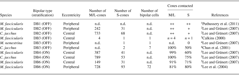
Cone contacts of diffuse bipolar (DB) cells in primate retina
1 The study suggests that individual M/L- and S-cones make similar numbers of contacts with DB1 cells, but a quantitative analysis is not provided.
2 The study analyzed the intensity of Glt-1 immunofluorescence (FMB and DB2 cells) opposite cone pedicles.
3 The results are based on electron microscopic reconstruction.
4 Bipolar cells were labeled with DiI, and S-cones were identified by immunohistochemistry. Note that most DB3 cells did not have S-cones in their dendritic field.
5 Bipolar cells were identified by immunohistochemistry. Cone pedicles were identified by PNA labeling.
M/L, medium/long wavelength sensitive; S, short wavelength sensitive; DB, diffuse bipolar; ++, high number of contacts; +, low number of contacts; n = number of cones; n.d., not determined.
In summary, despite the anatomical evidence for a blue-OFF midget bipolar and blue-OFF midget ganglion cell in the fovea of macaque retina, there is no direct physiological evidence for a small field blue-OFF ganglion cell. There maybe different types of blue-OFF ganglion cells, and a sign-inverting amacrine cell receiving input via the blue cone (ON) bipolar cell may exist, but further studies are needed to understand the synaptic circuitry of the blue-OFF pathway.
S-cone input to diffuse bipolar cells
Boycott and Wässle (Reference Boycott and Wässle1991) distinguished six types of diffuse (cone) bipolar (DB) cell in macaque retina, and comparable numbers have been found in marmoset retina (Chan et al., Reference Chan, Martin, Clunas and Grünert2001). In addition, a large field bipolar cell has been identified (named g-type cell by Polyak; GB by Rodieck, Reference Rodieck, Steklis and Erwin1988 and Boycott & Wässle, Reference Boycott and Wässle1991; and GBb by Kolb et al., Reference Kolb, Linberg and Fisher1992). More recently, Joo et al. (Reference Joo, Peterson, Haun and Dacey2011) analyzed this GB cell in more detail. The GB cell stratifies at the same level as the DB4 cell and thus presumably is an ON cell. The dendrites of GB cells on average contact about half of the cones in their dendritic field (Table 1). The connections with S-cones were not studied directly in the Golgi-impregnated retinas, but a nearest neighbor analysis between the cone contacts of individual GB cells and a comparison with the S-cone spacing at the same eccentricity as determined from blue cone bipolar cells revealed that even if the GB cell contacted S-cones, it would not do so exclusively. In addition, in that study, an example of a GB cell passing but not contacting S-cones in its dendritic field is shown. Whether the GB cell plays a role in color vision needs to be explored.
Diffuse bipolar types contact all cone types in their dendritic field (Hopkins & Boycott, Reference Hopkins and Boycott1997), but because S-cones make up only a low proportion of all cones, the dominant input to all diffuse bipolar types derives from M/L-cones. Moreover, there is evidence for all diffuse bipolar types that a proportion of each type does not have an S-cone in its dendritic field and thus does not receive S-cone input (see Table 1 in Chan et al., Reference Chan, Martin, Clunas and Grünert2001; see Fig. 9A-C in Lee & Grünert, Reference Lee and Grünert2007). The specific S-cone connectivity is now known for nearly all of the diffuse bipolar types and is summarized in Table 2. With the possible exception of the DB1 cell (Puthussery et al., Reference Puthussery, Gayet-Primo, Taylor and Haverkamp2011), diffuse bipolar contacts are biased against S-cones, that is, they make lower number of contacts with individual S-cones in comparison to individual M/L-cones (Lee & Grünert, Reference Lee and Grünert2007). Thus, the ganglion cell types postsynaptic to diffuse bipolar cells are expected to receive little S-cone input. For example, the DB2 and DB3 cells provide input to OFF parasol cells (Jacoby et al., Reference Jacoby, Wiechmann, Amara, Leighton and Marshak2000; Calkins & Sterling, Reference Calkins and Sterling2007), and the DB4 and DB5 cells presumably contact ON parasol cells (Boycott & Wässle, Reference Boycott and Wässle1991). Parasol cells project to the magnocellular layers of the lateral geniculate nucleus. Consistent with the anatomical data, electrophysiological recordings from the magnocellular layers as well as recordings from parasol cells have shown that S-cone inputs are absent or weak (Derrington et al., Reference Derrington, Krauskopf and Lennie1984; Reid & Shapley, Reference Reid and Shapley1992; Dacey & Lee, Reference Dacey and Lee1994; Shapley, Reference Shapley2006; Sun et al., Reference Sun, Smithson, Zaidi and Lee2006; Tailby et al., Reference Tailby, Szmajda, Buzas, Lee and Martin2008b; Field et al., Reference Field, Gauthier, Sher, Greschner, Machado, Jepson, Shlens, Gunning, Mathieson, Dabrowski, Paninski, Litke and Chichilnisky2010) but see also Chatterjee and Callaway (Reference Chatterjee and Callaway2002).
In summary, diffuse bipolar cells receive little input from S-cones and thus most likely do not play a significant role in S-cone pathways.
Nonprimate mammals
S-cone signals are particularly important for nonprimate mammals as they lack red-green opponency, and their color discrimination relies solely on S- and M-/L-cone contrast. Here we discuss how bipolar cells interact with amacrine and color-opponent ganglion cells in the inner plexiform layer, to propagate S-cone signals and to facilitate blue–green color discrimination. Initial studies of color pathways largely relied on staining and antibody labeling to explore the vast array of connections carrying S-cone signals through the inner plexiform layer. Recent progress has been made in identifying the cell types involved in relaying S-cone signals, not only by their stratification patterns but also by their electrophysiological responses to S-cone isolating stimuli.
S-cone contacts of bipolar cells
The S-cone pathways in nonprimate retinas have mainly been explored in rabbit, mouse, and ground squirrel. In rabbit retina, Famiglietti (Reference Famiglietti1981) identified a wide-field cone bipolar cell with sparse dendritic branching. These bipolar cells had axon terminals in sublamina b (upper region of stratum 5), characteristic of ON bipolar cells and were named wb cells. Famiglietti determined that unlike narrow- and medium-field bipolar cells which contact all cones in their field, wb cells only contacted 13% of the cones in their fields, leading him to propose that wb cells and their counterpart wa cells (OFF-type, bordering stratum 1 and 2) potentially made selective connections with S-cones. Famiglietti (Reference Famiglietti2008) offered the IIb2 ganglion cell as a potential postsynaptic destination of the wb cell and provided a camera lucida drawing depicting a wb cell in stratum 5 alpha costratifying and potentially contacting the IIb2 cell. Although informative, these results were nonetheless speculative in nature. MacNeil and Gaul (Reference MacNeil and Gaul2008) later confirmed by intraocular injections of biocytin that wb cells in the rabbit retina were indeed connected to S-cones. Also in the rabbit retina, Liu and Chiao (Reference Liu and Chiao2007) injected bipolar cells with Neurobiotin and examined their synaptic connections to immunostained S-cones. They identified seven wa-like cells (which they named BB and BB-like cells) that appeared to contact S-cones selectively and had axon terminals stratifying in sublamina 1—a possible blue-OFF bipolar cell. However, a similar cell type has not been reported in other species and more evidence is required to confirm the existence of blue-OFF bipolar cells.
The first direct evidence of blue (ON) cone bipolar cells in mammals other than primates came from work in the mouse retina. Serendipitously, it was found that in a transgenic mouse line that expresses Clomeleon (a chloride-sensitive fluorescent indicator, the expression of which was driven by Thy-1 promoter), blue cone bipolar cells were prominently labeled and made selective contacts with “true” S-cones that only express S-opsin (Haverkamp et al., Reference Haverkamp, Wässle, Duebel, Kuner, Augustine, Feng and Euler2005). These cells were later referred to as type 9 cone bipolar cells, which show S-cone preferred light responses (Breuninger et al., Reference Breuninger, Puller, Haverkamp and Euler2011).
The first direct electrophysiological evidence that blue cone bipolar cells have a sign-inverting ON-center response was presented by Li and DeVries (Reference Li and DeVries2006) who performed paired recordings of cones and cone bipolar cells in the ground squirrel retina. In addition to the blue cone bipolar cell that received exclusive S-cone input, they also identified two other types (OFF b2, ON b5) that receive mixed S/M-cone input. The remaining types of identified bipolar cells appeared to receive almost pure M-cone signal. Those chromatic-pure bipolar cells likely define the spectral properties of the ganglion cells that participate in blue-green color opponency, whereas those with mixed S/M-cone input likely convey luminance signals (Li & DeVries, Reference Li and DeVries2006). It remains to be shown whether the spatial opponency in blue cone bipolar cells matches the cospatial opponent signal found in blue-ON/yellow-OFF ganglion cells described in macaque by Crook et al. (Reference Crook, Davenport, Peterson, Packer, Detwiler and Dacey2009). Remarkably, despite recording from hundreds of bipolar cells, Li and DeVries (Reference Li and DeVries2006) did not encounter a homolog of the wa cell in the ground squirrel retina. Likewise Breuninger et al. (Reference Breuninger, Puller, Haverkamp and Euler2011) suggest that there is no blue-OFF bipolar pathway in mice.
S-cone amacrine cell and the blue-OFF pathway
In contrast to the blue ON pathway, the blue-OFF pathway is apparently more diverse and is less well understood. The blue-OFF signal is necessary for blue-green opponency in dichromats. However, evidence for a blue-OFF bipolar cell [as a counterpart to the blue cone (ON) bipolar cell] remains scarce (see above). An alternative to such a blue-OFF bipolar cell is suggested by the rod pathway, which lacks a rod-selective OFF bipolar cell. In the rod pathway, the AII amacrine cell inverts ON signals thereby obviating the need for a rod OFF bipolar cell (reviewed by Bloomfield & Dacheux, Reference Bloomfield and Dacheux2001). A similar sign-inversion mechanism for the blue-OFF signal has been proposed previously (Kolb et al., Reference Kolb, Goede, Roberts, McDermott and Gouras1997; Hemmi et al., Reference Hemmi, James and Taylor2002; McMahon et al., Reference McMahon, Packer and Dacey2004; Neitz & Neitz, Reference Neitz and Neitz2011).
Functionally, until recently, there has been no data regarding such an S-cone amacrine cell, largely due to experimental difficulties. Since S-cones represent only ∼6% of the total cone population (which are in turn outnumbered by rods in most mammalian retinas, Kryger et al., Reference Kryger, Galli-Resta, Jacobs and Reese1998; Li & DeVries, Reference Li and DeVries2004), neurons in the S-cone pathway are rarely encountered in recordings. At the same time, amacrine cells are the most diverse retinal neurons with over 30 morphological types (Masland, Reference Masland2001), making it even more challenging to identify a minority that participate in the S-cone pathway. Recently, two independent yet complementary studies provided strong evidence for the existence of an S-cone amacrine cell that inverts blue-ON signals and transmits them to the blue-OFF pathway (Chen & Li, Reference Chen and Li2012; Sher & DeVries, Reference Sher and DeVries2012). Chen and Li (Reference Chen and Li2012) conducted whole cell recordings from a subpopulation of amacrine cells in the ground squirrel retina that responded to S-cone isolating stimuli. These S-cone amacrines are bistratified with processes at the outer and inner boundaries of the inner plexiform layer (Fig. 3C). The inner tier, which contains prominent synaptic boutons, costratifies with the axon terminals of the blue cone bipolar cell (Fig. 3A and 3C). Immunohistochemical labeling of these cells using antibodies against the glycine transporter suggested that they were glycinergic amacrine cells. As anticipated, the S-cone responses were abolished by application of L-AP4 which blocked ON bipolar responses (Fig. 3B). The outer tier of dendrites arising from S-cone amacrines potentially contact a subpopulation of intrinsically photosensitive ganglion cells that have dendritic arbors that predominantly extend along the outer border of the inner plexiform layer (Berson et al., Reference Berson, Dunn and Takao2002; Dacey et al., Reference Dacey, Liao, Peterson, Robinson, Smith, Pokorny, Yau and Gamlin2005). In support of this, melanopsin-expressing ganglion cells in macaque have been shown to possess an S-OFF/(L + M)-ON color opponent receptive field (Dacey et al., Reference Dacey, Liao, Peterson, Robinson, Smith, Pokorny, Yau and Gamlin2005). S-cone signals traversing this pathway likely code luminance and participate in the entrainment of the circadian system, and the perception of brightness (Brown et al., Reference Brown, Tsujimura, Allen, Wynne, Bedford, Vickery, Vugler and Lucas2012).
(A) A blue cone bipolar cell (magenta) in a ground squirrel retinal slice labeled by dye injection after recording was shown to contact an S-cone (cyan) labeled by an antibody against S-opsin. (B) As illustrated in the proposed S-cone amacrine cell pathway (left panel), either blocking S-cone to blue cone bipolar cell synapse with 4 μM L-AP4 or blocking blue cone bipolar cell to S-cone amacrine cell synapse with 5-μM NBQX eliminated S-cone amacrine cell light response to S-cone isolating stimulus (right panel). (C) The S-cone amacrine cell has large synaptic varicosities coramifying at the bottom of the inner plexiform layer with blue bipolar cell axon terminals (A). (D) Superimposed spike triggers an average of four types of ganglion cells simultaneously recorded in the same piece of ground squirrel retina in four solutions: control, 75-μM LY341495 + 50-μM L-AP4 (LY/AP4), washout, 100-μM strychnine. Insets indicate sites of drug action. In keeping with the proposed pathway (left panel in A), either blocking blue cone bipolar cell input (LY + L-AP4) or blocking S-cone amacrine cell output (Strychnine) eliminated the blue-OFF response in blue-OFF/green-ON ganglion cells. (A–C) are modified from Chen and Li (Reference Chen and Li2012) and (D) is modified from Sher and DeVries (Reference Sher and DeVries2012), both with permissions. Scale bar in A = 10 µm.
Sher and DeVries (Reference Sher and DeVries2012) used a 512 electrode array to identify/classify color-opponent ganglion cells as either: S-OFF/M-ON or S-ON/M-OFF based on their spike-triggered average (STA) response to white noise stimuli. In keeping with the result of Chen and Li (Reference Chen and Li2012), a combination of L-AP4 and LY341495 that blocked the ON bipolar response in turn blocked not only S-ON but also S-OFF responses in ganglion cells, indicating that the S-OFF response is dependent on the ON pathway (Fig. 3D). Additionally, strychnine blocked the S-OFF response but spared the M-ON response suggesting the involvement of a glycinergic amacrine cell in relaying the ON signal to the S-OFF/M-ON ganglion cells (Fig. 3D). These results collectively depict a pathway in which a glycinergic S-cone amacrine cell receives input from the blue cone bipolar cell and in turn provides an inhibition to the putative blue-OFF ganglion cell either directly or indirectly.
Interestingly, another form of amacrine cell involvement in color processing has been demonstrated recently by Euler and colleagues (Chang et al., Reference Chang, Breuninger and Euler2013). They discovered that “alpha-like” ganglion cells in the mouse retina utilized the coexpression gradient of opsin along the dorsoventral axis to create chromatic opponency. This form of opponency required a large diameter stimulus (in mm range) and could be blocked by GABAzine and TMPA (GABAergic receptor blockers) or TTX (Na+ channel blocker), suggesting that GABAergic spiking amacrine cells were essential for the color opponent responses observed in “alpha-like” ganglion cells.
Thus, the dorsoventral gradient of S cone pigment creates an additional layer of color opponency whereby neighboring ganglion cell responses become color opponent without necessitating traditional cone type selective channels. This region known as the opsin transitional zone also imparts additional properties to neighboring direction selective retinal ganglion cells as a result of the chromatic and spatial information coded in the opsin gradient. Calcium imaging of direction selective retinal ganglion cells demonstrated that these cells responded to chromatic changes similarly to motion perceived by achromatic stimuli (Chang et al., Reference Chang, Breuninger and Euler2013). Such effects on ganglion cell chromatic processing illustrate how the retina can maintain chromatic opponency despite regional differences in cone density or structural wiring asymmetry.
Conclusions and future directions
In sum, there is abundant evidence that an S-cone selective ON cone bipolar cell (the blue cone bipolar cell) carries blue-ON signals to the inner plexiform layer in most mammals including primates. Although it has only been demonstrated in the ground squirrel retina so far, it is likely that a common blue-OFF pathway exists in mammals whereby an S-cone amacrine cell inverts blue-ON signals from the blue cone bipolar cell. A recent psychophysical study on the human S-cone pathways lends further support for this view. Shinomori and Werner (Reference Shinomori and Werner2012) characterized the temporal properties of the S-ON and S-OFF pathways and determined that the S-cone impulse responses to increases in light (increment threshold or ON response) were faster than decrement thresholds (OFF response), which is in line with the proposed pathway in which OFF signals are generated via one or more additional synapse(s).
For macaque, in the fovea region, where a midget bipolar cell makes a solitary contact with a single cone, there is evidence that a midget bipolar cell contacts an S-cone, providing an additional channel for blue-OFF signals. Species variation may exist, as this pathway has not been found in New World monkeys. In peripheral retina, this mechanism weakens as multiple midget bipolar cells provide input to midget ganglion cells and the S-cone amacrine cell may be the dominant conduit for blue-OFF signals. In agreement with the idea that there is a difference in the processing of blue-OFF signals between central and peripheral retina, patients with congenital stationary night blindness (CSNB), who lose their ON pathway completely, appear to have intact central, but impaired peripheral, blue-yellow color perception (Terasaki et al., Reference Terasaki, Miyake, Nomura, Horiguchi, Suzuki and Kondo1999), suggesting that the peripheral blue-yellow opponency relies solely on the ON pathway.
The prominence of the S-cone amacrine cell in conducting blue-OFF signals may explain the discrepancies attributed to early electroretinogram (ERG) studies which attempted to identify which pathways (ON or OFF) were involved in blue cone signaling (Evers & Gouras, Reference Evers and Gouras1986; Smith et al., Reference Smith, Harwerth, Crawford and Duncan1989). Evers and Gouras (Reference Evers and Gouras1986) reported that under Ganzfeld stimulation, the S-cone system in macaque monkey relied solely on the ON bipolar system, whereas the L- and M-cones employed both ON and OFF bipolar systems. They hypothesize that this difference may be due to the presence or absence of an OFF bipolar system and show that injection of 2-amino-4-phoshonobutyric acid (APB, a blocker of the ON response) into the vitreous completely eliminated the S-cone ERG. These results are consistent with a pathway involving an S-cone amacrine cell as the major contributor to the blue-OFF response under peripheral illumination, as outlined above. In a psychophysical study, however, Smith et al. (Reference Smith, Harwerth, Crawford and Duncan1989), who used a foveal stimulus (limited to 0.67–8.0°), did not observe an effect related to S-cone function upon injection of APB suggesting that in central retina, S-cone OFF signals are mediated by OFF bipolar pathways insensitive to APB. Taken together, these studies as well as the observed phenotype in patients with CSNB lend support to the notion that in Old World primates including humans: (1) central blue-yellow color opponency is mediated by midget OFF bipolar cells, whereas in the periphery (2) S-cone amacrine cells comprise the dominant pathway through which the blue-OFF signal is generated.
There are several outstanding questions that remain in order to understand how S-cone signals are carried through the retina and are involved in color discrimination. Recent results in macaque monkeys suggest that S-cones themselves are already color and spatial opponent since the H2 horizontal cell provides feedback (L + M cone sensitivity) onto the S-cones (Packer et al., Reference Packer, Verweij, Li, Schnapf and Dacey2010). Thus, it could be expected that the blue cone bipolar cell would also show color opponency; however, this has not yet been directly demonstrated in recordings from blue cone bipolar cells. The functional significance of S-cone opponency resulting from horizontal cell feedback also remains unclear as it appears to be redundant to the chromatic opponency produced by the inner retina. It has been suggested that horizontal cell feedback may act as a form of presynaptic inhibition that results in optimal processing of S-cone signals by adjusting the synaptic gain depending on the spectral background (Packer et al., Reference Packer, Verweij, Li, Schnapf and Dacey2010). Interestingly, the spatial opponency identified in S-cones resulting from horizontal cell feedback is lost at the level of the ganglion cells where opposing surrounds likely cancel.
In contrast, in other mammals that possess an opsin gradient such as mice, chromatic stimuli also inherently carry spatial information. It remains to be seen how the higher visual areas processes this information to correctly represent the visual scene.
The recent evidence supporting the S-cone amacrine cell’s involvement in generating the blue-OFF response has also led to new questions regarding the structural identification of its output cell, the blue-OFF ganglion cell. It will be necessary to determine if the blue-OFF ganglion cell is functionally conserved across different species and whether it serves an evolutionary role in hue perception and/or an additional input for regulating the circadian clock.
Last, it will be interesting to see, if the S-cone amacrine cell, like the blue cone bipolar cell, is a widely conserved feature of color vision.
Acknowledgments
We would like to thank our colleagues and co-authors for their help. For this review T. Chan, K. Ghosh, S. Lee, P. Martin, I. Telkes and H. Wässle made important contributions. We would like to thank Heinz Wässle, Paul Martin, Alexander Sher, and Steven DeVries for providing images and Paul Martin for critically reading an earlier version of this manuscript. This work was supported by Project Grants from the National Health & Medical Research Council of Australia 632640 and 1022609 to UG, and the National Eye Institute (USA) intramural research program to KJM & WL.