The isolated ventricular septal defect belongs to the group of simple congenital heart diseases, divided into large defects repaired early in life or those thought to be so trivial that they do not merit repair or intervention. Patients with either repaired or trivial, unrepaired defects have been thought to have a normal life expectancy with excellent prognosis, and accordingly, consensus guidelines did not recommend regular follow-up of these patients.Reference Baumgartner, Bonhoeffer and De Groot1,Reference Silversides, Dore and Poirier2 In the later years, however, a growing number of papers report on events in these adults that cannot be considered normal, suggesting we should reconsider the current management of these adults.Reference Videbaek, Laursen, Olsen, Hofsten and Johnsen3–Reference Gabriels, De Backer and Pasquet7 Even in the young adult patients in their 20s, recent clinical studies describe various surprising findings of lower functional capacity,Reference Heiberg, Laustsen, Petersen and Hjortdal8,Reference Maagaard, Heiberg and Hjortdal9 disrupted ventricular contractilityReference Heiberg, Schmidt, Redington and Hjortdal10 and morphology,Reference Maagaard, Heiberg and Eckerstrom11 abnormal pulmonary function,Reference Rex, Eckerstrom and Heiberg12 and reduced cardiac output during exerciseReference Asschenfeldt, Heiberg, Ringgaard, Maagaard, Redington and Hjortdal13 when they are compared with their healthy peers. Interestingly, in a new set of guidelines, updated recommendations now advise all of these patients should be seen every 3 years at a clinic with expertise in adults with congenital heart disease.Reference Stout, Daniels and Aboulhosn14
A number of different theories have been offered for the various abnormal findings in this patient group with one of the most frequent explanations being post-surgical effects in the repaired patients. Nevertheless, the similarities in reduced clinical outcomes between repaired and unrepaired ventricular septal defects suggest otherwise. One central question still remains to be answered – how is the congenital shunt affected during physical activity? A clarification of this may help us further in explaining the reduced clinical parameters measured in young patients with unrepaired defects compared with healthy peers. Additionally, the possible increasing incompetence of the semilunar valves and the resulting retrograde flow has also been suggested to play an important role. Therefore, with the current MRI study, we set out to describe the shunt properties and possible changes in retrograde flow during exercise in young adults with unrepaired, small ventricular septal defects who had previously demonstrated lower exercise capacityReference Maagaard, Heiberg and Hjortdal9 and larger right ventricular volume.Reference Maagaard, Heiberg and Eckerstrom11 Furthermore, cardiac output during exercise was assessed and compared with a group of healthy adults matched on age and gender.
Methods
Ethics
Written informed consent was obtained from all patients. The protocol of the study conforms to the ethical standards of The Regional Committee on Biomedical Research Ethics of the Central Denmark Region (chart: 1-10-72-74-14), The Danish Data Protection Agency (chart: 2007-58-0010), and with the Helsinki Declaration of 1975, revised in 2008.
Study population
Young adults with a congenital, small, and unrepaired ventricular septal defect were included along with healthy peers who were matched on gender and age. Inclusion criteria for patients were age between 18 and 40 years and an unrepaired, congenital ventricular septal defect. Exclusion criteria were spontaneous defect closure since last follow-up visit, other congenital cardiac defects than a ventricular septal defect, associated syndromes, for example, Down’s syndrome, documented arrhythmia other than right bundle branch block, cardiac or pulmonary disease including any valve pathology, or missing patient chart. Healthy controls were recruited through the official webpage www.forsoegsperson.dk and included ad hoc to match with the patients on gender and age. Control patients were excluded on the same grounds as the patients. Patients and controls were included in random order throughout the study. On the same day as the exercise MRI, all patients also underwent upright exercise testing, echocardiography, and cardiac MRI at rest, where reduced exercise capacity and an altered right ventricular morphology were demonstrated in patients when compared with controls.Reference Maagaard, Heiberg and Hjortdal9,Reference Maagaard, Heiberg and Eckerstrom11,Reference Maagaard, Heiberg, Asschenfeldt, Ringgaard and Hjortdal15
Clinical data
Demographics such as height, weight, and resting blood pressure were recorded under standardised conditions for all patients. Impedance analysis was performed using the ImpediMed Ltd Model SFB7 (ImpediMed Ltd, Brisbane, Queensland, Australia) prior to the exercise test to assess body composition. Body surface area was calculated using Dubois formula. Daily exercise habits were determined by using the International Physical Activity Questionnaire,Reference Craig, Marshall and Sjostrom16 in which patients filled in their weekly amount of physical activity, as divided in levels of high, moderate, and low intensities.
Echocardiography
Before the MRI, patients underwent transthoracic echocardiography at rest using a GE Vivid 7 (GE Healthcare, Horten, Norway) with a 2.5 MHz probe. Echocardiographic focus points were recently described.Reference Maagaard, Heiberg and Eckerstrom11 Defects were visualised and measured by continuous-wave Doppler, any existing valvular insufficiencies were measured, and ejection fraction was estimated.
Magnetic resonance imaging
The MRI protocol has previously been described in detail, and the applied methodology for cardiac output assessment was validated.Reference Asschenfeldt, Heiberg, Ringgaard, Maagaard, Redington and Hjortdal13,Reference Heiberg, Asschenfeldt, Maagaard and Ringgaard17 Briefly, MRI at rest and during exercise was performed using a 1.5 T Philips Achieva dStream whole-body scanner, equipped with 33 mT/m gradients with a slewrate of 180 mT/m per ms and software release R517 using an 18-cm surface coil and spine coil arrays. Real-time phase-contrast MRI sequence was used to measure blood flow in the ascending aorta and main pulmonary artery. All data on blood flow in the great vessels were collected at rest and during the exercise test in real time with free breathing. Flow was measured in a transversal view approximately 2 cm above the aortic valve in the ascending aorta and in a second measurement plane above the pulmonary valve but proximal to the bifurcation, resulting in a transversal view of the pulmonary trunk. Real-time scans comprised 170 consecutive, phase-contrast flow acquisitions without electrocardiographic triggering (each lasting 81 ms, giving a frame rate of 12.2 frames/second) with velocity encoding varying from 170 to 290 cm/second depending on exercise level. Order of scans was randomised, ensuring equal amounts of scans starting with the aorta, then the pulmonary and vice versa.
Bicycle protocol
Patients were positioned on an MRI-compatible ergometer bicycle (MRI cardiac ergometer, Lode, Groningen, The Netherlands), mounted on the MRI table, as previously described.Reference Hjortdal, Emmertsen and Stenbog18 The patients’ feet were strapped to the ergometer pedals, while ensuring maximum knee joint extension at 30 degrees and shoulder restraining in order to minimise movement during exercise. Heart rate was monitored by standard electrocardiography monitoring system and by pulse oximetry (Nonin 7500FO pulse oximeter with a fibre optic sensor cable 8000FC-30). Workload protocol started at 25 W for 2 minutes and 15 seconds and gradually increased by 25 W every 75th second. Patients were instructed to keep pedalling at a speed of 60–70 rounds/minute until exhaustion was reached. A clinical study by Asschenfeldt et al between a standard cardiopulmonary breath-by-breath exercise test, performed in the supine position, and the current MRI set-up was recently performed. The comparative study showed good agreement when estimating cardiac output during exercise by the standard cardiopulmonary exercise test and the MRI exercise test, respectively (currently submitted).
Data analyses
The MRI analyses were performed offline, using an in-house produced software (Siswin, version 0.9, by Ringgaard, Aarhus, Denmark, 2008). The contours of the ascending aorta and the pulmonary artery were traced manually with an elliptical, four-point, region-of-interest tool, performed by one blinded observer. From each flow sequence, cardiac output, heart rate, and stroke volume were calculated as mean values. Cardiac output was defined as mean blood flow across the scanning plane and calculated by mean forward flow subtracted mean retrograde flow. Cardiac output and retrograde flow measurements were adjusted for body surface area. The shunt ratio was calculated as the mean cardiac output from the pulmonary artery relative to the mean cardiac output from the ascending aorta. The amount of blood flow through the shunt was calculated as the difference in mean flow between the pulmonary artery and ascending aorta. Transverse vessel areas of the ascending aorta and pulmonary artery were acquired on the basis of the traced vessel wall regions of interest, described previously.Reference Maagaard, Heiberg, Asschenfeldt, Ringgaard and Hjortdal15 Exercise intensities were divided into five intervals, defined by the percentage of each participant’s obtained maximal workload: very low (>0 to <25%); low (≥25 to <50%); moderate (≥50 to <75%); submaximal (≥75 to <100%); and maximal (=100%).
Inter- and intra-observer variability
Two observers, blinded to clinical data, independently measured 41 MRI scans of six randomly chosen patients. The main observer was tested for intra-observer variability by repeating measurement in 37 scans during real-time MRI from five randomly selected patients.
Correlation analyses
Correlation analyses were applied for aortic cardiac index and mean pulmonary flow at maximal exercise and the previously measured peak oxygen uptake.Reference Maagaard, Heiberg and Hjortdal9 Furthermore, correlation was tested between aortic retrograde flow and left ventricular end-diastolic volume index as well as between pulmonary retrograde flow and right ventricular end-diastolic volume index.Reference Maagaard, Heiberg and Eckerstrom11
Endpoints
Our primary endpoint was shunt ratio during exercise. Secondary endpoints were cardiac indices and retrograde flow of the ascending aorta and pulmonary artery, all measured during exercise.
Statistical methods
Before study start, a sample size was calculated based on a previous conducted exercise study on operated ventricular septal defects.Reference Heiberg, Laustsen, Petersen and Hjortdal8 This was chosen, as the same two groups of patients of the current study were also tested with upright bicycle exercise within the same time period, as previously published.Reference Maagaard, Heiberg and Hjortdal9 We expected a difference in peak oxygen uptake that was 80% of the previously found difference in surgically closed patients with the same standard deviations. A statistical power of 90% and a significance level of 5% were set, indicating that at least 19 patients were needed in each group. Continuous, normally distributed data were reported as means with standard deviations, and non-parametric data were reported as median with 95% confidence interval. Normally distributed data were tested using the unpaired Student’s t-test with either equal or unequal variance as appropriate. Curves of cardiac indices, retrograde flows, and shunt ratio during exercise between patients and controls were tested by applying two-way ANOVA. Correlation analyses were made using Pearson’s sample correlation r. Inter- and intra-observer agreements were assessed with intraclass correlation coefficient using a two-way mixed model.Reference Shrout and Fleiss19 A number of single measures were chosen from random patients from both groups, with two pre-defined raters, looking for absolute agreement. p-values ≤ 0.01 were considered statistically significant. Statistical analyses and drawing of plots were performed using StataIC 11.2 (StataCorp LP, College Station, Texas, United States of America) and GraphPad Prism 6 (GraphPad Software, La Jolla, California, United States of America).
Results
Study population
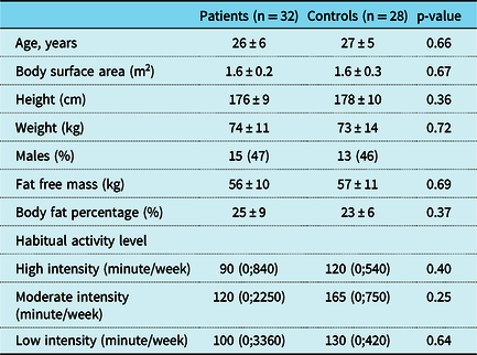
A total of 32 adults with unrepaired ventricular septal defects and 28 healthy controls were included in the MRI study from May, 2014 to May, 2016 at Aarhus University Hospital, Denmark. Daily activity levels and demographics of patients and controls are displayed in Table 1, demonstrating no differences between groups. Exercise studies, performed in patients earlier in the day, revealed lower peak oxygen uptake in patients, 36.8 ± 9 ml/kg/m2, compared with controls, 43.8 ± 9 ml/kg/m2 (p < 0.01), and comparable blood pressures, respiratory exchange ratios, and heart rates, all previously published.Reference Maagaard, Heiberg and Hjortdal9 Furthermore, the heart rate reserve, calculated as the difference between resting and maximal heart rate, was comparable between patients (99 ± 15 beats per minute) and controls (100 ± 11 beats per minute), p = 0.79. Considering the type of defect, nine were muscular and 23 were perimembranous. None of the patients dropped out of the study.
Demographics and clinical data on patients with unrepaired ventricular septal defects and healthy controls

Data presented as mean with ±standard deviation, total numbers (with percentages), or median with (total ranges)
Echocardiography
All patients had normal ventricular ejection fractions at rest as evaluated by echocardiography. None of the patients demonstrated measurable aortic valve regurgitation at rest. Tricuspid annular plane systolic excursion was 21 ± 3 mm in patients and 22 ± 3 mm in controls, p = 0.42, with none of the patients demonstrating measurements <17 mm. Right ventricle outflow tract diameter was 31 ± 1 mm in patients and 28 ± 1 mm in controls, p = 0.28. Regarding the pulmonary valve, insufficiency was noted in 10 patients with a median of 13 mmHg (total ranges 3–34 mmHg) and in nine controls with a median of 9 mmHg (total ranges 5–12 mmHg), p = 0.58. None of the patients had significant peak tricuspid regurgitation velocities ≥2.8 m/s, suggesting normal estimated pulmonary pressures at rest. Other echocardiographic values have previously been described.Reference Maagaard, Heiberg and Eckerstrom11
Magnetic resonance imaging
At rest, aortic cardiac index was 3.4 ± 0.8 L/minute/m2 in patients and 3.5 ± 0.8 L/minute/m2 in controls, p = 0.34, whereas pulmonary mean flow was higher in patients (4.0 ± 1.0 L/minute/m2) compared with controls (3.4 ± 0.9 L/minute/m2), p = 0.01. The mean shunt ratio of the patients was 1.2 ± 0.2. At rest, retrograde flow index in the pulmonary artery was higher in patients (0.5 ± 0.3 L/minute/m2) compared with controls (0.3 ± 0.2 L/minute/m2), p = 0.01. Retrograde aortic flow index at rest did not differ between patients (0.4 ± 0.4 L/minute/m2) and controls (0.3 ± 0.1 L/minute/m2), p = 0.03.
Exercise test
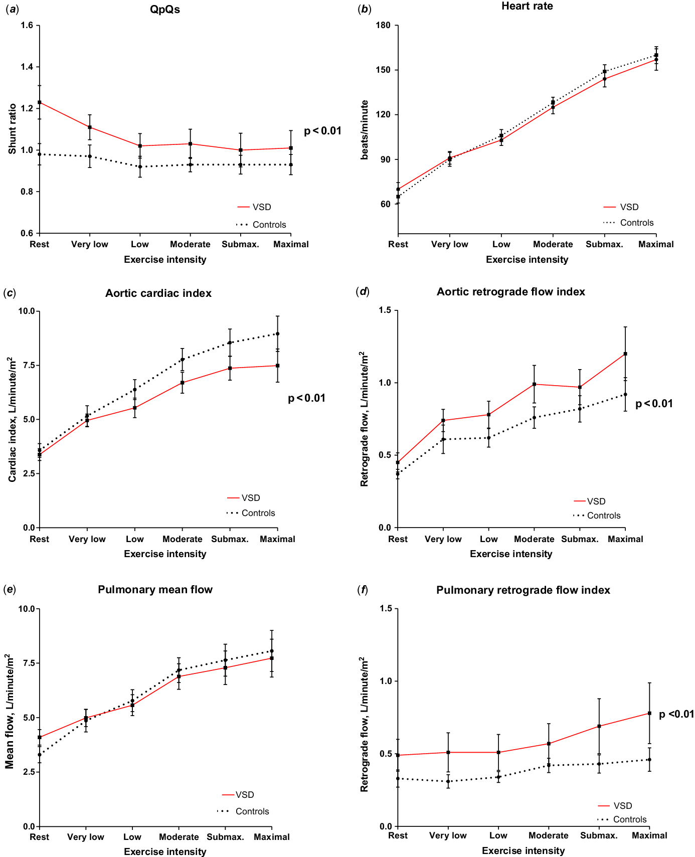
Blood flow measurement and corresponding heart rates during the exercise test are shown in Figure 1. At the end of the test, the patients reached a workload of 151 ± 7 W and the healthy controls reached 165 ± 8 W, p = 0.11. During exercise, the shunt ratio decreased (p < 0.01), and at peak exercise the shunt ratio was 1.0 ± 0.2. The absolute of blood flow through the defect was 0.5 ± 1.6 L/minute/m2 at rest and unchanged (0.4 ± 2.0 L/minute/m2, p = 0.50) at peak exercise. Aortic cardiac index was lower in patients than controls during the exercise test, p < 0.01 and was also lower at peak exercise in patients (7.5 ± 2.0 L/minute/m2) compared with controls (9.0 ± 2.0 L/minute/m2), p < 0.01. Aortic and pulmonary retrograde flow indices were higher in the patients compared with controls during the exercise test, both p < 0.01. At peak exercise, the retrograde flow index in the ascending aorta was 1.1 L/minute/m2 in patients and 0.8 ± 0.6 L/minute/m2 in controls (p = 0.01), and the pulmonary retrograde flow index was 0.7 ± 0.5 L/minute/m2 in patients and 0.4 L/minute/m2 in controls, p = 0.01. Heart rate did not differ between groups during the bicycle test.
Blood flow in the ascending aorta and pulmonary artery with corresponding heart rates at rest and different exercise intensities in patients and controls. (a) Flow-ratio between mean flow through pulmonary artery and ascending aorta (QpQs) (p < 0.01). (b) Heart rate at rest and during exercise. (c) Cardiac index of the ascending aorta (p < 0.01). (d) Retrograde flow index of the ascending aorta (p < 0.01). (e) Cardiac index of the pulmonary artery. (f) Retrograde flow index of the pulmonary artery (p < 0.01). VSD = ventricular septal defect. Intensity levels: very low (>0 to <25%); low (≥25 to <50%); moderate (≥50 to <75%); submaximal (≥75 to <100%); and maximal (=100%).

Correlation analyses
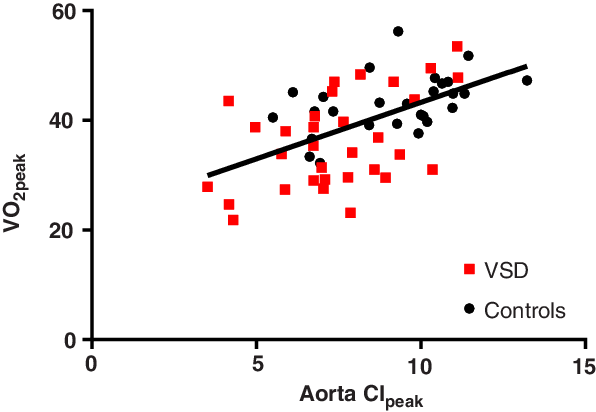
The peak cardiac index of the ascending aorta was correlated with the peak exercise capacity, which revealed a positive correlation for all participants (r = 0.5, p < 0.01) as shown in Figure 2. There was no correlation between biventricular end-diastolic volume indices and the retrograde flow indices in either of the two groups.
Correlation analyses between the maximal cardiac index of the ascending aorta and peak oxygen uptake for all participants. Positive correlation between VO2peak and aorta CImax for all participants (r = 0.5, p < 0.01), specifically with patients (r = 0.5, p < 0.01) and controls (r = 0.5, p = 0.01). VO2peak = peak oxygen uptake; CImax = maximal cardiac index.

Inter- and intra-observer variability
The observer variability demonstrated good agreements between inter- and intra-observer measurements with an intraclass correlation coefficient of 0.79 for the inter-observer and 0.77 for the intra-observer variability.
Discussion
Three important findings are presented in this exercise MRI study on young adults with unrepaired, small ventricular septal defects: Firstly, the shunt ratio decreased with increasing exercise intensities. Secondly, patients demonstrated a 17% lower aortic cardiac index at highest exercise levels. And thirdly, further flow analysis revealed a larger amount of retrograde flow both in the ascending aorta and the pulmonary artery in patients compared with controls during exercise.
Apart from the current study, only one other study has previously assessed the haemodynamic properties of the shunt ratio in unrepaired ventricular septal defects during dynamic exercise. In 1984, Bendien et al studied a group of 18 children, aged 14, with moderate defects, and a group of 17 children, aged 13, with small defects using cardiac catheterisation during moderate exercise.Reference Bendien, Bossina and Buurma20 The absolute left-to-right shunt flow remained unchanged from rest to exercise, but the shunt ratio decreased during exercise, reflecting the results of our study. In another exercise study from 1985, Otterstad et al investigated unrepaired and repaired defects at rest and during mild supine exercise while determining cardiac output.Reference Otterstad, Simonsen and Erikssen21 During exercise, the patients did not demonstrate a sufficient increase in cardiac output as expected, which reflect the current results of a lower aortic cardiac index during exercise. Apart from an MRI study on surgically closed ventricular septal defects,Reference Asschenfeldt, Heiberg, Ringgaard, Maagaard, Redington and Hjortdal13 no other studies have previously evaluated the retrograde flow in the unrepaired defects during exercise.
In order to better understand these remarkable findings, we propose one main hypothesis: The patients with unrepaired defects are not able to decrease pulmonary vascular resistance during exercise similarly as in healthy controls. As in the systemic circulation, the normal response to increasing exercise in the pulmonary system is a dilatation as well as vessel recruitment in the pulmonary vasculature which results in a reduced pulmonary vascular resistance.Reference Merkus, de Beer, Houweling and Duncker22 We postulate that patients with unrepaired defects are unable to properly dilate and recruit additional vessels in the pulmonary system.
In a significantly large shunt, the vast pulmonary hyperperfusion results in endothelial vascular damage and vessel dysfunction characterised by hypertrophic vessels with smaller lumen and thereby increased resistance. Lung biopsies have also revealed bronchial smooth muscles hypertrophy in patients with increased pulmonary artery pressure.Reference Schindler, Bohn, Bryan, Cutz and Rabinovitch23 In line with this, a subgroup of surgically treated ventricular septal defects revealed a pathologically increased resistance in the small airways in young adulthood.Reference Rex, Eckerstrom and Heiberg12 The systemic peripheral resistance is most likely not playing a role in the current results in these young adults, as previously published data on the same cohort from the same study period have noted no differences in continuously measured blood pressures during incremental exercise.Reference Maagaard, Heiberg and Hjortdal9 Whether this will develop with age and further affect exercise results remains speculative for now, as studies on the older patient population are scarce.
In the current study, patients demonstrated a decreasing cardiac index in the ascending aorta as compared with controls during exercise. Also, although demonstrating a higher mean flow in the pulmonary artery at rest, they were not able to increase pulmonary flow similarly as controls and ended with a slightly lower mean flow at peak exercise. Both the pattern of aortic cardiac index and pulmonary flow during exercise could be a reflection of an abnormal response in the pulmonary vasculature with an inability to lower the resistance, while still lowering the systemic vascular resistance in response to exercise. Likewise, the decrease in shunt ratio observed in our patients could also be interpreted as a reflection of the differing resistances in the pulmonary and systemic vasculature during exercise. Interestingly, in the haemodynamic exercise study on shunt ratio from 1984,Reference Bendien, Bossina and Buurma20 Bendien et al also noticed that pulmonary vascular resistance remains unchanged during exercise in both moderate and small defects, in contrast to the systemic vascular resistance which decreases with exercise. Their invasive measurements of the open defects are comparable to our findings of a decreasing shunt fraction. These findings support our theory of a more fixed pulmonary vascular resistance.
In 2010, Möller et al used echocardiography to discover increasing right ventricular systolic pressure during exercise both in patients with open and closed ventricular septal defects.Reference Moller, Brun and Fredriksen24 The authors suggest that the increasing systolic pressure in the right ventricle is an indication of an increasing pulmonary vascular resistance in response to exercise. The same group of authors furthermore investigated surgically closed defects in a placebo-controlled trial.Reference Brun, Moller, Fredriksen, Thaulow, Pripp and Holmstrom25 These patients did not experience the expected reduction in pulmonary vascular resistance in response to sildenafil during exercise, and the authors concluded that the pulmonary vascular bed may be dysfunctional as patients do not seem able to achieve proper pulmonary vascular dilatation. Another indication of this proposed inability to decrease pulmonary resistance during exercise may be seen in the current MRI study, where unrepaired defects demonstrate increased pulmonary retrograde flow during exercise as compared with controls. It is possible that the proposed inability to lower pulmonary vascular resistance during exercise is a coincidental finding in these patients and not associated with the ventricular septal defect. However, the group of healthy, matched controls did not display the same flow pattern during exercise. Also, the patients in this study were randomly included from a large group of small, unrepaired ventricular septal defects followed in outpatient clinics. Finally, similar to our results, young adults with surgically closed defects have also been found to display increased pulmonary retrograde flow during exercise in an MRI study from 2017.Reference Asschenfeldt, Heiberg, Ringgaard, Maagaard, Redington and Hjortdal13 This study further emphasised the previous notion that although the ventricular septal defect is successfully closed, the pulmonary hyperperfusion during the first few years of life has comparable effects on the pulmonary vascular bed as in adults who have a small, but long-standing pulmonary hyperperfusion through their unrepaired defect.
Limitations
In some of the scans at higher workload, the flow measurements revealed aliasing because the flow velocities became too high for the currently set velocity encoding, most often due to turbulent blood flow. If the aliasing was present centrally in the vessel where the flow current was clearly part of the rest of the forward flow, the aliasing was manually corrected so that it counted as part of the forward flow. However, careful measures were taken not to include too much in the forward flow as the amount of turbulent flow also increased with the higher levels of heart rates during exercise. Another limitation to be considered is the lack of information on potential semilunar valve regurgitation during increasing exercise. This was considered in the pilot phase but found to be impossible to assess with the current set-up. One way of learning more on potential valve incompetence during exercise could be to evaluate this by using echocardiography during a supine bicycle test. This was however not part of the current set-up.
Conclusion
Young adults with small, unrepaired ventricular septal defects revealed declining shunt ratio as well as an impaired aortic cardiac index during exercise. Patients also demonstrated larger retrograde flow through both the pulmonary artery and the ascending aorta during exercise. As the latest in a growing number of studies reporting on impaired cardiac and pulmonary function, this paper supports the view that the long-term outcome in these patients can no longer be considered completely benign and regular follow-up visits in specialist centres are advised.
Acknowledgements
The authors warmly acknowledge research secretary Jette Breiner for her appreciated contribution and medical student Christine Bauer-Jensen for her assistance in the data analysis of the phase-contrast MRI.
Financial support
This study was supported by Aarhus University, Lægeforeningens Forskningsfond, and Snedkermester Sophus Jacobsen and Hustru Astrid Jacobsen’s Fund.
Conflict of interest
None.