Case report
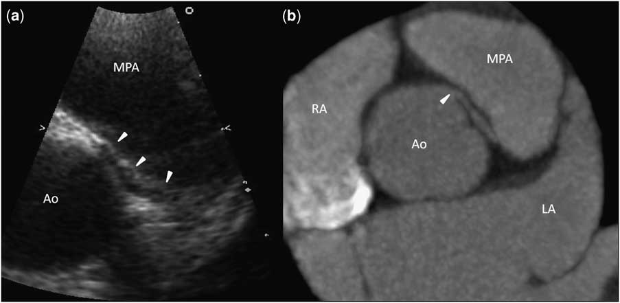
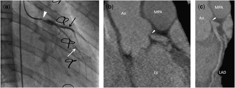
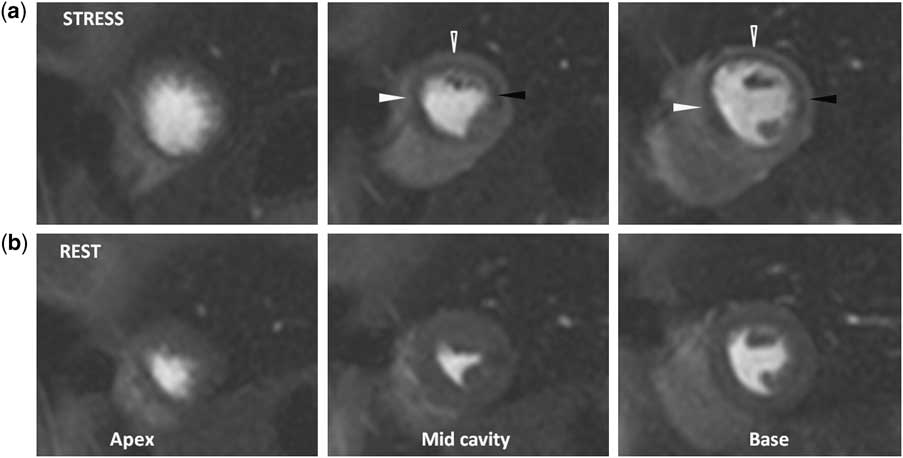
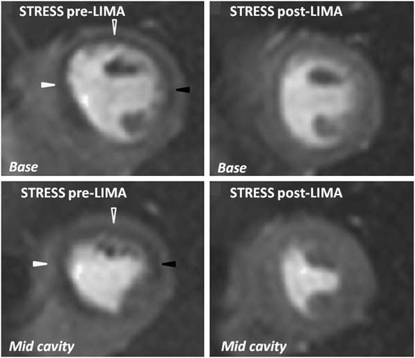
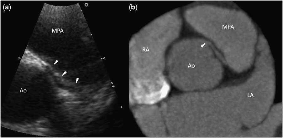
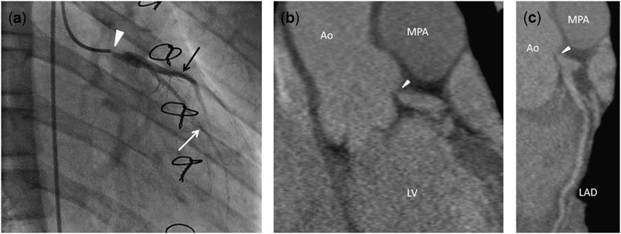
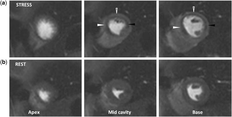
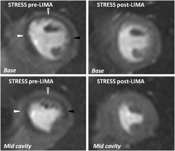
A previously healthy 14-year-old boy presented with a 6-month history of episodes of exercise-induced, central crushing chest pain, dizziness and light-headedness, with two recent episodes of collapse without loss of consciousness. Clinical examination was unremarkable and his resting electrocardiogram was normal. His troponin I level rose from 0.3 microgram per litre on admission to 16.4 micrograms per litre 12 hours later. A transthoracic echocardiogram showed normal regional wall motion, but the left coronary artery was seen to arise from the right coronary sinus and take an interarterial course between the right ventricular outflow tract and the aortic root (Fig 1a). A cardiovascular magnetic resonance scan demonstrated no oedema or late gadolinium enhancement, but the coronary arteries were not well visualised. A computed tomography coronary angiogram was performed on a Siemens Sensation 64 scanner (Siemens, Erlangen, Germany). It demonstrated the left main stem arising anomalously from the right sinus of Valsalva, close to, but separate from, a normal right coronary artery origin. The left main stem arose at an acute angle in relation to the aortic root and passed between the aortic root and right ventricular outflow tract. The proximal 2 centimetres of the left main stem appeared concentrically narrowed, suggesting a probable intramural course (Fig 1b). The patient underwent surgical reimplantation of the left main stem into left coronary sinus. The post-operative recovery was good with no evidence of cardiac ischaemia, and the patient was discharged with no concerns. After 3 months, the patient reported occasional dull left-sided chest ache unrelated to exercise. An exercise electrocardiogram demonstrated lateral ST segment depression, whereas transthoracic echocardiography revealed mild aortic regurgitation and a moderate pericardial effusion. The aortic regurgitation does not appear to have been present pre-operatively and the pericardial effusion resolved in the weeks following surgery and was felt to be post-surgical. Left ventricular size and function remained normal, with no resting regional wall motion abnormality. Invasive coronary angiography yielded severe ostial stenosis of the left main stem (Fig 2a). A non-gated computed tomography angiogram (Siemens FLASH, Siemens, Erlangen, Germany) confirmed the severe ostial stenosis (Fig 2b and c), and a subsequent perfusion cardiac magnetic resonance scan demonstrated extensive and profound inducible ischaemia in the left main territory (Fig 3) but no late gadolinium enhancement. Subsequently, a coronary artery bypass graft was performed, and a repeat perfusion cardiac magnetic resonance scan showed complete resolution of the inducible myocardial ischaemia (Fig 4).
Axial views from the initial echocardiogram (a) and axial maximum intensity projection image from the pre-operative gated computed tomography coronary angiogram (b) showing the anomalous course of the left coronary artery from the right coronary sinus (arrowheads) and its course between the aorta (Ao) and the main pulmonary artery (MPA). RA = right atrium; LA = left atrium.
Post-operative coronary catheter angiogram (a) and coronal maximum intensity projection (b) and a curved planar reformat (c) images from the post-operative non-gated computed tomography coronary angiogram demonstrating a tight stenosis at the origin of the left main stem (arrowheads) at the site of implantation onto the left coronary sinus. There is contrast opacification of the left anterior descending (white arrow) and the left circumflex (black arrow) arteries. Ao = aorta; LAD = left anterior descending artery; LV = left ventricle; MPA = main pulmonary artery.
Magnetic resonance images from a scan performed following left main stem reimplantation surgery. The images, obtained during stress (top row) and rest (bottom row), are displayed through the left ventricular axis following intravenous gadolinium showing a large area of reduced enhancement (arrowheads) in the left ventricular myocardium during stress testing. This is indicative of inducible ischaemia, in the anterior (white open arrowhead), lateral (black arrowhead), and septal walls (white arrowhead) of the left ventricle, particularly at the levels of the mid-cavity and base.
Contrast-enhanced magnetic resonance images from stress perfusion scans performed before and following left internal mammary artery to left anterior descending bypass grafting. The left ventricular axial images through the base (top row) and mid-cavity (bottom row) show that the inducible ischaemia (arrowheads) in the anterior (white open), lateral (black), and septal (white) walls resolves following graft surgery.
Discussion
The incidence of aberrant coronary anatomy is estimated to be about 1% in the general population. Although the majority of anomalous coronary artery anomalies are of no clinical significance, when the coronary artery arises from the opposite coronary sinus and passes between the aorta and the right ventricular outflow tract the anomalous course of the vessel is often termed “malignant”.Reference Baltaxe and Wixson 1 – Reference Yamanaka and Hobbs 3
More recently, it has been acknowledged that “left from right” origins are likely to be associated with sudden cardiac death.Reference Mumtaz, Lorber and Arruda 4 This is partly based on a large American Armed Forces Institute of Pathology population study in which a third of the 64 sudden cardiac death cases – among 6.3 million army recruits aged 18 to 35 years – were related to anomalous origin of a coronary artery from opposite sinus, the commonest cardiac abnormality in this series.Reference Eckart, Scoville and Campbell 5 As in this case, in each of the cases of anomalous coronary artery of that series, the left coronary arose from the right (anterior) coronary sinus and coursed between the aorta and the right ventricular outflow tract.Reference Eckart, Scoville and Campbell 5 In the largest angiographic study conducted to date, 1686 of 126,595 (1.3%) patients who underwent invasive coronary angiography had coronary anomalies, of whom 22 (0.017%) had a left abnormal origin of a coronary artery from the opposite sinus, that is, 1% of all coronary anomalies.Reference Yamanaka and Hobbs 3 Of the latter, 75% had a “malignant” interarterial course.
The higher incidence of sudden cardiac death is thought to be attributable to the acute angle of the proximal left main stem to the ostium, the intussuscepted and hypoplastic intramural segment, compression between the commissure of the right and left coronary cusps and lateral compression of the intramural segment and proximal vessel between the aorta and right ventricular outflow tract.Reference Lipsett, Cohle and Berry 6 , Reference Kim, Seo and Do 7
Imaging aberrant coronary anatomy
Although cardiovascular magnetic resonance imaging was a sensible initial choice to attempt to delineate the proximal coronary arteries in an adolescent because it does not use ionising radiation, the limited spatial resolution of cardiac magnetic resonance means that it is less ideal for full delineation of the coronary arteries than computed tomography coronary angiography. With sub-millimetre isotropic spatial resolution, computed tomography coronary angiography is now regarded as the gold standard for delineation of coronary artery anomalies. Computed tomography coronary angiography provides complete three-dimensional data sets, as opposed to two-dimensional image acquisitions of during invasive coronary angiography, and allows full assessment of the course of aberrant vessels and their relationship to other cardiovascular structures. More recent scanners are now also able to perform computed tomography coronary angiography at low radiation doses (1–3 millisieverts), and these advances have somewhat alleviated the concern over the use of computed tomography coronary angiography in young people.
Since the advent and establishment of computed tomography coronary angiography as a non-invasive imaging tool for coronary artery assessment, many coronary anatomy studies have been performed. Cademartiri et alReference Cademartiri, La Grutta and Malago 8 evaluated, using 64-slice computed tomography coronary angiography, 543 consecutive patients with suspected or known coronary artery disease in whom catheter coronary angiography suspected variants or anomalies of coronary arteries. This study demonstrated 43 separate anomalies of origin and course, of which abnormal origin of a coronary artery from the opposite sinus was found in eight patients (1.5%). Of these eight patients, five (63%) cases had a malignant course, whereas four were high-risk “left from right” abnormal origin of a coronary artery from opposite sinus.
In our own institution, 54 (1.4%) of the 3790 standard computed tomography coronary angiographies performed between May 2005 and August 2010 demonstrated cases of coronary artery anomalies. Of these 54 cases, 38 demonstrated abnormal origin of a coronary artery from the opposite sinus, of which 25 were “left from right”. However, only one, that of an anomalous origin of the left coronary artery from the right coronary sinus, presented with an acute coronary syndrome (unpublished data).
Who to operate on?
In this case in which the patient was symptomatic with demonstrable myocardial ischaemia, surgical management was warranted and initially involved surgical reimplantation of the “malignant” anomalous coronary artery. Unfortunately, this led to a subsequent stenosis of the reimplanted left main stem ostium and eventual left internal mammary artery graft. In symptomatic patients, surgery is often indicated, but in asymptomatic patients, surgery is more controversial. There is a growing advocacy for surgery to be performed on those asymptomatic patients with “left from right” origins who are deemed at highest risk from sudden cardiac death.
With the advent of computed tomography coronary angiography, anomalous coronary anatomy is increasingly seen, often as an incidental finding in patients undergoing investigation for chest pain, and often in patients in their fifth or sixth decade. These patients have often been asymptomatic and, even if they have a “left from right” abnormal origin of a coronary artery from opposite sinus, are unlikely to be at high risk for sudden cardiac death from this anomaly. There is anecdotal evidence that sudden cardiac death is most likely in those aged between 10 and 30 years,Reference Mumtaz, Lorber and Arruda 4 probably associated with the myocardial demands of these young and physically active years, and this would suggest that reimplantation or surgical de-roofing may be less relevant in those incidental cases detected on routine computed tomography coronary angiography for angina assessment.
Surgical options
Surgical strategies proposed to treat this defect include coronary reimplantation, unroofing of the intramural segment, and coronary artery bypass grafting.Reference Mumtaz, Lorber and Arruda 4 , Reference Jaggers and Pal 9 – Reference Gulati, Reddy and Culbertson 12 In anomalous coronary artery with aortic origin and course between great arteries, reimplantation of the coronary artery to its correct aortic sinus and de-roofing of the intramural malignant segment has been used successfully with good short-term results.Reference Mumtaz, Lorber and Arruda 4 However, not much data are available regarding the long-term outcome of surgically repaired cases, regardless of the technique used.Reference Davis, Cecchin and Jones 13
Summary
We present a case of a young patient with symptomatic malignant aberrant left coronary artery anatomy who underwent corrective surgery to reimplant the left coronary artery into the left coronary sinus. However, he re-presented with symptoms owing to a subsequent tight ostial left main stem stenosis following surgery. In order to correct his symptoms and myocardial ischaemia, coronary artery bypass grafting was undertaken.
Acknowledgements
We are grateful to Dr Eliana Reyes of Royal Brompton Hospital for her help with the cardiac magnetic resonance images. We are also grateful to Drs Michael Rubens and Simon Padley for their invaluable assistance with this case.