Implications
Neonatal lamb mortality causes considerable economic loss and reduced animal welfare in sheep production. Most research, including postmortem studies, has been performed in outdoor lambing flocks. Relatively high mortality rates have been reported, to a great extent caused by exposure to adverse weather conditions. In some countries, lambing takes place indoors under close supervision, and assistance is provided to lambs when needed. In such flocks, neonatal mortality is generally lower than in outdoor lambing flocks and it would be expected that main causes would be different. To introduce appropriate preventive measures, postmortem studies should be carried out to ascertain the causes of death under such management conditions.
Introduction
Perinatal mortality contributes significantly to reduced farm income in sheep production worldwide (Nash et al., Reference Nash, Hungerford, Nash and Zinn2000; Østerås et al., Reference Yapi, Boylan and Robinson2007). Furthermore, neonatal mortality is a welfare issue when being the endpoint of disease, hunger or pain in young lambs (Mellor and Stafford, Reference Mellor and Stafford2004; Dwyer, Reference Dwyer2008b).
In most countries, lambing takes place outdoors with varying degree of surveillance during the peri-parturient period. In flocks practicing outdoor lambing, neonatal mortality rates ranging from 6% to 14% have been reported (Wiener et al., Reference van den Brom, Luttikholt, Lievaart-Peterson, Peperkamp, Mars, van der Poel and Vellema1983; Cloete et al., Reference Cloete, Van Halderen and Schneider1993; Everett-Hincks and Dodds, Reference Everett-Hincks and Dodds2008). Exposure to adverse weather conditions and insufficient intake of colostrum are important risk factors for neonatal mortality, and hypothermia and starvation were main causes of death in these studies. Bacterial infections and trauma are other important causes of death (Huffman et al., Reference Huffman, Kirk and Pappaioanou1985; Rad et al., Reference Østerås, Gjestvang, Vatn and Sølverød2011; Kjelstrup et al., Reference Kjelstrup, Arnesen, Granquist and L’Abée-Lund2013). One study indicated that the majority of neonatal deaths in lambs up to 5 weeks of age was a consequence of prolonged birth (Hinch et al., Reference Hinch, Davis, Crosbie, Kelly and Trotter1986). Most neonatal deaths occur during the first 3 days of life (Wiener et al., Reference van den Brom, Luttikholt, Lievaart-Peterson, Peperkamp, Mars, van der Poel and Vellema1983; Huffman et al., Reference Huffman, Kirk and Pappaioanou1985; Binns et al., Reference Binns, Cox, Rizvi and Green2002).
In the Nordic countries, lambing typically occurs indoors under close supervision. Apart from the protection against poor weather conditions, such management practices increase the possibility of early detection of cases of dystocia and of lambs that do not suck their dam. If timely intervention is provided, the risk of neonatal deaths decreases and risk of mortality is usually lower than in flocks in outdoor lambing flocks. Data from the Norwegian Sheep Recording System (NSRS) showed that the annual rate of liveborn lambs that died within 5 days of age varied between 2.2% and 3.2% in the period from 2000 to 2010 (Holmøy and Waage, Reference Holmøy, Waage and Gröhn2015).
Few reports have been published on the postmortem findings in neonatal lambs dying in indoor lambing flocks. Studies from the United States (Yapi et al., Reference Wiener, Woolliams and Macleod1990) and the United Kingdom (Green and Morgan, Reference Green and Morgan1993) both found that starvation was an important cause of death in such flocks; additional major causes were bacterial infections and trauma.
In Norway, close surveillance is practiced on most sheep farms in the lambing season. According to a comprehensive survey, every lambing was supervised on 83% of the farms (Holmøy et al., Reference Holmøy, Kielland, Stubsjøen, Hektoen and Waage2012). To provide evidence-based recommendations on the prevention of neonatal mortality to sheep farmers with a similar management system, knowledge of the causes of death and their relative importance is crucial. Therefore, a postmortem study was undertaken in sheep flocks located in different parts of Norway to identify causes of neonatal lamb mortality.
Material and methods
Study population
Approximately one-third of all sheep flocks in Norway are enrolled in the NSRS. The NSRS is a national register that contains information on sheep pedigree, and production, health and mortality of individual animals in enrolled flocks. In all, 14 of the 17 study flocks were enrolled in the NSRS.
Selection of flocks
Postmortem examinations were performed on lambs from 17 flocks in 2010 and in a subset of 12 of the original 17 flocks in 2011. Selection of participating flocks was a convenience sample, based on proximity to the Norwegian School of Veterinary Science (NSVS) in Oslo or Sandnes and expected farmer compliance. Farmers were identified by the Norwegian Sheep Health Service and the project manager contacted them by phone. A total of six flocks were located in the western coastal areas, six flocks in the eastern inland area and five flocks distributed across the southern parts of Norway were included. All farmers filled in a questionnaire that provided information on housing conditions and management during the housed period and specific management practices pertaining to the lambing season.
Flock visit periods
The NSVS and a member of the project group, each owned and run one of the selected flocks in Western Norway. Other members of the project group did not visit these two flocks; dead lambs that were eligible for necropsy were submitted throughout the lambing in 2010 and in 2011 independent of flock visit periods. In 2010, the rest of the study flocks in Western Norway (n=4) were visited daily during two periods, from 23 to 26 March and from 5 to 18 April, due to Easter break. The first lambing in these flocks occurred on 23 February, the last lambing occurred on 14 June (10% percentile on 26 March, 50% percentile on 8 April, 90% percentile on 24 April). Flocks in Eastern Norway (n=11) were visited daily from 19 April to 16 May 2010. The first lambing in these flocks occurred on 5 March, the last lambing occurred on 6 June (10% percentile on 17 April; 50% percentile on 25 April; 90% percentile on 6 May).
In 2011, flocks in Western Norway (n=4) were visited daily from 4 to 15 April. The first lambing in 2011 occurred on 9 March, the last lambing occurred on 2 June (10% percentile on 29 March, 50% percentile on 10 April, 90% percentile on 25 April).
Flocks in Eastern Norway were visited daily from 25 April to 8 May. The first lambing in 2011 occurred on 10 March, the last lambing occurred on 29 May (10% percentile on 19 April, 50% percentile on 20 April, 90% percentile on 6 May). Lambs that died outside visiting periods were not examined.
Collection of lambs
At flock visits, all dead lambs (stillborn lambs and lambs that died during the first 5 days after birth) were collected for necropsy. The lambs were transported directly by car or shipped overnight by postal services to the site of examination. Information on ewe identification, date of lambing, litter size, course of birth for each lamb and date of death was collected on a submission form. Course of birth was defined by the farmer using the system they are familiar with: ‘normal’ (no help), ‘moderate assistance’ (light traction), ‘prolonged assistance’ (dystocia) or caesarean section. Whether the farmer thought the lamb was stillborn, died within 3 h or died more than 3 h after birth was also recorded on the form. Liveborn lambs were finally distinguished from stillborn lambs by the presence of partly or fully inflated lungs at postmortem examination. Results of postmortem examination of stillborn lambs will be reported in a subsequent paper.
Postmortem examination
Postmortem examinations were performed by trained pathologists at the NSVS. Examinations were performed according to a standard protocol compiled from methods used at the NSVS and McFarlane’s technique (McFarlane, Reference McFarlane1965). The protocol included the recording of BW, sex and details of the external appearance of the lamb and findings at gross examination of thoracic and abdominal organs. The brain and meninges were assessed grossly for lesions, including congestion and epidural or subdural haemorrhages. Specimens for histopathological examination were routinely sampled from the liver, lung, heart, kidney and spleen. Brain samples were collected after splitting the cranium in two equal halves. Placentas were sampled when available. Specimens from the liver, lung and abomasal content were routinely sampled for bacteriological analyses. Other organs were sampled for histopathological and/or bacteriological examinations when indicated.
Histopathological examination
Tissue samples were fixed in 4% buffered formalin and paraffin-embedded; tissue sections were cut at 4 µm and stained with haematoxylin and eosin. When indicated, sections were stained with appropriate stains, for example gram stain or Giemsa stain to visualize infectious agents.
Bacteriological analyses
Each specimen was streaked onto two bovine blood-agar (BA) plates and one bromothymol blue (BTB) lactose plate. One of the BA plates was streaked with a β-haemolysin producing Staphylococcus pseudintermedius on top of the inoculum to ensure growth of factor V-dependent bacteria within Haemophilus spp. The S. pseudintermedius top-streaked BA plate and the ordinary streaked BA plate were incubated in 5% CO2 for 24 h and anaerobically for 48 h, respectively, both at 37°C. The BTB plate was incubated aerobically at 37°C for 24 h. BTB plates and CO2-incubated BA plates with no visible growth after 24 h were incubated further for additional 24 to 48 h. A gram stain examination (1000× magnification with oil immersion) was performed on all specimens before streaking.
Molecular detection of Toxoplasma gondii
Samples of brain, cotyledon or both were subjected to analysis for the detection of T. gondii by real-time PCR. Brain and cotyledon samples were kept at −20°C until thawed at room temperature. From the brain samples two spatially separated 20 to 25 mg pieces were excised from the frontal lobe. Equally sized samples of cotyledon were prepared when available. The MagNA Pure LC DNA Isolation Kit II (Tissue) was applied for DNA extraction, according to the manual provided in the kit (Roche Diagnostics, Penzberg, Germany). The PCR was performed in a LightCycler® 480 instrument (Roche) according to the manual provided in the T. gondii LightMix-kit (TibMolBiol, Berlin, Germany). The reaction utilized 2.5 µl template and 2.0 µl T. gondii LightMix-kit in a mix with 2.0 µl of the LightCycler 480 ProbesMaster kit (Roche) and 3.5 µl sterile water in 96-well plates. The 134 base pair gene fragment was quantified using the standard curve provided in the kit (TibMolBiol) and thereafter verified by melting curves. Positive control was the T. gondii strain T45 from brain tissue of experimentally inoculated mice which were confirmed positive by microscopy before DNA extraction. Negative control was sterile water.
Data storage and description
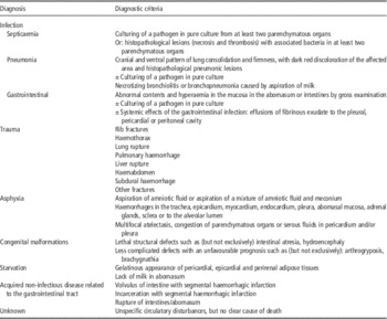
Based on the written protocols (one per lamb) data files were created for analysis with SAS 9.2 (SAS Institute Inc., Cary, NC, USA). Based on lesions found during gross and histopathological examination coupled with results from bacteriological and PCR analyses, lambs were assigned to one of seven main causes of death: infection, trauma, asphyxia, congenital malformation, starvation, acquired non-infectious disease related to the gastrointestinal tract and unknown. Diagnostic criteria are shown in Table 1. Cumulative mortality on the time scale for each primary cause of death was calculated based on times of birth and death. Frequency distributions were used to describe categorical data. BW was reported as mean±standard deviation.
Case definitions

Results
Characteristics of the flocks
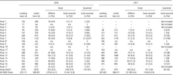
Table 2 shows the number of lambing ewes, number of lambs born, number of stillborn lambs, stillbirth rate, number of liveborn lambs that died before 5 days of age, early neonatal mortality rate, number of liveborn lambs submitted for postmortem examination and the proportion of early neonatal deaths examined in each study flock that were enrolled in the NSRS, for each study year. Table 2 also includes a summary for study flocks enrolled in the NSRS and, for comparison, a summary including all flocks enrolled in the NSRS.
The number of lambing ewes, number of lambs born, number of stillborn lambs, stillbirth rate, number of liveborn lambs that died between 0 and 5 days of age, early neonatal mortality rate, number of liveborn lambs submitted for postmortem examination and the proportion of early neonatal deaths examined in the study from each flock each study year

NSRS=Norwegian Sheep Recording System.
1 Not enrolled in the NSRS, number and rates not applicable, the number of lambing ewes=the number of breeding ewes.
2 Study flocks enrolled in the NSRS.
3 Most likely due to registration of liveborn lambs that died shortly after birth as stillborn in the registry.
The Norwegian White breed was predominant in 16 flocks; one flock of 27 ewes consisted solely of Spæl ewes. Study flocks were housed during pregnancy and lambing, either in insulated or non-insulated buildings. Pregnant ewes were kept in groups according to age, the number of expected lambs and/or body condition score before lambing. A standard vaccination programme against Clostridium spp. or Clostridium spp. and Pasteurella spp. was applied in all flocks. Yearling ewes were vaccinated twice, 8 to 12 weeks and 4 to 6 weeks before lambing. Older ewes were vaccinated 4 to 6 weeks before lambing. In the majority flocks ewes lambed for the first time as yearlings. Ewes and lambs were under close supervision during the lambing season, with at least one person present at all times. All flocks used claiming pens at lambing and most flocks practiced daily cleaning of claiming pens. In the majority of flocks, lambs were routinely checked for the ability to suckle after birth, and assisted if necessary. Number of breeding ewes and details of management factors for each flock are shown in Supplementary Table S1.
Characteristics of examined lambs
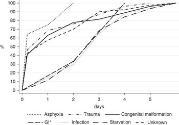
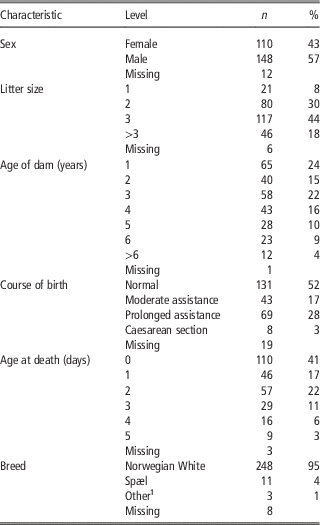
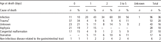
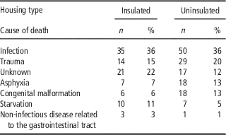
A total of 562 dead lambs (stillborn and liveborn) were examined. In 270 of these lambs, lungs were partially or fully inflated and thus characterized as liveborn and reported in this paper. The 270 examined lambs were from 250 different litters. Distribution of the examined lambs by various characteristics is shown in Table 3. In 81% of twin or higher order litters, one liveborn lamb died. In 12% of the litters there were also stillborn lambs, whereas in 7% of the litters more than one lamb died and all lambs that died were alive at birth. Overall, of the liveborn lambs, 27% died within 3 h, 41% within 24 h and 80% within day 2 after birth. In 70% of the lambs that died on the day of birth, the soft membrane covering the sole at birth was still present, indicating that they had not walked. The mean BWs (SD) at necropsy were 5.9 kg (2.2), 4.4 kg (1.1), 3.6 kg (1.0) and 3.5 kg (1.1), in lambs from singleton, twin, triplet and quadruplet or higher order litters, respectively. In the NSRS, surviving lambs of the Norwegian White breed from single, twin, triplet and quadruplet litters weigh on average 5.5 kg (1.2), 4.9 kg (1.0), 4.5 kg (0.9) and 4.0 kg (0.8), respectively (Hektoen L., personal communication). Table 4 shows the primary cause of death by age at death for all liveborn lambs examined. Figure 1 displays the cumulative mortality by age at death for each primary cause of death. The distribution of causes of death were reasonably similar for lambs born in insulated and uninsulated farm buildings (Table 5).
Cumulative mortality by age of the lambs for each primary cause of death. *GI=acquired non-infectious disease related to the gastrointestinal tract.

Distribution of the 270 liveborn lambs examined in 2010 and 2011 by various characteristics

Percentage distribution does not include missing values.
1 Includes blazed sheep and crossbreeds.
Distribution of the 270 liveborn lambs examined in 2010 and 2011 by age at death (days) and primary cause of death found at postmortem examination

1 Two lambs were euthanized due to low vitality.
Distribution of the 270 liveborn lambs examined in 2010 and 2011 by housing type and primary cause of death found at postmortem examinationFootnote 1

1 Flock 7 had both housing types and were not included as it was not known where each lamb was born.
Infection
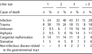
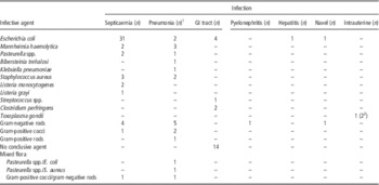
Infection was the primary cause of death in 36% (n=97) of the lambs; 48% (n=47) of these died from septicaemia, 25% (n=24) from pneumonia, 22% (n=21) from gastrointestinal infections and 5% (n=5) from other infections. The proportion of lambs dying from infection was largest in twin and triplet litters (Table 6: 40% and 37%, respectively v. 24% in single litters). Table 7 shows the number of postmortem examinations diagnosed with an infectious disease, ordered by the type of infection and infectious agent. Escherichia coli was the most frequently isolated agent (42%) and in total 14% of neonatal deaths were caused by this bacterium. Depletion of coronary groove fat was found in half of the lambs diagnosed with an infection (n=48); of these, coexisting traumatic lesions were found in 12% (n=6) of the lambs. Of the total number of lambs diagnosed with infection, 20% (n=19) had coexisting traumatic lesions, and multiple rib fractures were most frequently encountered (n=13). Of the lambs that died on the day of birth, only one was confirmed as being infected in utero with T. gondii. The majority of lambs diagnosed with septicaemia (80%) or gastrointestinal infections (71%) died within 3 days of birth, whereas the age at death for cases diagnosed with pneumonia was equally distributed from day 1 to day 4 after birth (54% before 3 days of age). The proportion of lambs that died of infection in flocks where daily cleaning of claiming pens was or was not practiced, did not differ substantially (34% v. 37%, respectively).
Distribution of the 270 liveborn lambs examined in 2010 and 2011 by litter size and primary cause of death found at postmortem examination

Number of cases diagnosed with infectious disease by type of infection and infectious agent (cultured or, in cases where culturing were not successful, detected in gram-stained tissue specimen)

GI tract=gastrointestinal tract.
The table includes three cases of T. gondii infection detected by real-time PCR.
1 Not included: two cases of aspiration pneumonia.
2 T. gondii was considered a subsidiary cause of death in one lamb which was euthanized due to brachygnatia and in one lamb that died of E. coli abomasitis/enterotoxaemia.
Trauma
Trauma was recognized as the primary cause of death in 20% (n=53) of the cases. Of these, 46% died within 3 h after birth and 70% (n=37) within 24 h. Of these, 65% (n=23) required prolonged assistance at delivery, while 25% (n=9) were assisted moderately. The proportion of lambs dying from trauma was highest in singleton lambs (Table 6: single litters 38% v. 15% to 24% in litters >1). In 35% (n=13) of lambs that died on the day of birth, moderate to severe localized subcutaneous oedema was found, most frequently localized in the head. Asphyxia – haemorrhages in trachea, aspiration of amniotic fluid and/or meconium – was found in nine cases. Major lesions in cases where trauma was recognized as the primary cause of death are shown in Supplementary Table S2. Two of the lambs included were euthanized due to low vitality (hypothermic, unable to stand, absent sucking reflex); thus, in these cases trauma was not the direct cause of death. In most cases of injuries involving rib fractures, an extensive number of ribs were broken (mean 10 ribs, range 1 to 20).
Asphyxia
Findings indicative of asphyxia were present in 10% (n=27) of the lambs and included aspiration of amniotic fluid or aspiration of a mixture of amniotic fluid and meconium, haemorrhages in the trachea, epicardium, myocardium, endocardium, pleura, abomasal mucosa, adrenal glands, sclera or to the alveolar lumen. A total of 42% (n=11) of these lambs required prolonged assistance at delivery, while 19% (n=5) were assisted moderately. Concurrent lesions frequently found in asphyxiated lambs were multifocal atelectasis, congestion of parenchymatous organs or serous fluids in pericardium and/or pleura. Minor meningeal haemorrhages were found in three lambs. Concurrent non-lethal traumatic lesions, intramuscular or subcutaneous haemorrhage, and small amounts of blood in the abdomen were found in six lambs. In one lamb, aspiration of large amounts of meconium to the alveolar lumen had resulted in multifocal pneumonia.
Congenital malformations
Congenital malformations were found in 10% (n=27) of the lambs; major lesions are shown in Supplementary Table S3. In 55% (n=15) of the cases, malformations were gross defects visible to the farmer. These included lambs born with umbilical hernias, various skeletal malformations and three lambs with multiple external and additional internal defects. Cases with multiple malformations included one lamb with micropthalmia, palatoschisis and atresia ani, a second lamb with brachygnathia inferior, pulmonary hypoplasia, cardiac atrial and ventricular septal defects, and atresia ani, and a third lamb with brachygnathia inferior and superior, palatoschisis, cheilioshisis, micropthalmia, hydrocephalus internus and ventricular septal defect. Most of the less complicated defects would not immediately lead to death per se; however, due to an unfavourable prognosis they were euthanized shortly after birth. Congenital malformations not detectable or not so obvious to the farmer included six cases of atresia ani or atresia jejuni, one case of malposition of the duodenum causing obstruction of the intestine, one case of laryngo-oesophageal fistula and two cases of diaphragmatic hernia. Most of these defects would result in lamb death. The lambs with congenital malformations originated from 12 flocks.
Starvation
Depletion of perirenal and coronary groove fat was the sole pathological finding in 6% (n=17) of the lambs. Lack of milk in the abomasum and/or the intestinal tract in all but two lambs further suggested that they had not fed adequately. Amongst these lambs, only one was from a singleton litter. Of 17, 10 cases were from a single flock; excluding this flock reduced the prevalence of lambs that died of starvation to 3%.
Acquired non-infectious disease related to the gastrointestinal tract
Acquired non-infectious diseases related to the gastrointestinal tract were found in 2% (n=6) of the cases. Volvulus of the small intestine with segmental haemorrhagic infarction was present in two lambs and haemorrhagic incarceration of the small intestine through a rupture in the mesentery was present in one lamb. In the three remaining lambs, the abomasum was ruptured along the major curvature. Case history in lambs that suffered from ruptured abomasum did not indicate specific events such as tube feeding or trauma before death.
Unknown cause of death
In 16% (n=43) of the lambs, no cause of death was obtained. A total of 76% (n=33) of these lambs were from triplet or higher order litters and 56% died within 24 h of birth. Lesions found indicated unspecific circulatory disturbances and included congestion of internal organs, pulmonary oedema and serous fluid in pericardium and pleural cavity. Fibrinous effusion to pericardium, fibrinous perihepatitis and multifocal acute alveolar injury, respectively, were found in three cases. These subtle lesions may indicate an acute systemic infection. However, tissues from all three cases were negative on bacteriological examination and did not exhibit other gross or histopathological lesions to support infectious disease.
Discussion
In line with other studies (Huffman et al., Reference Huffman, Kirk and Pappaioanou1985; Binns et al., Reference Binns, Cox, Rizvi and Green2002), most liveborn lambs died during days 0 to 2, indicating that events related to lambing and the immediate post-lambing period are essential for lamb survival. The most frequent cause of early neonatal death in our study was infection, which accounted for 36% of all deaths. Infection caused fewer deaths in the 1st week of life in two UK studies (10.5%, Wiener et al., Reference van den Brom, Luttikholt, Lievaart-Peterson, Peperkamp, Mars, van der Poel and Vellema1983; 12.1%, Green and Morgan, Reference Green and Morgan1993). However, a US study of 60 neonatal deaths found that infectious diseases were responsible for 28% of deaths occurring before 3 weeks of age (Huffman et al., Reference Huffman, Kirk and Pappaioanou1985). In the American study, E. coli septicaemia and enterotoxaemia were most prevalent causes of infection (11 out of 17 cases), while Pasteurella spp., Salmonella typhimurium and Salmonella dublin were responsible for four deaths. In the present study, E. coli accounted for 42% of deaths caused by infection and 14% of all early neonatal deaths, thus must be considered as an important infectious agent in this selection of cases. In comparison, the two second most frequently isolated agents (Mannheimia haemolytica and Staphylococcus aureus) each accounted for 5% of deaths caused by infection and 2% of all early neonatal deaths. Providing new bedding for lambing pens daily has been associated with lower flock level mortality rates the first 24 h (Binns et al., Reference Binns, Cox, Rizvi and Green2002), probably mediated through prevention of build-up of high pathogen burdens in the environment. However, in our study there were minimal differences in the proportion of lambs dying from infection in flocks where daily cleaning of claiming pens was or was not practiced. Depletion of coronary groove fat in half of the cases indicated that lambs had sucked poorly before death, which might have been either a predisposing factor or a consequence of the infection acquired. Poor sucking may also result from low vitality due to trauma, which was found in 10% of lambs with depletion of coronary groove fat. Moreover, the proportion of lambs dying from infection was largest in twin and triplet litters. Mastitis, which is associated with reduced milk production and quality, or poor milk production per se, might result in inadequate milk supply to the lambs and increase the risk of lamb mortality, particularly in lambs from litters of multiples (Nash et al., Reference Nash, Hungerford and Nash1996; Arsenault et al., Reference Arsenault, Dubreuil, Higgins and Bélanger2008).
The sampling performed in the present study was completed before the earliest reports of Schmallenberg virus in central Europe (Hoffmann et al., Reference Hoffmann, Scheuch, Höper, Jungblut, Holsteg, Schirrmeier, Eschbaumer, Goller, Wernike, Fischer, Breithaupt, Mettenleiter and Beer2012; van den Brom et al., Reference Speijers, Carson, Dawson, Irwin and Gordon2012) and thus this virus is unlikely to have caused congenital malformations detected in this study. All defects observed in this study have been described previously (Dennis, Reference Dennis1975). The proportion of affected lambs (10%) was similar to the findings in previous studies of causes of neonatal lamb mortality (Wiener et al., Reference van den Brom, Luttikholt, Lievaart-Peterson, Peperkamp, Mars, van der Poel and Vellema1983; Green and Morgan, Reference Green and Morgan1993). Other infectious agents that are recognized as sources of reproductive losses in sheep in Europe, but not specifically screened for in this study are Chlamydophila abortus, Coxiella burnetii and pestivirus causing border disease (Nettleton et al., Reference Nettleton, Gilray, Russo and Dlissi1998; Aitken and Longbottom, Reference Aitken and Longbottom2007; Agerholm, Reference Agerholm2013). Enzootic ovine abortion caused by C. abortus has never been diagnosed in Norway (Kampen A., Norwegian Veterinary Institute, personal communication) and there is no indication that C. burnetii infections are present in farmed ruminants in Norway (Kampen et al., Reference Kampen, Hopp, Grøneng, Melkild, Urdahl, Karlsson and Tharaldsen2012). Bovine viral diarrhoea virus was eradicated from Norway in 2004 (Løken and Nyberg, Reference Løken and Nyberg2013) and there have been no verified clinical cases of border disease in sheep following the eradication of bovine viral diarrhoea virus.
Traumatic injuries accounted for one-fifth of the total deaths in the current study, which is a moderately higher proportion than previously reported by Maund et al. (Reference Maund, Duffell and Winkler1980). Injuries may be immediately fatal. Alternatively, pain and physical dysfunction may reduce mobility and vitality and thereby interrupting the mother–young bonding (Mellor and Stafford, Reference Mellor and Stafford2004). Traumatic injuries to the lamb may occur during birth, particularly in cases of fetopelvic disproportion. Lambs confined indoors may be trapped in pen hurdles, and small or inappropriately shaped lambing pens may increase the risk of injuries inflicted by ewes. The prevalence of injuries in neonatal deaths reportedly ranges from 11% to 14% in flocks housed during lambing (Yapi et al., Reference Wiener, Woolliams and Macleod1990; Green and Morgan, Reference Green and Morgan1993). Differentiating between the two types of injuries at postmortem examination is difficult, if not impossible, so we chose to report all traumas unified, although age at death and case history could have given an indication of the causal relation. Nearly 70% of lambs diagnosed with traumatic injuries as the primary cause of death died on the day of birth. Furthermore, 90% of these required assistance at birth either by moderate or prolonged human intervention. The proportion of lambs dying from trauma was highest in singleton lambs. When dystocia occurred, the risk of lamb loss increases more in ewes with single lambs than in ewes with larger litters (Holmøy et al., Reference Holmøy and Waage2014). Dystocia is reported as a more frequent cause of death in single lambs than in lambs in larger litters (Dalton et al., Reference Dalton, Knight and Johnson1980; Hall et al., Reference Hall, Fogarty and Gilmour1995). In cases of dystocia associated with litters of multiples, often due to malpresentation of lambs (Speijers et al., Reference Rad, Movassaghi, Sharifi, Naseri and Seifi2010), successful obstetric delivery may be achieved with less trauma because of the smaller foetuses.
The overall proportion of lambs dying from starvation in the present study (6%) was substantially lower than that reported from the United Kingdom, both in an outdoor lambing flock and in housed flocks (15.4%, Wiener et al., Reference van den Brom, Luttikholt, Lievaart-Peterson, Peperkamp, Mars, van der Poel and Vellema1983; 48.6%, Green and Morgan, Reference Green and Morgan1993), respectively. Less monitoring and exposure to varying weather conditions usually contributes to hypothermia–starvation as a more important cause of lamb death in flocks lambing outdoors (e.g. Wiener et al., Reference van den Brom, Luttikholt, Lievaart-Peterson, Peperkamp, Mars, van der Poel and Vellema1983) than in flocks housed at lambing. An indoor system might also make it easier to foster surplus lambs. In the housed lambs studied by Green and Morgan (Reference Green and Morgan1993), nearly half of the neonatal deaths were attributable to starvation. Sparse information was given about management routines during lambing in the flocks investigated; thus, monitoring of newborn lambs might have differed substantially from those practiced in flocks in the present study. Altered or absent maternal behaviour may be an underlying factor for starvation as a cause of death. Moreover, maternal behaviour have been reported to vary between breeds (Nowak and Poindron, Reference Nowak and Poindron2006; Dwyer, Reference Dwyer2008a). Studies comparing grooming behaviour and acceptance of sucking attempts have not been carried out in Norwegian breeds.
The overall prevalence of meningeal haemorrhages in the current study (4.5%) was substantially lower than previously reported. In two studies that performed both gross and histological evaluation of brains of liveborn lambs that died during the first 3 days and the 1st week of life, respectively, the overall prevalence of meningeal haemorrhages was 42.5% and 47.1% (Haughey, Reference Haughey1973; Holst et al., Reference Holst, Fogarty and Stanley2002). Some of the discrepancy between this and previous studies might be due to different sensitivity in scoring systems used. Ischaemic injuries to the nervous system have been recognized as a contributor in lambs that die with signs of dystocia, that is swollen tongue, injected sclera, subcutaneous oedema or haemorrhage (Lashley et al., Reference Lashley, Roe, Kenyon and Thompson2013). In a study by Dutra et al. (Reference Dutra, Quintans and Banchero2007), 14% of lambs displayed histological evidence of hypoxic-ischaemic neuronal injury; however, concurrent gross lesions were not reported. Histological examination of the brain in the present study may have elucidated the cause of death in some of the cases with unknown cause of death, but such evaluation was not performed.
Lambs that died within the predefined study periods, corresponding to peak lambing in each flock, were examined. However, all lambs that died during the entire lambing season were not examined. If some causes of death were more or less likely to occur at the start or end of the season than at peak lambing, this could have biased the distribution of the causes that we recorded within each flock. For example, lambs born at peak lambing might be exposed to higher pathogen burdens than lambs born at the start of lambing season due to the rapid increase in the number of animals housed. Consequently, infectious diseases could be relatively more prevalent at peak lambing than at the start of lambing. Three flocks contributed to around 40% of the examined lambs. However, apart from the proportion of lambs that died of starvation, the exclusion of these flocks did not substantially change the distribution of causes of death.
This study was performed in a small sample of Norwegian flocks; thus, the sample cannot be considered representative of Norwegian sheep flocks in general. Selection of participating flocks was a convenient sample, based on proximity to the NSVS and expected compliance from the farmer. However, flocks included were all considered to have typical features characteristic of Norwegian sheep flocks; they were housed during pregnancy and lambing and closely monitored during the lambing season.
Conclusion
In this study, the main causes of early neonatal lamb mortality were infection and traumatic lesions. Most deaths occurred shortly after birth, suggesting that events related to lambing and the immediate post-lambing period are critical for lamb survival.
Acknowledgements
The authors wish to thank participating farmers who welcomed them on their farms and veterinary colleagues willing to participate in the field teams. Torfinn Moldal, the Norwegian Veterinary Institute and Professor emeritus Thor Landsverk, Section for anatomy and pathology thanked for their significant contribution in performing postmortem examinations. Tore Engen, Aga Zakoscielna, Siri B. Hamre and the staff at the histopathology lab in Oslo are acknowledged for technical assistance. Professor Henning Sørum, Aud Kari Fauske and Gaute Skogtun at Section for microbiology, immunology and parasitology are acknowledged for providing bacteriological diagnostics. Wenche Okstad at Section for small ruminant research is thanked for her contribution to Toxoplasma diagnostics. The Agricultural Agreement Research Fund, the Foundation for Research Levy on Agricultural Products, Animalia Norwegian Meat and Poultry Research Centre and the Norwegian Sheep and Goat Association are acknowledged for funding this study.
Supplementary Material
To view supplementary material for this article, please visit http://dx.doi.org/doi:10.1017/S175173111600152X