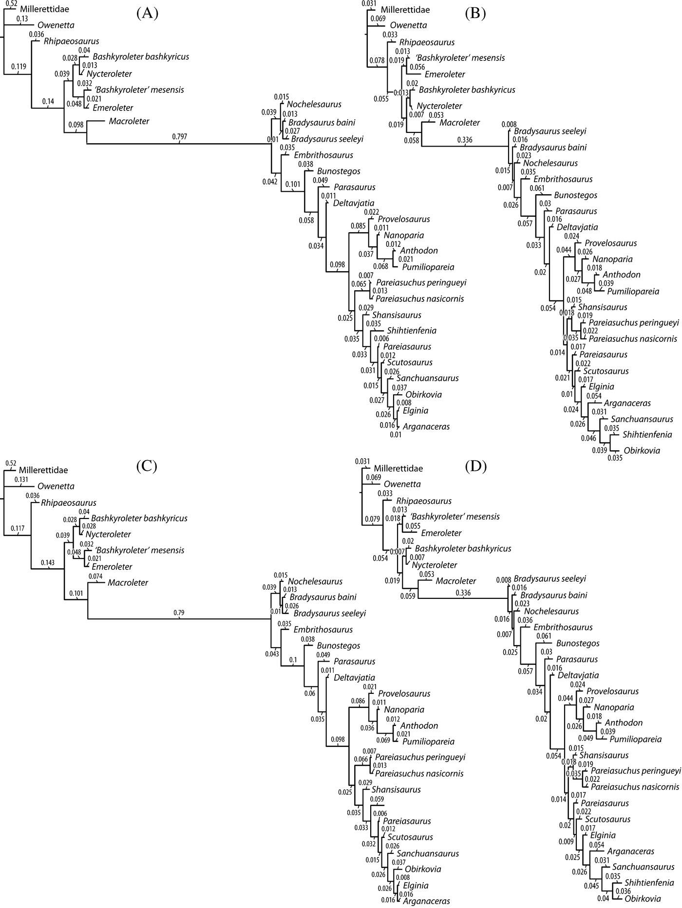
Pareiasaurs were conspicuous members of Middle and Late Permian terrestrial ecosystems, but despite their seeming ubiquity, their anatomy and relationships are surprisingly poorly known. Lee (Reference Lee1997a) undertook a complete alpha taxonomic revision of all pareiasaurian taxa, managing to reduce the 49 named species to a more rational 17. Around this time, Lee completed the first phylogenetic analysis of the group (Lee Reference Lee1997b), also asserting that a monophyletic Pareiasauria included turtles. Since this analysis, several new pareiasaurs have been named (Arganaceras vacanti Jalil & Janvier, Reference Jalil and Janvier2005; Bunostegos akokanensis Sidor et al., Reference Sidor, Blackburn and Gado2003; Obirkovia gladiator Bulanov & Yashina Reference Bulanov and Yashina2005). There are currently 22 accepted pareiasaur taxa, including the “Welgevonden pareiasaur” from South Africa and, in addition, there are two unnamed taxa from the Argana Basin of Morocco (Jalil & Janvier Reference Jalil and Janvier2005).
Although they are morphologically distinct, few pareiasaurs are known in detail, severely hampering the understanding of the anatomy and evolutionary trends in the clade (Lee Reference Lee1995a, Reference Lee1997a, Reference Leeb; Lee et al. Reference Lee, Gow and Kitching1997). Unfavourable preservation plays a significant role in this lack of information, as does the highly ossified nature of the pareiasaurian skull. More detailed descriptions do exist for Elginia mirabilis (Maxwell Reference Maxwell1991), Pareiasuchus nasicornis (Lee et al. Reference Lee, Gow and Kitching1997), Scutosaurus karpinskii (Lee Reference Lee1995a; Bystrov Reference Bystrov1957), Arganaceras vacanti (Jalil & Janvier Reference Jalil and Janvier2005) and Parasaurus geinitzi (Tsuji & Müller Reference Tsuji and Müller2008). These descriptions, however, involve only partial skeletons and primarily cranial material, and no complete description has been published for a single taxon in the last 50 years. This situation results primarily from a paucity of well-prepared specimens belonging to a single pareiasaur taxon that include both cranial and postcranial material.
The South African Karoo Basin contains the most continuous set of Middle and Late Permian strata and also holds the greatest taxonomic diversity and absolute abundance of pareiasaur fossils. Correspondingly, most of the existing literature concerns these taxa and this basin. Pareiasaurs, however, were widely distributed in the Middle and Late Permian, and are also found in Brazil, China, Germany, Morocco, Niger, Russia, The United Kingdom (Scotland), Tanzania and Zambia. After the South African fauna, Russia is the second most productive region for pareiasaur fossils. Three Russian genera are currently recognised, two of which are represented by a large number of specimens. Scutosaurus karpinskii Amalitzky, Reference Amalitzky1922 is the most numerous of the Russian pareiasaurs and is found in the area of North Dvina, considered part of the Vyatskian Gorizont (Upper Tatarian) sensu Modesto & Rybczynski (Reference Modesto, Rybczynski, Benton, Shishkin, Unwin and Kurochkin2000). Obirkovia gladiator, one of the most recently discovered pareiasaurs, is found in the uppermost S. karpinskii tetrapod assemblage zone, which correlates roughly with the upper part of the Cistecephalus or lower part of the Dicynodon Assemblage Zone of the Karoo Basin (Bulanov & Yashina Reference Bulanov and Yashina2005). Obirkovia is only known from a single quadratojugal, an element whose morphology suggests a close relationship to the Scottish pareiasaur Elginia.
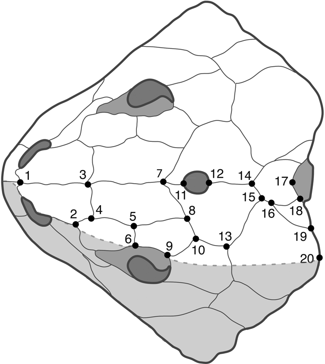
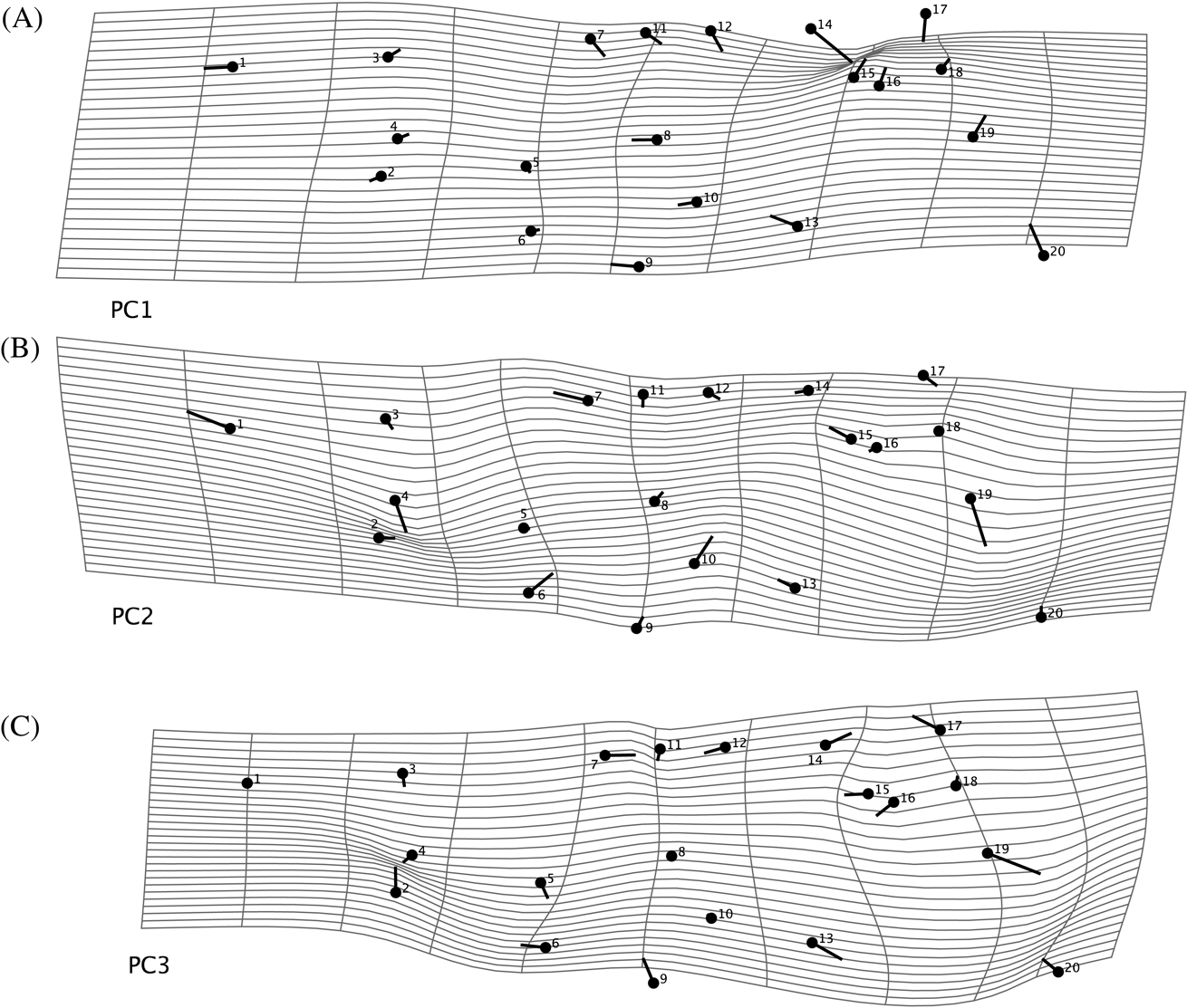
Specimens of the third Russian pareiasaur taxon, Deltavjatia rossicus (Hartmann-Weinberg Reference Hartmann-Weinberg1937), were first found along the Vjatka River near the town of Kotel'nich in the Kirov Province of Russia between 1933 and 1935. The specimens of Deltavjatia found in the Kotel'nich locality are often exceptionally well preserved (see Benton et al. Reference Benton, Newell, Khlyupin, Shumov, Price and Kurkin2012). In addition, a large number of specimens of varying sizes – many of which include postcranial material – have been excavated and prepared in the last decade. The wide variation in size suggests that a number of ontogenetic stages are present among the samples, providing an ideal opportunity to evaluate growth patterns within a single pareiasaur species. Due to the often exceptional preservation of the material from the Kotel'nich locality, it is relatively easy to discern sutures in the skull of Deltavjatia. This condition is atypical in pareiasaurs, which usually have a highly ossified skull roof, and allows for the identification and placement (in multiple specimens) of appropriate landmarks for the completion of a geometric morphometric analysis of skull allometry in Deltavjatia.
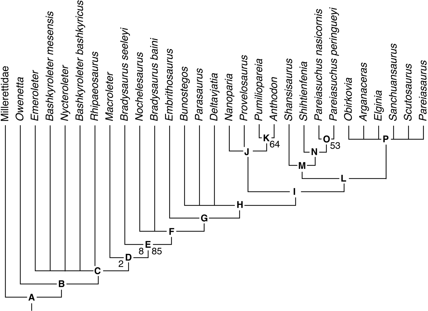
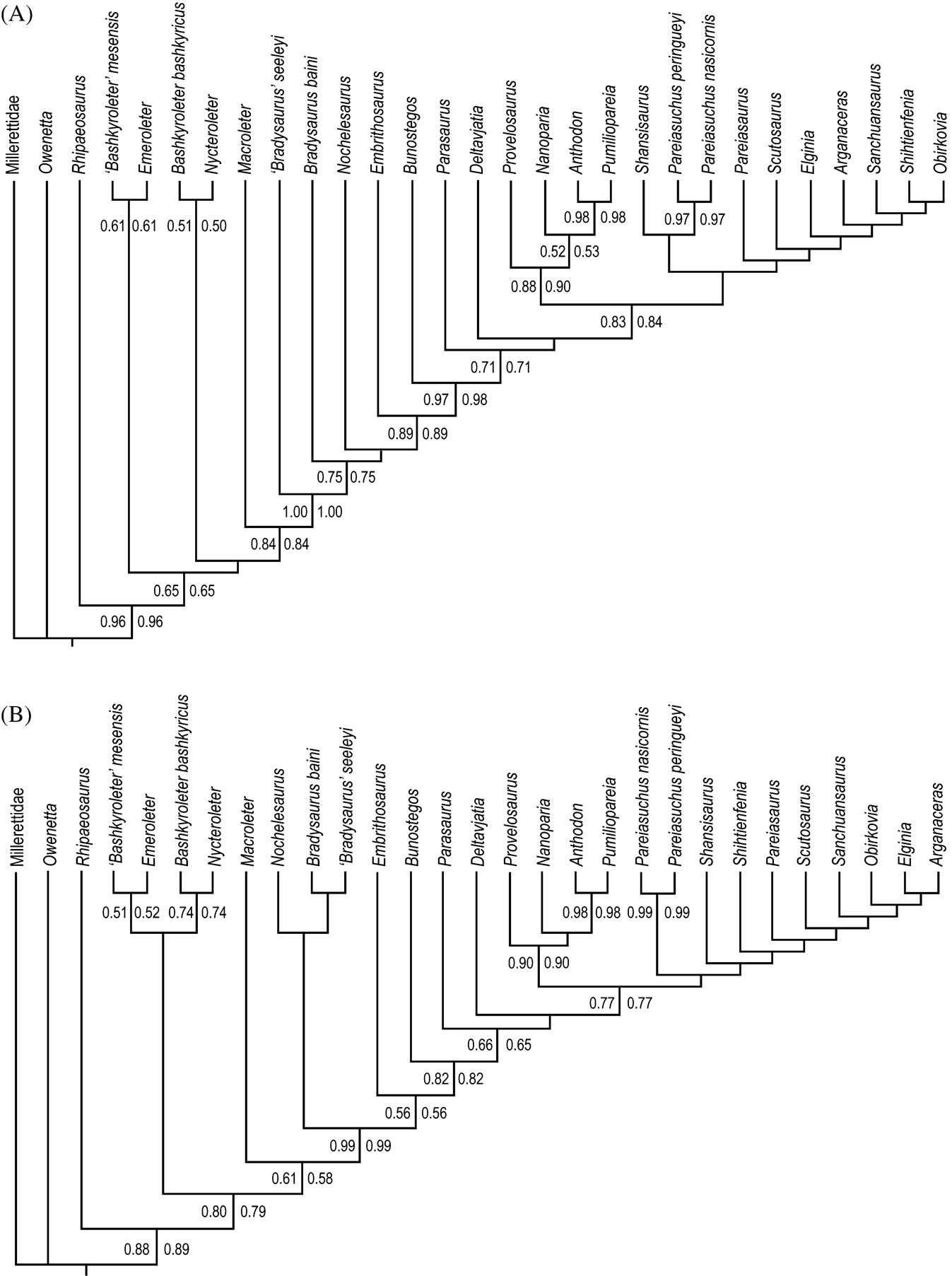
This paper describes in detail the cranial and postcranial anatomy of the pareiasaur Deltavjatia rossicus and undertakes the first geometric morphometric analysis of skull ontogeny in a parareptile. It also presents a revised phylogeny of the pareiasauromorphs, incorporating new taxa and new morphological information.
Institutional abbreviations. KPM, Kotel'nich Paleontological Museum, Kotel'nich, Russia; PIN, Paleontological Institute, Moscow, Russia; PMU, Paleontological collections of the Museum of Evolution, Uppsala, Sweden; ROM, Royal Ontario Museum, Toronto, Canada; SMNS, Staatliches Museum für Naturkunde Stuttgart, Germany; TMP, Royal Tyrrell Museum of Paleontology, Drumheller, Canada; UMZC, University Museum of Zoology Cambridge, Cambridge, UK.
1. Systematic Palaeontology
Reptilia Laurenti, Reference Laurenti1768
Parareptilia Olson, Reference Olson1947
Pareiasauria Seeley, Reference Seeley1888
Deltavjatia Ivakhnenko, Reference Ivakhnenko1987
Deltavjatia rossicus (Hartmann-Weinberg Reference Hartmann-Weinberg1937)
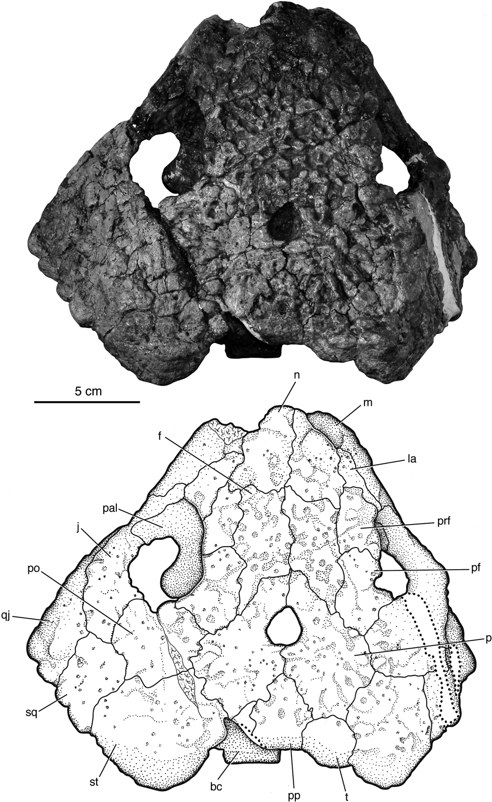
Deltavjatia rossicus PIN 2212/2 (Holotype), photograph and drawing of skull in dorsal view. Abbreviations: bc=braincase; f=frontal; j=jugal; la=lacrimal; m=maxilla; n=nasal; p=parietal; pal=palatine; pf=postfrontal; po=postorbital; pp=postparietal; prf=prefrontal; qj=quadratojugal; sq=squamosal; st=supratemporal; t=tabular.

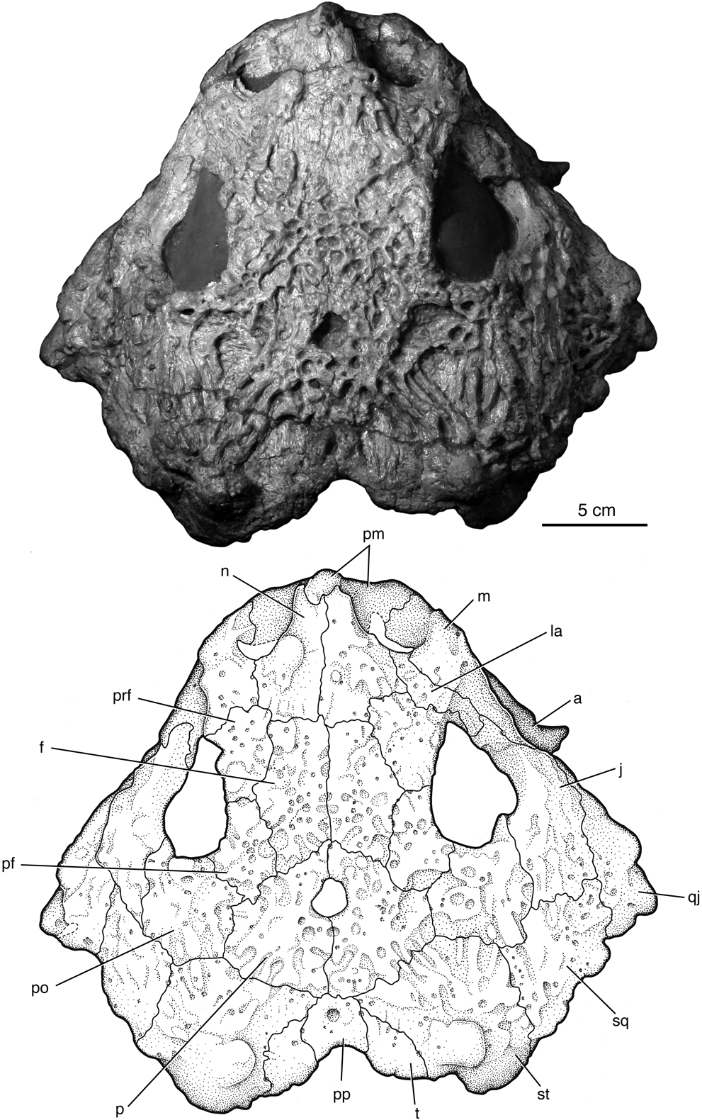
Deltavjatia rossicus KPM 232, photograph and drawing of skull in dorsal view. Abbreviations: a=angular; f=frontal; j=jugal; la=lacrimal; m=maxilla; n=nasal; p=parietal; pf=postfrontal; pm=premaxilla; po=postorbital; pp=postparietal; prf=prefrontal; qj=quadratojugal; sq=squamosal; st=supratemporal; t=tabular.

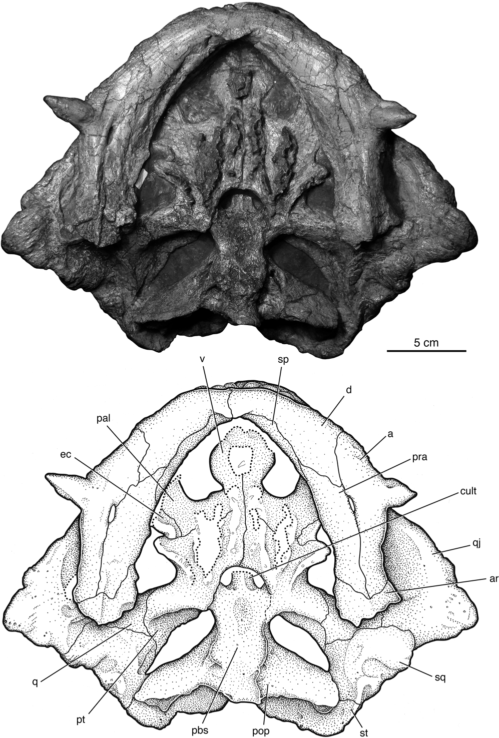
Deltavjatia rossicus KPM 232 photograph and drawing of skull in ventral view. Abbreviations: a=angular; ar=articular; cult=cultriform process; d=dentary; ec=ectopterygoid; pal=palatine; pbs=parabasisphenoid; pop=paroccipital process; pra=prearticular; pt=pterygoid; q=quadrate; qj=quadratojugal; sp=splenial; sq=squamosal; st=supratemporal; v=vomer.

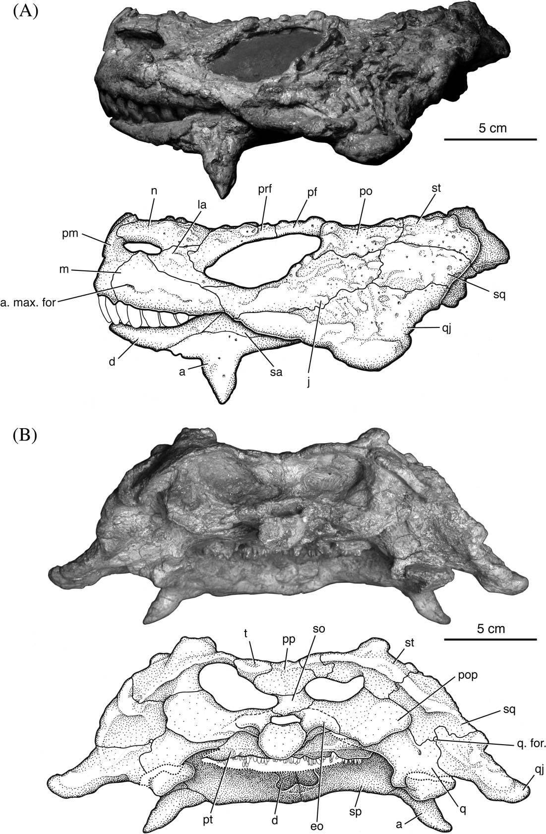
Deltavjatia rossicus KPM 232, photograph and drawing of the skull in left lateral (A) and posterior (B) views. Abbreviations: a=angular; a. max. for.=anterior maxillary foramen; d=dentary; eo=exoccipital; j=jugal; la=lacrimal; m=maxilla; n=nasal; pf=postfrontal; pm=premaxilla; po=postorbital; pop=parocciptal process; pp=postparietal; prf=prefrontal; pt=pterygoid; q=quadrate; q. for.=quadrate foramen; qj=quadratojugal; sa=surangular; so=supraoccipital; sp=splenial; sq=squamosal; st= supratemporal; t=tabular.

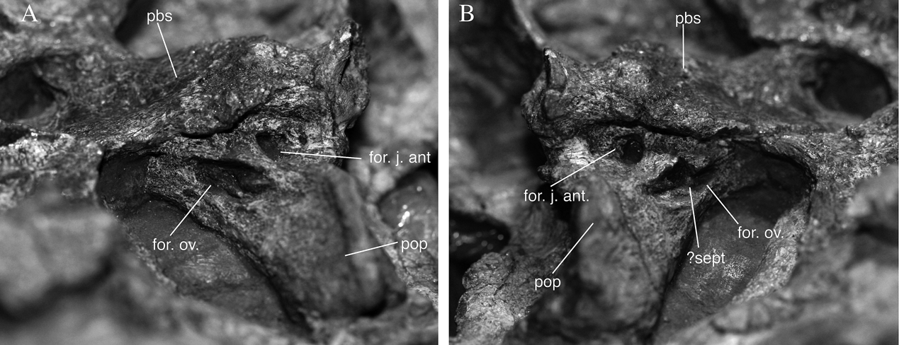
Deltavjatia rossicus KPM 232, photographs of braincase in left (A) and right (B) lateral views. Abbreviations: for. j. ant.=foramen jugulare anterius; for. ov.=foramen ovalis; pbs=parabasisphenoid; pop=paroccipital process; sept=bony septum.

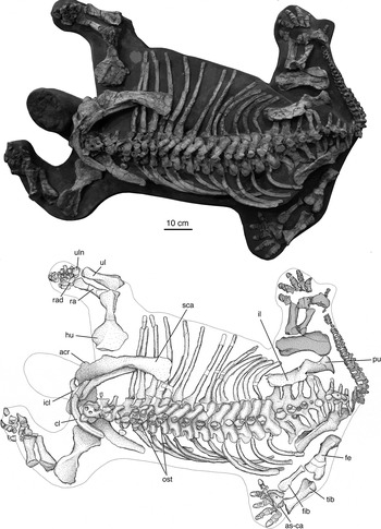
Deltavjatia rossicus KPM 232, photograph and drawing of postcranial skeleton in dorsal view. Greyed elements belong to another skeleton. Abbreviations: acr=‘acromion-like’ process; as-ca=astragalocalcaneum complex; cl=clavicle; fe=femur; fib=fibula; hu=humerus; icl=interclavicle; il=ilium; ost=osteoderms; pu=pubis; ra=radius; rad=radiale; sca=scapula; tib=tibia; ul=ulna; uln=ulnare.

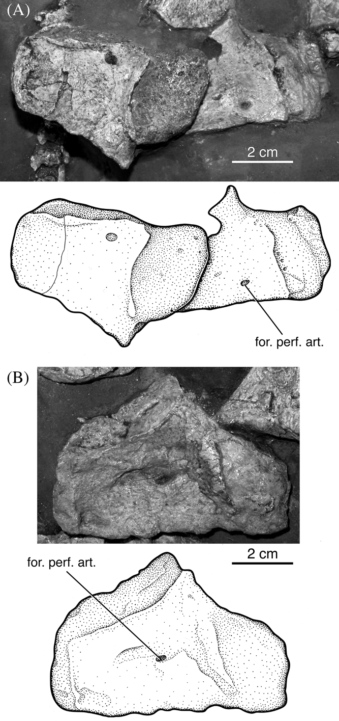
Deltavjatia rossicus KPM 232, photograph and drawing of astragalocalcaneum complex in dorsal (A) and ventral (B) views. Abbreviation; for. perf. art.=foramen for the perforating artery.

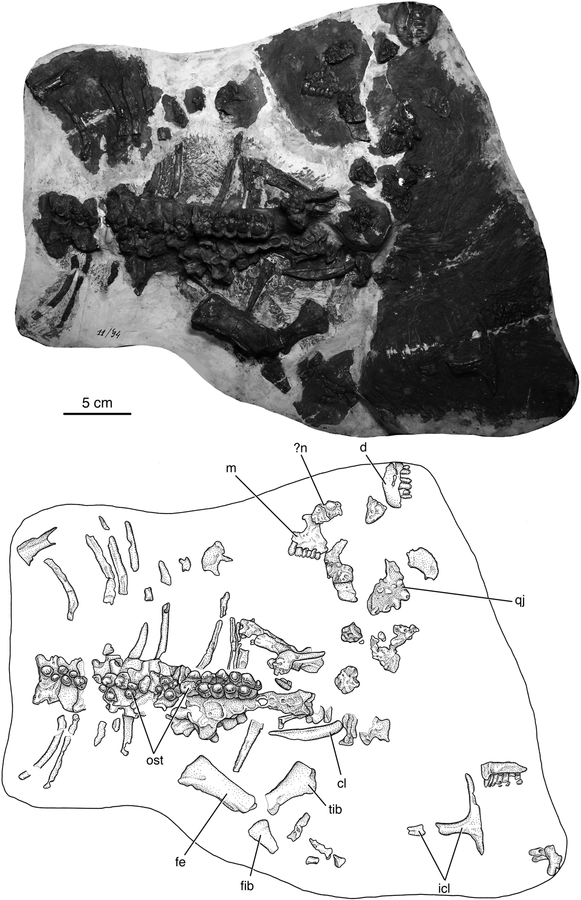
Deltavjatia rossicus KPM 11/94, photograph and drawing of specimen. Abbreviations: cl=clavicle; d=dentary; fe=femur; fib=fibula; icl=interclavicle; m=maxilla; n=nasal; ost=osteoderms; qj=quadratojugal; tib=tibia.

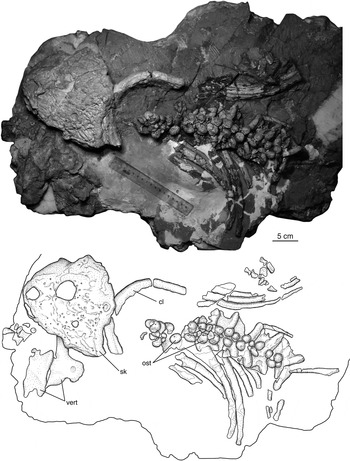
Deltavjatia rossicus KPM uncat P1, photograph and drawing of specimen. Abbreviations: cl=clavicle; ost=osteoderm; sk=skull; vert=vertebra.

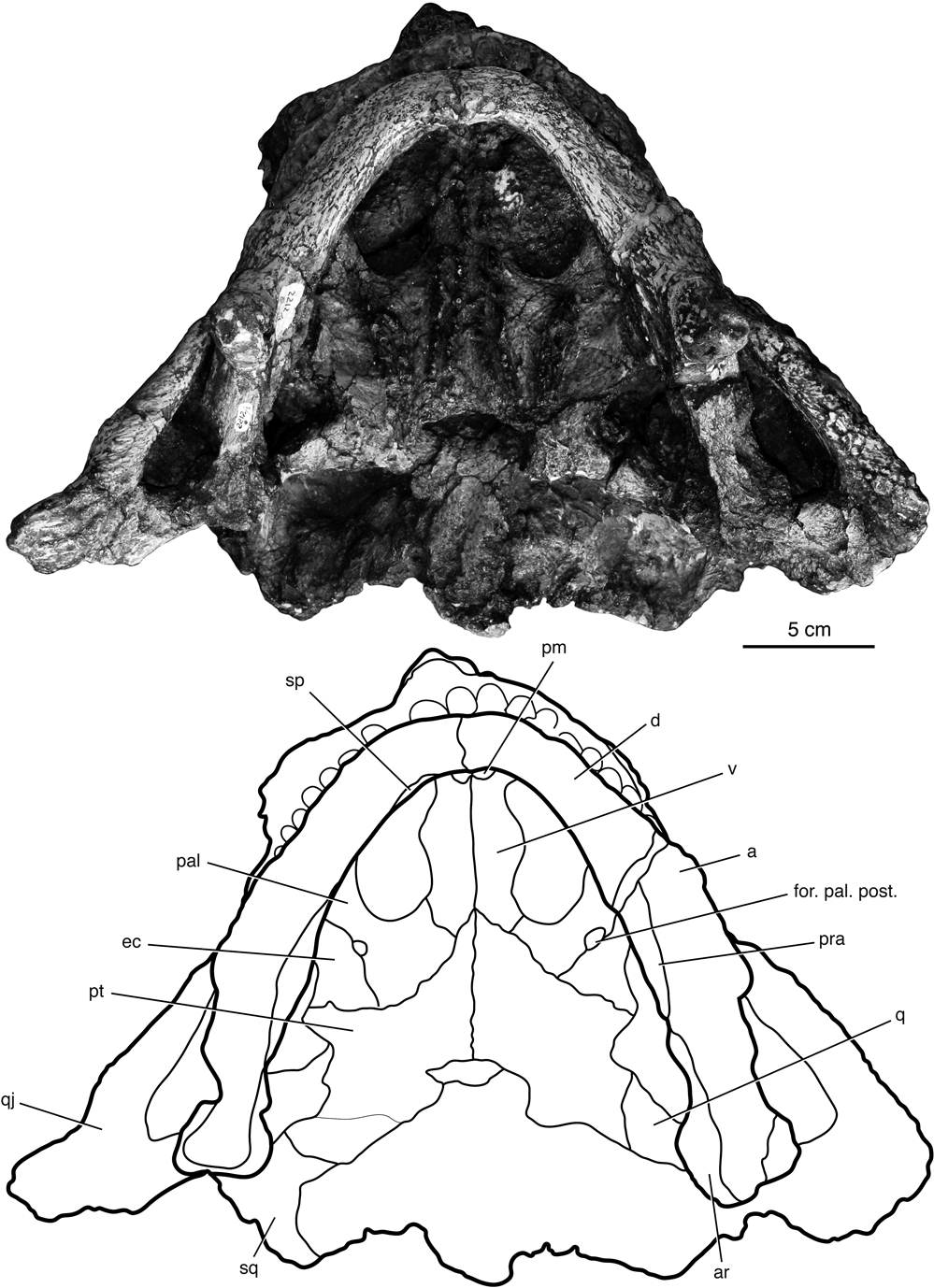
Deltavjatia rossicus PIN 2212/1 (Formerly the holotype of Pareiasuchus vjatkensis and Deltavjatia vjatkensis), photograph and outline of the skull in ventral view. Abbreviations: a=angular; ar=articular; d=dentary; ec=ectopterygoid; for. pal. post.=foramen palatinum posterius; pal=palatine; pm=premaxilla; pra=prearticular; pt=pterygoid; q=quadrate; qj=quadratojugal; sp=splenial; sq=squamosal; v=vomer.

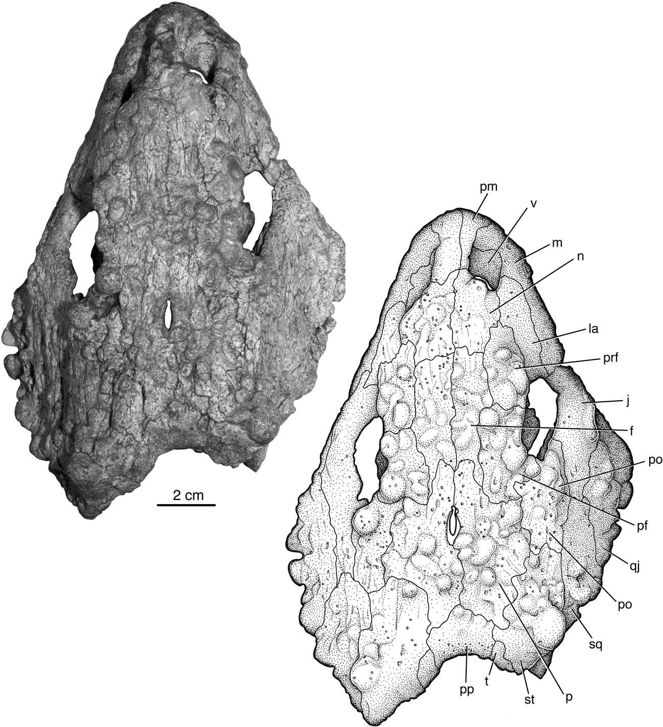
Deltavjatia rossicus PIN 2212/6, photograph and drawing of skull in dorsal view. Abbreviations: f=frontal; j=jugal; la=lacrimal; m=maxilla; n=nasal; p=parietal; pf=postfrontal; pm=premaxilla; po=postorbital; pp=postparietal; prf=prefrontal; qj=quadratojugal; sq=squamosal; st=supratemporal; t=tabular; v=vomer.

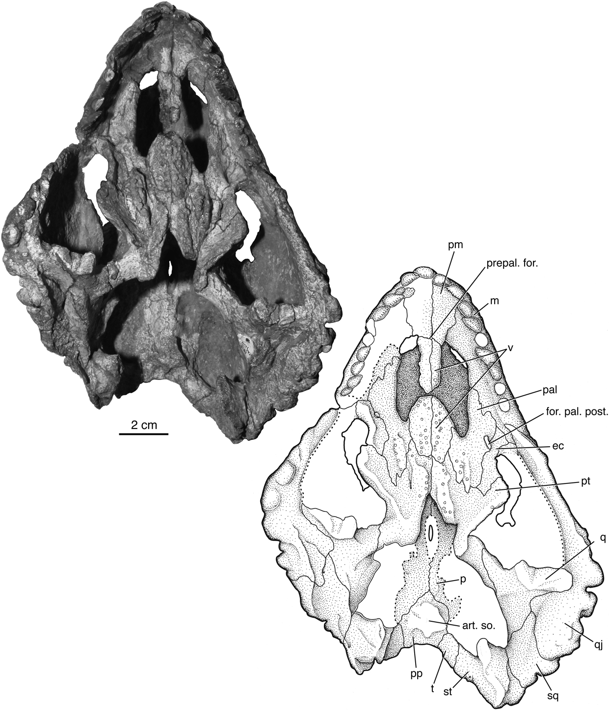
Deltavjatia rossicus PIN 2212/6, photograph and drawing of skull in ventral view. Abbreviations: art. so.=articulation for the supraoccipital; ec=ectopterygoid; for. pal. post.=foramen palatinum posterius; m=maxilla; p=parietal; pal=palatine; pm=premaxilla; pp=postparietal; prepal. for.=prepalatal foramen; pt=pterygoid; q=quadrate; qj=quadratojugal; sq=squamosal; st=supratemporal; t=tabular; v=vomer.

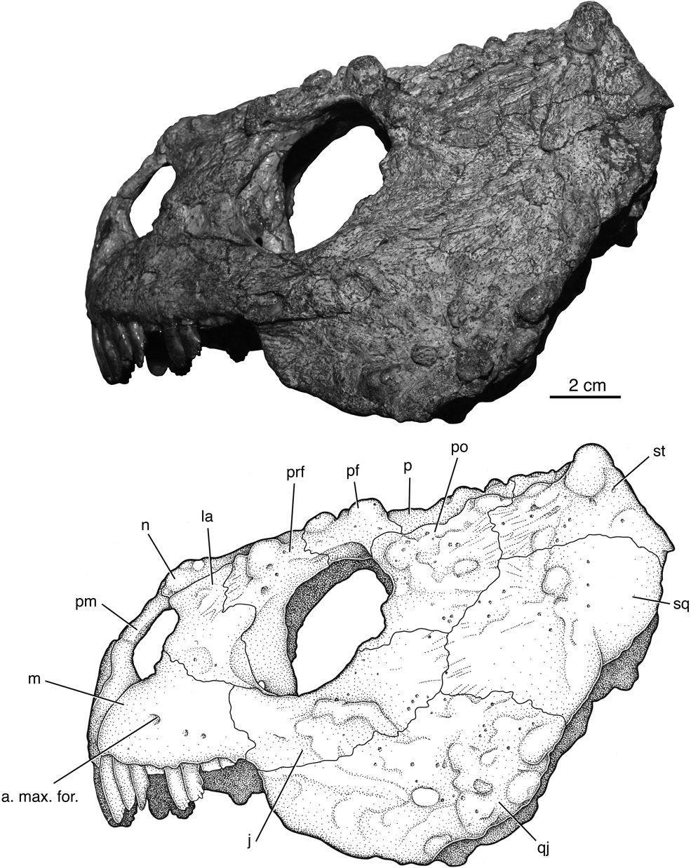
Deltavjatia rossicus PIN 2212/6, photograph and drawing of skull in left lateral view. Abbreviations: a. max. for.=anterior maxillary foramen; j=jugal; la=lacrimal; m=maxilla; n=nasal; p=parietal; pf=postfrontal; pm=premaxilla; po=postorbital; prf=prefrontal; qj=quadratojugal; sq=squamosal; st=supratemporal.

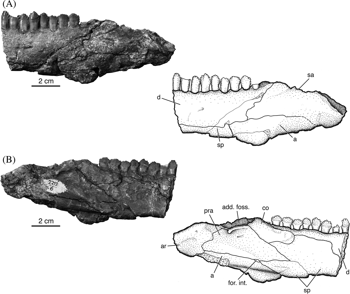
Deltavjatia rossicus PIN 2212/6, photograph and drawing of mandible in external (A) and internal (B) views. Abbreviations: a=angular; add. foss.=adductor fossa; ar=articular; co=coronoid; d=dentary; for. int.=intermandibular foramen; pra=prearticular; sa=surangular; sp=splenial.

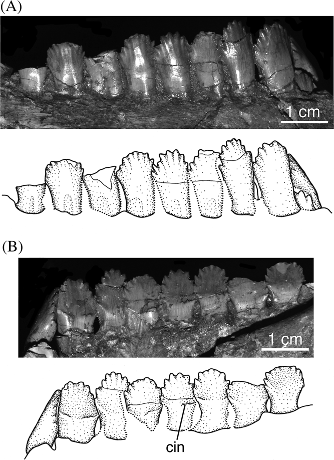
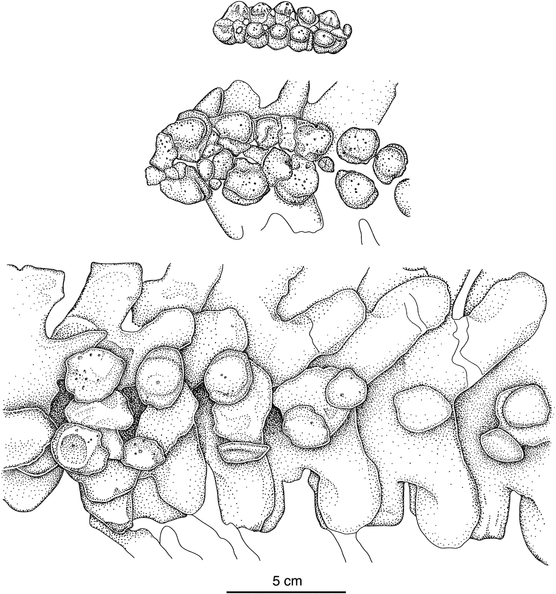
Deltavjatia rossicus PIN 2212/6, photograph and drawing of teeth in labial (A) and lingual (B) views. Abbreviation: cin=cingular.

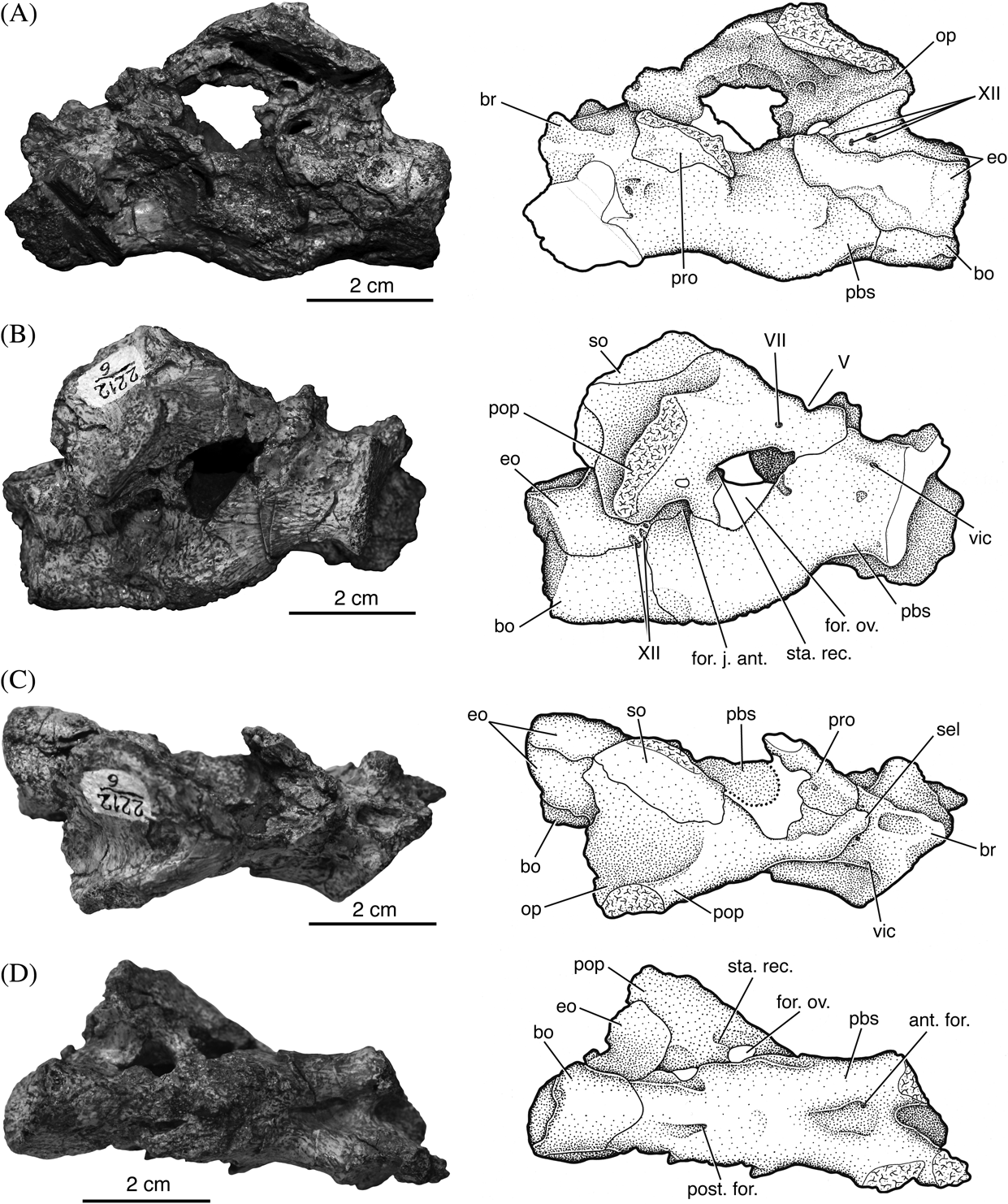
Deltavjatia rossicus PIN 2212/6, photograph and drawing of braincase in left (A) and right (B) lateral, dorsal (C) and ventral (D) views. Abbreviations: ant. for.=anterior foramen; bo=basioccipital; br=basisphenoid rostrum; eo=exoccipital; for. j. ant.=foramen jugulare anterius; for. ov.=foramen ovalis; op=opisthotic; pbs=parabasisphenoid; pop=paroccipital process; post. for.=posterior foramen; pro=prootic; sel=sella turcica; so=supraoccipital; sta. rec.=stapedial recess; vic=vidian canal; V, VII, XII=foramen for cranial nerves V, VII, XII.

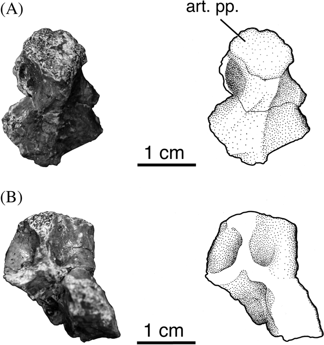
Deltavjatia rossicus PIN 22126/6, photograph and drawing of supraoccipital in anterior (A) and posterior (B) views. Abbreviation: art. pp.=articulation for the postparietal.

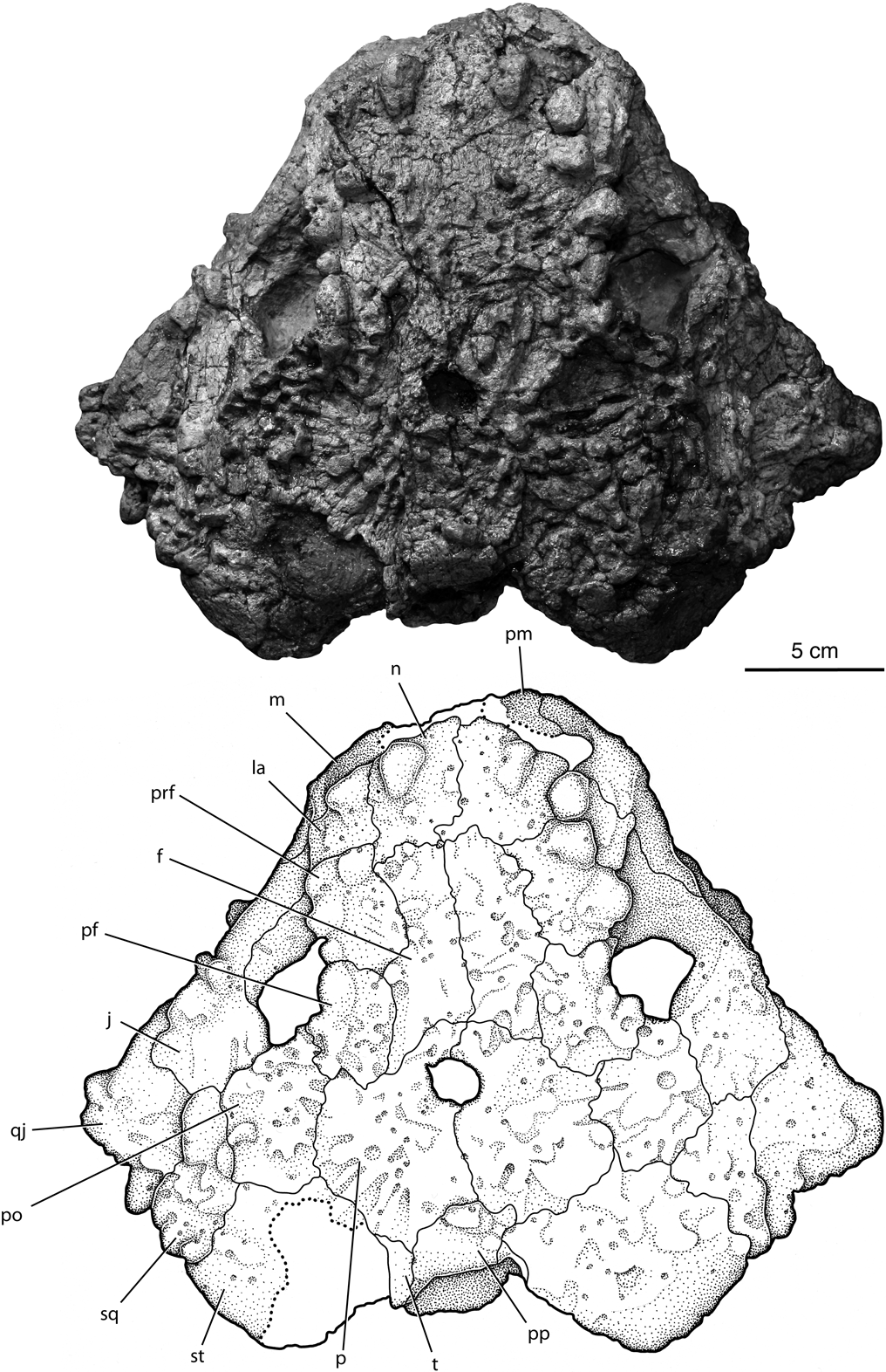
Deltavjatia rossicus PIN 2212/8, photograph and drawing of skull in dorsal view. Abbreviations: f=frontal; j=jugal; la=lacrimal; m=maxilla; n=nasal; p=parietal; pf=postfrontal; pm=premaxilla; po=postorbital; pp=postparietal; prf=prefrontal; qj=quadratojugal; sq=squamosal; st=supratemporal; t=tabular.

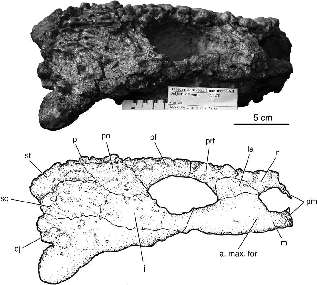
Deltavjatia rossicus PIN 2212/8, photograph and drawing of skull in right lateral view. Abbreviations: a. max. for.=anterior maxillary foramen; j=jugal; la=lacrimal; m=maxilla; n=nasal; p=parietal; pf=postfrontal; pm=premaxilla; po=postorbital; prf=prefrontal; qj=quadratojugal; sq=squamosal; st=supratemporal.

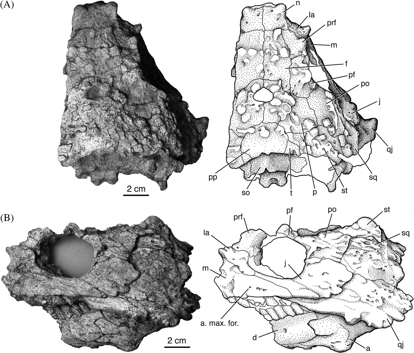
Deltavjatia rossicus ROM 44764, photograph and drawing of skull in dorsal (A) and left lateral (B) views. Abbreviations: a=angular; a. max. for.=anterior maxillary foramen; d=dentary; f=frontal; j=jugal; la=lacrimal; m=maxilla; n=nasal; p=parietal; pf=postfrontal; po=postorbital; pp=postparietal; prf=prefrontal; qj=quadratojugal; so=supraoccipital; sq=squamosal; st=supratemporal; t=tabular.

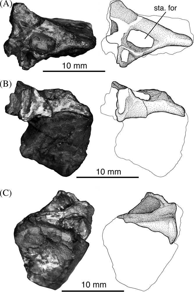
Deltavjatia rossicus SMNS 58317, photograph and drawing of stapes in ventral (A), anterior (B) and posterior (C) views. Abbreviation: sta. for.=stapedial foramen.

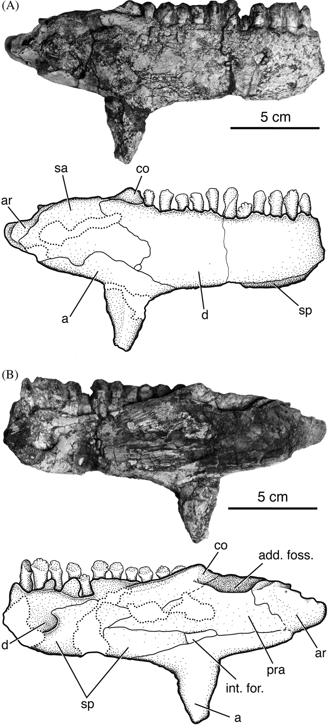
Deltavjatia rossicus SMNS 58317, photograph and drawing of mandible in external (A) and internal (B) views. Abbreviations;: a=angular; add. foss.=adductor fossa; ar=articular; co=coronoid; d=dentary; int. for.=intermandibular foramen; pra=prearticular; sa=surangular; sp=splenial.

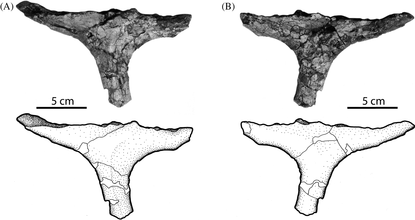
Deltavjatia rossicus SNMS 58317, photograph and drawing of interclavicle in ventral (A) and dorsal (B) views.

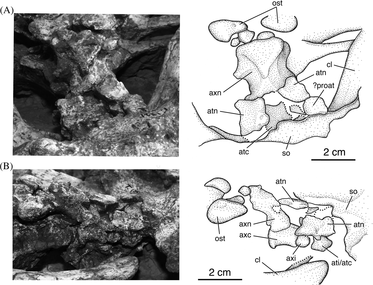
Deltavjatia rossicus UMZC T1321, photograph and drawing of atlas–axis complex in dorsal (A) and right lateral (B) views. Abbreviations: atc=atlas centrum; ati=atlas intercentrum; atn=atlas neural arch; axc=axis centrum; axi=axis intercentrum; axn=axial neural arch; cl=clavicle; ost=osteoderm; proat= proatlas; so=supraoccipital.

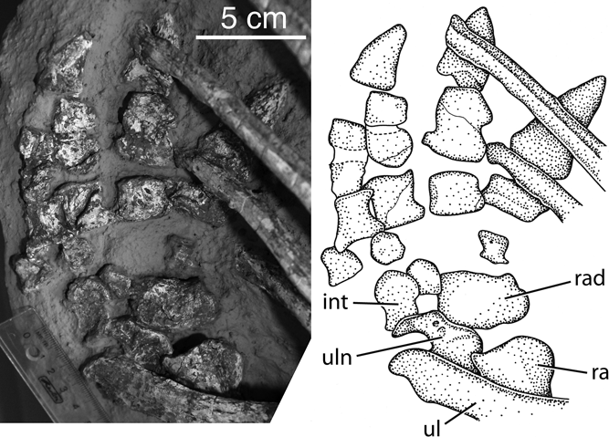
Deltavjatia rossicus UMZC T1321, photograph and drawing of right manus (inverted) in ventral view. Abbreviations: int= intermedium; ra=radius; rad=radiale; ul=ulna; uln=ulnare.

1937 Pareiasuchus vjatkensis Hartmann-Weinberg, p. 664; Taf. 1; Taf. 2; Taf. 3.
1937 Anthodon rossicus Hartmann-Weinberg, p. 676; Taf. 4; Taf. 5.
1987 Deltavjatia vjatkensis Lebedev in Ivakhnenko, p. 75, txt-fig. 23.
Holotype. PIN 2212/2 (Fig. 1), complete skull (the original holotype of “Anthodon rossicus”).
Type locality and horizon. Vjatka River, Kotel'nich, Kirov Province, Russia. Tatarian (Severodvinskian Gorizont), Upper Capitanian to lowermost Wuchiapingian. Specimens found in red mudstone in a series of localities that extend for approximately 20 km on the western bank of the Vjatka River south of the town of Kotel'nich. Skeletons often preserved in upright posture as if mired. Roughly correlated with the Pristerognathus and lowermost Tropidostoma Assemblage Zones of the South African Karoo Basin (Benton et al. Reference Benton, Newell, Khlyupin, Shumov, Price and Kurkin2012).
Referred material. KPM 232 (Figs 2–7), complete skull and lower jaw; complete postcranial missing posterior caudals and right hindlimb, (these features are replaced in the in-situ mount by elements from another specimen) as well as some phalanges from the right manus; KPM 11/94 (Fig. 8), some disarticulated skull elements including maxilla and quadratojugal; partially articulated dorsal vertebrae with osteoderms; disarticulated limb elements and interclavicle; KPM 11/99, partial ‘neonate’ skull; KPM 15/00, left half of skull; KPM uncat/P1 (Fig. 9), complete skull and lower jaw; vertebral column missing posterior dorsal and caudal vertebrae; partial shoulder girdle; ribs; osteoderms; PIN 2212/1 (Fig. 10), complete skull and mandible (the original holotype of “Pareiasuchus vjatkensis”); PIN 2212/6 (Figs 11–17), complete skull and lower jaw; PIN 2212/8 (Figs 18, 19), complete skull; PMU R1259, complete skeleton missing most of tail; ROM 44764 (Fig. 20), complete skull and lower jaw; SMNS 58317 (Figs 21–23), complete skull and lower jaw; incomplete postcranial including articulated presacral vertebrae, partial shoulder girdle including clavicle, humerus, partial pelvic girdle; TMP 95.44.10, complete skull and lower jaw; partially articulated and mostly complete postcranial; missing posterior caudals, right and left manus and some ribs; UMZC T1321 (Figs 24, 25), complete skull and lower jaw; vertebral column missing posterior sacrals and caudals; ribs; complete shoulder girdle and both forelimbs; partial left hindlimb; osteoderms.
Diagnosis (Lee Reference Lee1997b). Ventrolateral cheek flange terminates in a long, flattened rectangular boss; occipital condyle projects far behind posterior border of the postparietal; boss on postfrontal enlarged into a conical horn.
Revised diagnosis. Medium-sized pareiasaur, with snout-vent length approximately 1.5 m, distinguished by the presence of three clear autapomorphies: ventrolateral cheek flange terminates in a long, flattened rectangular boss; occipital condyle projects behind posterior border of the postparietal; distinctive dermal sculpturing consisting of lozenge-shaped and inconsistently-placed tubercles interspersed with round pits and raised bosses. Some of the teeth have a cusped cingulum on their lingual surface. There appears to be no consistent horn-like boss on the postfrontal, so this has been removed as an autapomorphy of the taxon.
Nomenclatural notes. The taxonomic history of this species is complicated. Multiple pareiasaurs were named from the Kotel'nich locality, but only Deltavjatia rossicus, a new combination, is currently valid. Pareiasaur fossils from the Vjatka River, Kirov Province, Russia, were first described by Hartmann-Weinberg as belonging to two new taxa; a new species of Pareiasuchus (Pareiasuchus vjatkensis), and a new species of Anthodon (Anthodon rossicus) (Hartmann-Weinberg Reference Hartmann-Weinberg1937). These two have since been synonymised and assigned to a new genus, but there has been significant confusion about the correct nomenclature for this taxon. A footnote in Kalandadze et al. (1968) suggests that P. vjatkensis could belong to A. rossicus, but no formal designation is made. Kuhn (Reference Kuhn and Kuhn1969, pp. 80–81) did synonymize P. vjatkensis with “A.” rossicus. In 1987, Ivakhnenko (Reference Ivakhnenko1987) independently synonymised A. rossicus with “P.” vjatkensis and, recognising that the taxon was not closely related to Pareiasuchus, he designated a new genus, Deltavjatia. Lee (Reference Lee, Benton, Shishkin, Unwin and Kurochkin2000) agreed that all pareiasaurs from the Vjatka River localities belonged to one taxon, but declared Kuhn's synonymisation illegal, asserting “P. vjatkensis” had page priority over A. rossicus. However, the ICZN no longer recommends page priority, instead enforcing the rule of first reviser (although the code does recommend that the first reviser apply the principle of page precedence). Because Kuhn (Reference Kuhn and Kuhn1969) was the first reviser, the correct combination, therefore, should be Deltavjatia rossicus (Hartmann-Weinberg Reference Hartmann-Weinberg1937).
This change in nomenclature also requires a reassignment of the type specimen of Deltavjatia. PIN 2212/2, the designated type specimen of “Anthodon rossicus” is now the type of the species.
2. Anatomical description of Deltavjatia rossicus
2.1. General description
Deltavjatia rossicus is a medium-sized pareiasaur, with a snout–vent length of approximately 1.5 m. Deltavjatia has a moderately sculptured skull, with enlarged bosses on skull roof elements in the larger specimens. When the braincase is present, Deltavjatia can be clearly identified by the distinct, exaggerated emargination of the posterior skull roof, such that much of the dorsal surface of the braincase, including most the supraoccipital, is exposed dorsally. It is a sparsely armoured form, with offset rows of simple osteoderms associated with the neural spines of the anterior dorsal vertebrae.
2.2. Cranial description
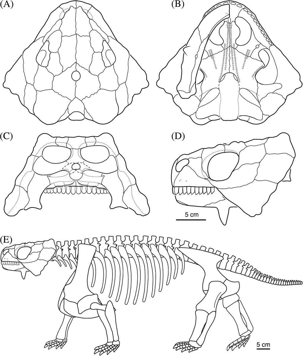
There is ample and well-preserved skull material of Deltavjatia available for study. All of its autapomorphies lie in the cranium. Skull reconstruction in dorsal, ventral, lateral and occipital views (Fig. 26A, B, C, D) are based on all available specimens, but primarily KPM 232, PIN 2212/6 and PIN 2212/8 for the dorsal, and PIN 2212/1, PIN 2212/6 and SMNS 58317 for the ventral views.
Reconstruction of the skull of Deltavjatia rossicus in dorsal (A), ventral (B), right lateral (C) and occipital (D) views; and full skeletal reconstruction (E).

2.2.1. Skull roof
In Deltavjatia, as in all pareiasaurs, the skull roof in mature individuals is fused to the palate and braincase. The skull in pareiasaurs is particularly deep, with wide, flaring cheek flanges.
The cranial sculpture (Figs 1, 2, 4A, 11, 13, 18–20) of Deltavjatia is distinctive. It involves a combination of large, irregularly rounded bosses interspersed with sparse, randomly distributed and roughly circular pits. There are no consistent raised radiating ridges as seen in Pareiasaurus and Pareiasuchus (Bystrov Reference Bystrov1957) and, unlike in Bradysaurus, there are flat areas often punctuated by round pits in between bosses. Often there are very fine radiating grooves emanating from the centre of the bone, the latter of which is usually demarcated by a prominent rounded boss on the larger specimens. The bosses themselves are not consistently circular in outline as seen in many other pareiasaur taxa, but are instead often subcircular, or even lozenge-shaped, imparting an inconsistent, pustule-like appearance to the bones of the skull roof.
The premaxilla (Figs 2, 4A, 11–13) of Deltavjatia is a triradiate structure, with a dorsal process that sutures with the nasal, a lateral process that sutures with the maxilla, and a posterior process on the palate that contacts the vomer. It also forms the anterior edge of the skull and, unlike the three premaxillary teeth illustrated by Lee (Reference Lee, Benton, Shishkin, Unwin and Kurochkin2000), the premaxillae of Deltavjatia bear two teeth (Fig. 12). The dorsal process, or internarial bar, formed by the premaxilla is relatively long and thin, so the nares face anteriorly rather than laterally. This bar is oriented vertically, consistent with the condition present in most other pareiasaurs. Dorsally, the dorsal process meets the nasal, slightly overlapping the medial process of this latter element. The flat palatal process of the premaxilla extends posteriorly to meet the anteriormost portion of the vomer. The prepalatal foramen is located posteriorly along the suture between the two premaxillae on the palatal surface (Fig. 12), near the confluence of the premaxillae and the vomers, as illustrated for Pareiasuchus nasicornis (Lee et al. Reference Lee, Gow and Kitching1997).
Posterolateral to the premaxilla, the maxilla (Figs 2, 4A, 12, 13, 19) is the other tooth-bearing bone of the skull. The maxillae of the larger specimens bear ten teeth, whereas there are fewer in the more juvenile specimens (PIN 2212/6 has nine teeth in the maxilla). The posterior spur of the maxilla and the anterior process of the quadratojugal do make contact for a short distance below the jugal, in contrast to previous descriptions of Deltavjatia (Lee Reference Lee1997b), although the prominence of this feature tends to vary between specimens and the contact is sometimes obscured by the ventral extension of the jugal. The maxilla is short compared to other closely related (non-pareiasaurian) parareptiles, the latter of which possess a maxilla that often extends past the posterior border of the orbit (see Emeroleter; Tsuji et al. Reference Tsuji, Müller and Reisz2012). In contrast, the maxilla of Deltavjatia forms a relatively long, vertical suture with the jugal, and barely extends past the anterior border of the orbit. The maxilla bears a series of foramina running parallel to the tooth row, the largest and most prominent of which is the anteriormost (Figs 4A, 13); the “anterolateral maxillary foramen” sensu Laurin & Reisz (Reference Laurin and Reisz1995), a feature that is a parareptilian synapomorphy. The element itself is relatively smooth and does not bear the sculpturing seen on the other dermal roof elements. Although there is a small boss on the maxilla close to the maxilla–lacrimal suture in some specimens, there is no prominent horn or boss as seen in Pareiasaurus. On the palatal surface, the maxilla forms the anterolateral border of the choana, and posteriorly it forms a medial suture with the palatine and the ectopterygoid.
In contrast to the closely related nyteroleters (Tsuji et al. Reference Tsuji, Müller and Reisz2012), the lacrimal (Figs 2, 4A, 11, 13, 18, 20A) of pareiasaurs participates in the posterior border of the external naris, perhaps related to their relatively shortened snout. The majority of the external surface of the lacrimal in Deltavjatia is overlapped by the dorsal process of the maxilla, thus appearing shorter externally than its full internal height. In the larger specimens, the lacrimal bears a very prominent boss just posterior to the external naris (Fig. 18). The dorsal edge of the element is bordered anteriorly by the nasal and posteriorly by the prefrontal. A process of the lacrimal extends posteroventrally, running just anterior to the ventrally directed process of the prefrontal. Posteriorly, the lacrimal bears one lacrimal foramen towards the upper half of the orbit (Fig. 13). Forming the anteroventral corner of the orbit, the ventral process of the lacrimal projects lightly posteriorly to meet the anterior process of the jugal.
The nasal (Figs 1, 2, 4A, 11, 18) meets its partner in a midline suture. It is a roughly rectangular bone that tapers towards the anterior end, where there is a slight medial excavation on the dorsal surface for the reception of the premaxilla. The nasal forms the majority of the dorsal edge of the naris. Posterior to the naris, the nasal sutures with the lacrimal anteriorly and the prefrontal posteriorly. The posterior border of the nasal is formed by a relatively straight transverse suture with the frontal. The nasal bears a large, rounded boss at the anterior end of the bone directly behind the naris.
The prefrontal (Figs 1, 2, 4A, 11, 13, 18, 20) forms the anterodorsal part of the orbital rim and, along with the nasal, makes up the dorsal border of the lacrimal. It contacts the nasal anteromedially, and the remainder of its medial limit is formed by a suture with the frontal. Posteriorly there is a short transverse suture with the postfrontal, excluding the frontal from the orbit. The ventral process of the prefrontal runs ventrally, posterior to the lacrimal, and forms a solid connection with the palate (Fig. 13). Sculpturing on the bone consists primarily of a large boss that lies close to the lateral edge (overtop of the orbit), and another, smaller boss close to the area where the prefrontal approaches the nasal-frontal suture.
The frontal (Figs 1, 2, 11, 18, 20) is a rectangular bone, meeting its pair in a straight midline suture. Anteriorly, it forms a roughly transverse suture with the nasal. Laterally, the element is bounded on its anterior half by the prefrontal and posteriorly by the postfrontal. In contrast to the condition seen in other parareptiles such as Procolophon (Laurin & Reisz Reference Laurin and Reisz1995), the frontal in pareiasaurs is excluded from making contact with the orbit. Posteriorly, the frontal appears to 'wrap around' the parietal; the fronto–parietal suture originates directed laterally, perpendicular to midline, but more laterally it is angled posterolaterally, forming a very thin tapering posterior process on each side (Fig. 11). This morphology contrasts with the reconstruction of Lee (Reference Lee, Benton, Shishkin, Unwin and Kurochkin2000), which shows this suture as straight and directed laterally. The dorsal surface of the frontal is characterised by a cluster of bosses towards the centre of the element, where the frontal, prefrontal and postfrontal meet. The rest of the bone is less sculptured, with the anterior portion bearing only shallow ridges that are directed antero-posteriorly.
The parietal (Figs 1, 2, 11, 18, 20) is a large bone that makes up most of the posterior section of the skull roof. The parietal, or pineal, foramen is contained entirely within the parietal and is positioned slightly anterior to the midpoint of the full antero–postero length of the bone, closer to the fronto–parietal suture. A posterolateral continuation of the fronto–parietal suture forms the suture with the postfrontal. The parietal also sutures laterally with the postorbital, and its posterolateral border is formed by a suture with the supratemporal. Posteriorly, the element is slightly embayed where it meets the single median postparietal. The parietal has several bosses clustered near the centre of the bone.
In some specimens of Deltavjatia the postparietal (Figs 1, 2, 4B, 11, 18, 20) features a small median boss close to the anterior edge of the bone where it sutures to the parietal (Fig. 11), but the remainder of the element is smooth and does not bear the sculpture characteristic of the other skull roof bones. As in all pareiasaurs (Lee Reference Lee1997a), the postparietal of Deltavjatia is a single, fused element, and is relatively large – over one-third of the length of the parietal itself. Ventrally, the postparietal receives the dorsal process of the supraoccipital, forming the main dorsal attachment surface of the braincase with the skull roof. This articulation takes the form of a short ventral process that is diamond-shaped in cross-section (Fig. 12). The postparietal forms a very short lateral suture with the large supratemporal, and the remainder of the lateral border is formed by the tabular or ‘supernumerary element’. The slightly concave posterior edge of the postparietal is not even with the posterior edge of the supratemporal, but is positioned more anteriorly, approximately halfway along the length of the supratemporal. Thus the median segment of the posterior edge of the skull roof is embayed significantly. This feature is so pronounced that in specimens where the braincase is present, some of the supraoccipital and the posterior part of the basioccipital can be seen in dorsal view (Fig. 20), an autapomorphy of the taxon.
The tabular (Figs 1, 2, 4B, 11, 18, 20) can be observed in all specimens of Deltavjatia where the posterior edge of the skull table is preserved, although in the larger specimens the sutures are more difficult to discern, and the element is often partially fused with the supratemporal and postparietal. In PIN 2212/6, there are clearly two smaller closely sutured, but still discrete elements on either side of the postparietal (Fig. 11). The postparietal and the tabular are strictly occipital elements in the majority of the more basal parareptiles (see Tsuji Reference Tsuji2006), which have been subsequently ‘dragged’ up onto, and integrated into, the skull roof in pareiasaurs. The extent of the tabular of Deltavjatia is typical for the majority of pareiasaurs, with the exception of Elginia, Arganaceras, and possibly Bunostegos, in which the tabulars form a median suture behind the postparietal, excluding the latter from the posterior edge of the skull table (Maxwell Reference Maxwell1991; Jalil & Janvier Reference Jalil and Janvier2005; Sidor et al. 2005).
The supratemporal (Figs 1, 2, 4A, 11, 13, 18–20) forms the posterolateral corner of the skull roof. It is a large element, bearing a very prominent round boss towards the posterior edge of the skull table, along with other smaller bosses clustered around it. The supratemporal is largely excluded from contact with the postparietal in Deltavjatia by the intervening tabular (Fig. 2), though a short suture between the former two is present in the majority of specimens. Anteriorly, the supratemporal forms a short suture with the postorbital, and ventrolaterally it sutures with the squamosal. The element also takes part in the attachment of the skull roof with the braincase, receiving the dorsal portion of the paroccipital process (Fig. 4B). The area of the supratemporal posterior to the large boss forms a noticeable posteroventrally-directed slope, and is mostly unsculptured, a morphology shared with the postparietal and tabular (Fig. 2).
The postfrontal (Figs 1, 2, 4A, 11, 13, 18, 20) of Deltavjatia is larger than reconstructed by Lee (Reference Lee, Benton, Shishkin, Unwin and Kurochkin2000), with its posterior edge extending even farther posteriorly. The postfrontal defines the posterodorsal edge of the orbit. It forms a short suture with the prefrontal anteriorly, and the majority of the median portion of the bone is delimited by the frontal. Posteromedially, the bone sutures with the parietal, and it is bounded laterally (and ventrally) by the postorbital. There is a large boss positioned at the centre of the bone along the edge of the orbit, with a number of smaller bosses clustered around the larger one (Fig. 18).
The postorbital (Figs 1, 2, 4A, 11, 13, 18, 20) is roughly triangular in shape, with its anterior edge forming the majority of the posterior rim of the orbit. It sutures with the jugal posteroventrally and the squamosal posteriorly. Sutures with the postfrontal and the frontal define the majority of the dorsal limit of the postorbital, but the element forms a short suture on its posteriormost edge with the supratemporal. The postfrontal bears a series of bosses along the orbital rim, with ridges and furrows radiating outwards from this point (Fig. 11).
The jugal (Figs 1, 2, 4A, 11, 13, 18, 20B) constitutes the majority of the ventral, and half of the posterior margin of the orbit. It is crescent-shaped in outline. The long anterior process thins anteriorly, and it forms a short suture with the lacrimal, with the majority of the anterior process bounded ventrally by the maxilla. Posteriorly, a gradually dorsally curving suture with the quadratojugal delineates the ventral edge of the bone. Posterodorsally, the jugal contacts the squamosal, and dorsally it forms an anteroventrally directed suture with the postorbital (Fig. 4A). There is a cluster of bosses located almost directly under the ventralmost extension of the orbit, a character shared with other pareiasaurs and Macroleter (Tsuji Reference Tsuji2006). In some specimens of Deltavjatia, the jugal forms a small portion of the ventral flange of the cheek, where the cheek flange meets the tooth-bearing region of the skull. This implies that the quadratojugal and the maxilla are separated, at least on the external surface of the skull in these specimens. Internally, however, these two latter bones indeed make contact, with the jugal forming a small overhang that obscures this suture externally (Fig. 4A).
The squamosal (Figs 1–4, 11–14, 18–20) is a large element, suturing dorsally and medially with the supratemporal, anteriorly with the postorbital and jugal, and ventrally with the quadratojugal. Internally, the squamosal sutures with the quadrate. A small posterior process of the pterygoid from the palate sutures with the squamosal at its dorsalmost contact with the quadrate. The squamosal makes up a large part of the cheek, and features a large oblong boss on the posterior edge towards the dorsal portion of the bone, where it sutures with the supratemporal, and a smaller, round boss just anterior to this (Fig. 19). Apart from these, the sculpturing of the squamosal consists primarily of ridges and furrows radiating outwards from the bosses towards the edges of the bone. Interiorly, the squamosal forms the ventral portion of the articulation with the paroccipital process of the braincase and is correspondingly thickened in this area, most likely to help reinforce the cheek. Although there is no apparent otic notch on the lateral (external) surface of the skull in pareiasaurs, in posterior view a small yet clear ‘notch’ facing internally can be seen enclosed primarily within the squamosal (Fig. 4B). This feature appears to be homologous to the otic notch that holds the large tympanum of the nycteroleters (Müller & Tsuji Reference Müller and Tsuji2007; Tsuji et al. Reference Tsuji, Müller and Reisz2012) but, corresponding to the more heavily ossified condition of the pareiasaur skull, it has been shifted considerably into what would have been a more protected position. The relative area occupied by this structure is smaller than the condition seen in the nycteroleters. The position of the otic notch is such that a stapes and cartilaginous extrastapes would have been directed slightly dorsolaterally from the braincase (see further discussion below).
The quadratojugal (Figs 1–4, 11–14, 18–20) comprises the entirety of the ventral margin of the cheek. As in all pareiasaurs, the element forms a prominent cheek flange, extending well below the level of the tooth row. In undistorted specimens, the anterior edge of this flange forms an angle of approximately 60 degrees with the tooth row. Although this angle is considerably lower than in pareiasaurs such as Pareiasaurus, the cheek flange of Deltavjatia is more pronounced than in Bradysaurus (Lee Reference Lee1997b). The quadratojugal delineates most of the posterior boundary of the jugal, and dorsally it forms a relatively straight horizontal suture with the squamosal. The dorsal border of the quadratojugal is approximately even with the ventral rim of the orbit. The stated autapomorphy of a long low boss on the quadratojugal (Lee Reference Lee1997b) is not consistently present, at least in the smaller two specimens (PIN 2212/6, ROM 44764). Instead, the quadratojugal ornamentation consists of two larger anterior conical spikes, with smaller bosses further posteriorly along the posterior edge of the element (Figs 4A, 20).
2.2.2. Palate
The palate of pareiasaurs is fairly conservative and, indeed, the palate of Deltavjatia differs little from the typical pattern. It is characterised by tall, denticle-bearing ridges and a reduced transverse flange of the pterygoid. Unlike in the nycteroleters, the palate, and especially the transverse flange of the pterygoid, is raised considerably above the level of the tooth row.
The vomer (Figs 3, 10, 12) of pareiasaurs is unique, typified by what is termed the ‘alar flange’ (Damiani & Modesto Reference Damiani and Modesto2001); a rounded lateral process defining the medial edge of the choana, resulting in a choana that is kidney-shaped in outline. It has been suggested that the vomer has a thin medial dorsal flange that forms a bony septum in the nasal chamber (Damiani & Modesto Reference Damiani and Modesto2001), but unfortunately none of the specimens of Deltavjatia examined for this study are preserved in a manner in which this feature could be observed.
Anteriorly, the vomer meets the premaxilla. The articulating surface of this contact is not straightforward, however. As described for an unidentified pareiasaur from the Tropidostoma Assemblage Zone of the Karoo (Damiani & Modesto Reference Damiani and Modesto2001), a channel or groove separates two articulating surfaces, with lateral processes that are larger than the median ones (Fig. 10). Posterior to the articulation with the premaxilla, a row of denticles runs along the median edge of the bone. The anteriormost denticle is the largest and is slightly recurved, and the size of the denticles decreases as the row continues posteriorly. At the level of the anteriormost extension of the vomer–palatine suture (slightly anterior to the posterior edge of the choana), the single row splits into two parallel rows. The vomer has very little sutural contact with the palatine, with this contact almost precluded by the long posterolaterally-directed suture with the pterygoid (Fig. 10).
The palatine (Figs 3, 10, 12) forms the posterolateral, the entire posterior edge, and a small portion of the medial edge of the choana. Laterally, the bone meets the maxilla in a gently sloping posterolaterally-directed suture. The posterolateral edge of the palatine sutures with the ectopterygoid, but this contact is interrupted by a large foramen, the ‘foramen palatinum posterius’ of Lee (Reference Lee1995b). This foramen is contained entirely within the suture between the palatine and the ectopterygoid (Fig. 10). It may be homologous to the ‘suborbital foramen’ of the nycteroleters (Tsuji et al. Reference Tsuji, Müller and Reisz2012); however, in nycteroleters and most other parareptiles, this foramen is contained between the ectopterygoid, maxilla and palatine. Thus, the term ‘foramen palatinum posterius’ is maintained here for pareiasaurs. Kordikova & Khlyupin (Reference Kordikova and Khlyupin2001) have suggested that the foramen palatinum posterius is proportionally much larger in neonates, almost completely separating the palatine from the maxilla. The palatine also sutures with the pterygoid posteromedially. A double row of denticles, which continues from the pterygoid, extends anterolaterally, reaching almost to the posterior edge of the choana.
The ectopterygoid (Figs 3, 10, 12) is a relatively small, roughly triangular element. The ventral surface of the bone is smooth and featureless. In ventral view, anteriorly the ectopterygoid sutures with the palatine, with the suture interrupted by the foramen palatinum posterius. The ectopterygoid sutures laterally with the maxilla, continuing the palatine–maxilla suture, and it also forms a short suture with the pterygoid posteriorly. In P. nasicornis, the ectopterygoid is reconstructed as forming a short suture with the quadratojugal (though in the description it is described as an ectopterygoid–jugal suture) (Lee et al. Reference Lee, Gow and Kitching1997), but this condition appears to be variable amongst pareiasaur taxa (Lee Reference Lee1997a). There is no contact between the ectopterygoid and the jugal or quadratojugal in the studied specimens of Deltavjatia. The lateral limit of the ectopterygoid is formed entirely by the maxilla (Fig. 12).
The pterygoid (Figs 3, 10, 12) is a large and complex bone, and is the dominant element of the palate. It is basically triangular, with an anterior process that terminates in a transverse suture with the vomer. Posterior to the suture with the vomer, the pterygoid forms a posterolaterally-directed suture with the palatine, which continues as a suture with the ectopterygoid. The pterygoid also takes part in the basipterygoid articulation, in which the basipterygoid process of the braincase fuses to the palate in mature individuals. The interpterygoid vacuity of Deltavjatia is very short and is rounded at its anterior end, more similar to the condition seen in the basal Bradysaurus than in the derived Anthodon (Lee Reference Lee1997a). The pterygoid also forms a long anterolateral suture with the quadrate, and may also form a short suture with the squamosal (Figs 3, 12).
Multiple rows of denticles run across the pterygoid. The double row of denticles continues posteriorly from the vomer and lines the edges of the interpterygoid vacuity. Another row of denticles begins approximately midway along the pterygoid–palatine suture. This set of denticles begins as a single row, but splits into two rows as it approaches the palatine. This double row, as with the other double rows on the palate of Deltavjatia, remains parallel throughout its length (Figs 3, 12, 26).
The transverse flange of the pterygoid is only slightly recurved. In outgroup parareptilian taxa, it generally points directly laterally and very closely approaches the cheek (Laurin & Reisz Reference Laurin and Reisz1995; see Macroleter, Tsuji Reference Tsuji2006). In Deltavjatia, as in other pareiasaurs, the transverse flange is directed anterolaterally and does not closely approach the cheek (Lee Reference Lee1997b). The transverse flange also bears a row of large denticles on a raised ridge on the ventral surface of the structure (Fig. 3).
No epipterygoids were found in the specimens of Deltavjatia examined in this study. The element is figured as a very small process running vertically from the cranial process of the pterygoid in Pareiasaurus serridens (Boonstra Reference Boonstra1930).
The quadrate (Figs 3, 4B, 10, 12) connects the posterior portion of the palate with the cheek and forms the upper half of the jaw articulation. The element is basically triangular and sutures with the pterygoid anteromedially, the quadratojugal ventrally, and the squamosal posteriorly. The quadrate forms an extensive suture with the quadrate flange of the pterygoid. This suture extends dorsally almost to the skull roof, close to where the paroccipital process of the braincase articulates with the squamosal and supratemporal (Fig. 12). The exact delineation of the suture between the quadrate and the pterygoid is difficult to trace in most specimens, however. On its posterior and posterolateral margins, the quadrate sutures with the quadratojugal. Further dorsally, a small quadrate foramen is found at the convergence between the quadrate, squamosal and quadratojugal (Fig. 4B). The suture between the squamosal and quadrate defines the posterior border of the otic notch in Deltavjatia.
The jaw articulation in Deltavjatia is medio-laterally broad and consists of a lateral and a medial condyle separated by a shallow groove (Fig. 12). The articulation occurs well anterior to the occiput – almost to the level of the basipterygoid processes – and ventral to the tooth row, a condition that is concordant with the herbivorous diet of pareiasaurs (Lee et al. Reference Lee, Gow and Kitching1997).
2.2.3. Braincase
The well-ossified condition of the pareiasaur skull applies in particular to the braincase, making it difficult, if not impossible, to discern the sutures between the constituent elements. However, the availability of well-preserved sub-adult Deltavjatia material allows a closer examination of the individual elements of the braincase. PIN 2212/6 includes an isolated braincase with partially disarticulated exoccipitals, allowing a detailed description of the braincase from all angles, in addition to insight into the structure of the individual elements. It is important to note, however, that this specimen is sub-adult, and thus certain features seen here may not be present in fully-grown specimens and vice-versa.
In pareiasaurs, the parasphenoid and the basisphenoid are fused into a single indistinguishable element, termed the parabasisphenoid (Figs 3, 10, 12, 16). Even in the relatively unfused braincase of the juvenile PIN 2212/6 (Fig. 16) the two elements cannot be distinguished from one another, indicating that this fusion probably occurred early in development. The parabasisphenoid has an hourglass silhouette in ventral view, with slightly flared anterior and posterior ends and a slightly constricted middle, though this constriction is less pronounced than seen in the outgroup taxa and other basal pareiasaurs such as Bradysaurus (Lee Reference Lee1997b). In pareiasaurs such as Scutosaurus, the hourglass shape is emphasised by the presence of large, paired basal tubera close to the suture with the basioccipital (Lee Reference Lee, Benton, Shishkin, Unwin and Kurochkin2000); however, these are absent in Deltavjatia. Even in the largest specimens, the ventral surface of the basisphenoid is smooth.
The basipterygoid processes project anterolaterally from the body of the parabasisphenoid, joining with the corresponding processes of the pterygoid on the palate. This joint between the braincase and the palate is fused and immobile, with no visible suture between the two. This morphology can be seen in all larger specimens of Deltavjatia, and is a pareiasaurian synapomorphy (Lee Reference Lee1997a), although an immobile basicranial articulation is also present in Macroleter (Tsuji Reference Tsuji2006) and Lanthanosuchus (deBraga & Reisz Reference deBraga and Reisz1996) within parareptiles. In the more juvenile PIN 2212/6, however, this fusion has not yet occurred, and the braincase can be completely removed from the skull. The articulating surfaces are slightly distorted and do not retain their original morphology. The ventral surface of the parabasisphenoid is relatively smooth, without the basal tubera mentioned above, with a noticeable circular indentation centered just posterior to the basicranial processes (Fig. 3). In PIN 2212/6, two small foramina can be seen towards the anterior portion of this pit (Fig. 16D), seemingly analogous to the “anterior foramen” identified by Lee et al. (Reference Lee, Gow and Kitching1997) on the ventral surface of the basisphenoid of Pareiasuchus nasicornis. This feature also appears to be present in well-preserved adult specimens, located far anteriorly on the basisphenoid, close to the cultriform process. There are two additional foramina just anterior to the suture with the basioccipital, situated within very deep, posteriorly directed grooves (Fig. 16B), possibly equivalent to the ‘posterior foramen’ of Lee et al. (Reference Lee, Gow and Kitching1997). However, these foramina are not present in larger specimens of Deltavjatia.
Due to the disarticulation of the dorsal elements of the braincase, the dorsal surface of the parabasisphenoid is exposed in PIN 2212/6. Just posterior to the basipterygoid processes, the median dorsal body, or parasphenoid rostrum, can be seen rising dorsally from the base of the parasphenoid, with a very small cultriform process projecting anteriorly (Fig. 16A, B, C). In Deltavjatia, the cultriform process is present, and extends at least as far anteriorly as the full length of the interpterygoid vacuity (Fig. 10). Its full anterior extent cannot be assessed from the specimens available, however, and in the juvenile braincase (PIN 2212/6), this feature is either very small or has been broken off. The sella turcica, a deep pit in which the pituitary body would have lain, can be seen immediately posterior to the basisphenoid rostrum (Fig. 16C). Just lateral to the sella turcica are the foramen and groove for the vidian canal (Fig. 16B, C).
The prootic (Figs 16, 17) and the opisthotic project dorsolaterally from the midline of the braincase and form the paroccipital processes, which connects the braincase with the skull roof. The prootic is sutured to the main body of the parabasisphenoid just posterior to the sella turcica. Anteriorly, the prootic is slightly embayed to accommodate the trigeminal nerve (V) (Fig. 16A, B). A small foramen for the facial nerve (VII) is present on the prootic just posterior to this embayment (Fig. 16B). The prootic then continues posteriorly, forming the anterior portion of the dorsal border of the foramen ovalis. Dorsally, it appears as if the two prootics meet at the midline dorsal to the parabasisphenoid, a morphology that differs from the condition portrayed in the procolophonid Leptopleuron, where the prootics do not meet and the parabasisphenoid is exposed dorsally (Spencer Reference Spencer2000). At its dorsalmost margin, the prootic contacts the supraoccipital. The exact length and orientation of this suture is not clear, however, as the entire suture between the prootic and the opisthotic cannot be discerned in PIN 2212/6. More posteriorly, the prootic then merges with the opisthotic to form the paroccipital process. The paroccipital process comprises the articulation of the braincase with the skull roof, but the prootic forms only the anteriormost portion of the paroccipital process and thus does not take part in the articulation.
The opisthotic (Figs 3, 4B, 16) originates posteriorly in a suture with the exoccipital, and these two bones together support the posterior part of the supraoccipital. Posteriorly, the opisthotic and the prootic form the paroccipital process, but it is the opisthotic alone that comprises the lateralmost extent of this structure, forming an articulation with the squamosal and supratemporal in the skull roof. In occipital view, the paroccipital process is u-shaped, dipping slightly ventrally before curving dorsally to fuse with the skull roof in the dorsolateral corner. The braincase in pareiasaurs is firmly fused with the skull, at both the palate and the paroccipital processes.
Anteriorly, the opisthotic contacts the parabasisphenoid via a ventral projection that delineates two large foramina (Fig. 16B). The larger, more anterior foramen can be identified as the fenestra ovalis. A semi-circular stapedial recess, which would have accommodated the footplate of the small stapes (described below), is present in the posterior edge of this larger fenestra (Fig. 16B). This feature does not appear in the braincase of Pareiasuchus nasicornis (Lee et al. Reference Lee, Gow and Kitching1997). The foramen ovalis is very open in the juvenile PIN 2212/6, but in KPM 232 there is some indication of a further division of the fenestra ovalis into a dorsal and ventral section, though they are not fully separate (Fig. 5). The lower of these can be interpreted as a ‘pressure relief window’ sensu Clack (Reference Clack1997), one of the indications of the presence of tympanic hearing, though this feature is not clearly defined on both sides of the skull. The smaller and more posterior of the two foramina is the foramen jugulare anterius of Haughton (Reference Haughton1929) and Lee et al. (Reference Lee, Gow and Kitching1997), a feature not present in the considerably less well-ossified procolophonid Leptopleuron (Spencer Reference Spencer2000).
The internal structure of the opisthotic in PIN 2212/6 consists of a very open network of pits (Fig. 16A), indicating that the otic capsule was not fully ossified in this specimen. A well-ossified state of the otic capsule is present in Macroleter (Müller & Tsuji Reference Müller and Tsuji2007), and the otic capsule of pareiasaurs is considered to be well-ossified (Lee Reference Lee1997a). The immaturity of PIN 2212/6 is likely the reason this region appears to be unossified in this case.
The dorsal exposure of the supraoccipital (Figs 4B, 17) is one of the autapomorphies of Deltavjatia. It is apparent, however, that this morphology is the result of the severe embayment of the posterior skull table, rather than the placement or morphology of the supraoccipital itself. In PIN 2212/6, the supraoccipital has been separated from the rest of the braincase and skull roof (Fig. 17) and the articular surface of the supraoccipital with this latter structure can be seen in the isolated element (Fig. 17A). The contact is roughly diamond-shaped, with the corners oriented along the axes of the skull. The ventral process of the supraoccipital then extends downwards as a thick column, before it flares to form the roof of the braincase and the dorsal edge of the foramen magnum. A tall, columnar surpaoccipital is one of the synapomorphies of pareiasaurs (Lee, Reference Lee1995b, Reference Lee1997a). Anteriorly, this column has a flattened indentation, in which is situated an anterior crest (Fig. 17A), and there is a small raised ridge on the posterior surface (Fig. 17B).
The exoccipital (Reference Benton, Newell, Khlyupin, Shumov, Price and KurkinFigs 4B, 16) is a paired element that projects dorsally and slightly laterally from the basioccipital. In pareiasaurs, it forms the dorsal half of the occipital condyle of the braincase and the lateral margins of the foramen magnum. The exoccipital consists of a basal process that sutures with the basioccipital, a vertical stem whose medial border forms the lateral edge of the foramen magnum, and a dorsal portion that connects with the opisthotic anteriorly and supports the supraoccipital dorsally. This dorsal portion consists of a medial and a lateral process. The medial process forms a significant part of the dorsal border of the foramen magnum, but does not extend to meet its pair at the midline (Fig. 4B). The long lateral process underlies the paroccipital process for a considerable distance, a character typical for pareiasaurs (Lee Reference Lee1995b). There is a series of foramina at the base of the dorsal process of the exoccipitals through which the branches of cranial nerve XII (hypoglossal) would have passed (Figs 16A, B). The base of the exoccipital meets its pair medially above the basioccipital, excluding this latter structure from forming part of the rim of the foramen magnum (Fig. 16), seemingly discordant with the condition described for Bradysaurus (Haughton Reference Haughton1929). In larger individuals of Deltavjatia, the exoccipital and basioccipital fuse indistinguishably.
The basiocciptial (Figs 4B, 16) constitutes the ventral half of the occipital condyle and the posteriormost part of the ventral surface of the braincase. In the larger specimens of Deltavjatia, the basioccipital has fused indistinguishably with the parabasisphenoid to form a single solid structure; but in the juvenile PIN 2212/6, the suture between the basioccipital and the parabasisphenoid can be discerned (Fig. 16D). The basioccipital makes up slightly more than half of the occipital condyle, with the fused exoccipitals forming the dorsal portion. The fusion of the exoccipitals excludes the basiocciptal from participation in the foramen magnum. The condyle itself is highly concave, with a relatively sharp (ie not swollen) rim in the specimens in which it is adequately preserved. This more closely resembles the condition described for Embrithosaurus than that for Bradysaurus, in which the condyle is not as concave and the rim is rounded (Haughton Reference Haughton1929).
The sphenethmoid or interorbital septum is acknowledged to be present in pareiasaurs (Romer Reference Romer1956). Primitively in tetrapods, the sphenethmoid is a large, complex bone (Clack Reference Clack2002). In amniotes, it is a solid plate that rises dorsally from the anterior portion of the parasphenoid (the cultriform process) to reach the frontal, and divides the anterior section of the skull into right and left halves (Romer Reference Romer1956). Its presence in the specimens of Deltavjatia examined in this study, however, is not certain. The area above the cultriform process (between the palate and the skull roof) is rarely prepared in complete skulls, and there is no evidence of its presence in PIN 2212/6, the only specimen in which this area is visible. The extremely short cultriform process in this specimen does not support a sphenethmoid. It is reasonable to infer, however, that PIN 2212/6 is not skeletally mature and thus the sphenethmoid is not yet ossified. This structure was certainly preformed in cartilage. Known pareiasaur sphenethmoids are described in well-ossified and presumably fully-grown individuals. The sphenethmoid of one such individual of Embrithosaurus from the Natural History Museum in London has been partially sectioned (Watson Reference Watson1914; Haughton Reference Haughton1929), and a specimen of Pareiasaurus is illustrated by Boonstra (Reference Boonstra1934a). A sphenethmoid is also present in a specimen of Bunostegos akokanensis (Tsuji et al. Reference Tsuji, Sidor, Steyer, Smith, Tabor and Ide2013).
The stapes (Fig. 21) of early tetrapods tends to be robust, with a role in helping to brace the braincase against the skull roof (Clack Reference Clack2002). In fully terrestrial animals with an acute sense of hearing, however, the structure of the stapes is quite different; instead becoming a small, relatively gracile bone that performs functions related to hearing with a tympanic ear (Clack Reference Clack2002; Müller & Tsuji Reference Müller and Tsuji2007). The stapes of pareiasaurs has been described and figured in two taxa – Arganaceras vacanti (Jalil & Janvier Reference Jalil and Janvier2005) and Scutosaurus karpinskii (Lee Reference Lee1995a) – but the morphology described varies considerably between these two accounts. Considering that both elements were found in isolation and that both of their morphologies are highly aberrant from the stapedial morphology seen in other parareptiles (Carroll & Lindsay Reference Carroll and Lindsay1985; Tsuji Reference Tsuji2006; Tsuji & Müller 2007), it is doubtful that either of these structures can be definitively identified as a stapes.
An element identified here as a stapes has been found in a specimen of Deltavjatia. In SMNS 58317, this element is visible on both sides of the skull in ventral view, and while the left element has been maintained in place, the right has been removed and prepared so it is visible in multiple views (Fig. 21). The element is approximately 1.5 cm in length; very small in comparison with the overall skull length of 25 cm. Proximally, the bone is flared, and there is a large stapedial foramen visible, best seen in ventral view. The proximal end – the footplate – possesses two articular surfaces separated by a sulcus. Distally, the stapes tapers to a point, imparting a triangular shape to the element, slightly longer than wide. This form is very similar to the stapes seen in other derived parareptile taxa such as Procolophon trigoniceps (Carroll & Lindsay Reference Carroll and Lindsay1985) and, in particular, Macroleter poezicus (Tsuji Reference Tsuji2006).
In addition to morphological similarity to the stapes of closely related parareptile groups, the stapes of SMNS 58317 is preserved in the area of the foramen ovalis (see description above) on both sides of the skull. It is clear that the stapes was only very loosely associated with the skull, and would have been preserved only in very well-preserved and undisturbed skeletons.
2.2.4. Mandible
The mandible of Deltavjatia does not deviate significantly from the typical pareiasaurian morphology. It is distinguished by a prominent angular boss extending from the ventral edge of the jaw that extends into a small, pointed horn, and by the single row of leaf-shaped teeth that mirror those on the maxilla. The lower jaw lacks the sculpturing typically seen on the external surface of the skull roof, but does bear a roughened texture on the external, or lateral, surface of the elements. It is composed of a number of overlapping sheet-like bones.
The dentary (Figs 4A, 14, 20B, 22B) is the tooth-bearing bone of the mandible and forms the majority of the anterolateral margin of this structure. It is exposed more on the external surface than the internal, where it is largely overlapped by the splenial. On the external surface of the mandible, the dentary meets the surangular posteriorly and forms a suture with the angular posteroventrally. Internally the dentary forms only the upper half of the mandibular symphysis, above the foramen intermandibularis medius (sensu Lee et al. Reference Lee, Gow and Kitching1997). The splenial and the coronoid overlap the dentary, ventrally and dorsally respectively. In the two specimens with isolated mandibles, there are 11 alveoli present in each dentary (Figs 14, 22). The bone has a number of small foramina on its external surface, but is otherwise unsculptured.
The splenial (Figs 3, 4B, 14, 22) is a platey bone that forms the anterior part of the medial surface of the jaw and, in contrast with most other parareptiles (Laurin & Reisz Reference Laurin and Reisz1995) such as Macroleter (Tsuji Reference Tsuji2006), the element does take part in the symphysis of the mandible (Figs 4B, 22B). The anterior part of the bone is slightly forked, with the upper and lower prongs forming the borders of a large foramen intermandibularis medius (Fig. 4B). The lower prong is significantly more robust than the upper. It is this ventralmost part of the splenial that meets its pair at the midline, taking part in the symphysis, as it does in stem amniotes such as Diadectes (Laurin & Reisz Reference Laurin and Reisz1995). The intermandibular foramen is formed at the confluence of the spenial, angular and the prearticular. It is almost directly dorsal to the angular boss.
Dorsal to the splenial, on the internal surface of the mandible, lies a single coronoid bone (Figs 14, 22). This element forms the dorsalmost extent of the mandible in the form of a relatively low coronoid process, which projects dorsally only to the height of the tooth crowns (Figs 14B, 22B). The anterior process of the bone does not extend much beyond the 5th tooth position. The coronoid forms the anterior rim of the adductor fossa, meeting the surangular on the external (lateral) side and the prearticular on the medial side of this structure. Anteriorly, the coronoid overlaps the posterodorsal process of the dentary and its anteriormost extent forms a short contact with the splenial.
The prearticular (Figs 14, 22) is also located on the inner (internal) surface of the mandible. The element overlaps the angular ventrally and the coronoid dorsally, where it forms the ventromedial border of the adductor fossa. Because the prearticular is very broad dorsoventrally, the adductor fossa of Deltavjatia faces predominantly dorsally. Anteroventrally, the prearticular is overlapped by the splenial.
The surangular (Figs 14, 22) lies dorsal to the angular and forms the majority of the posterodorsal portion of the mandible (on the external surface). It lies posterior to the dentary, extending to cover most of the articular laterally. It does not take part in the retroarticular process.
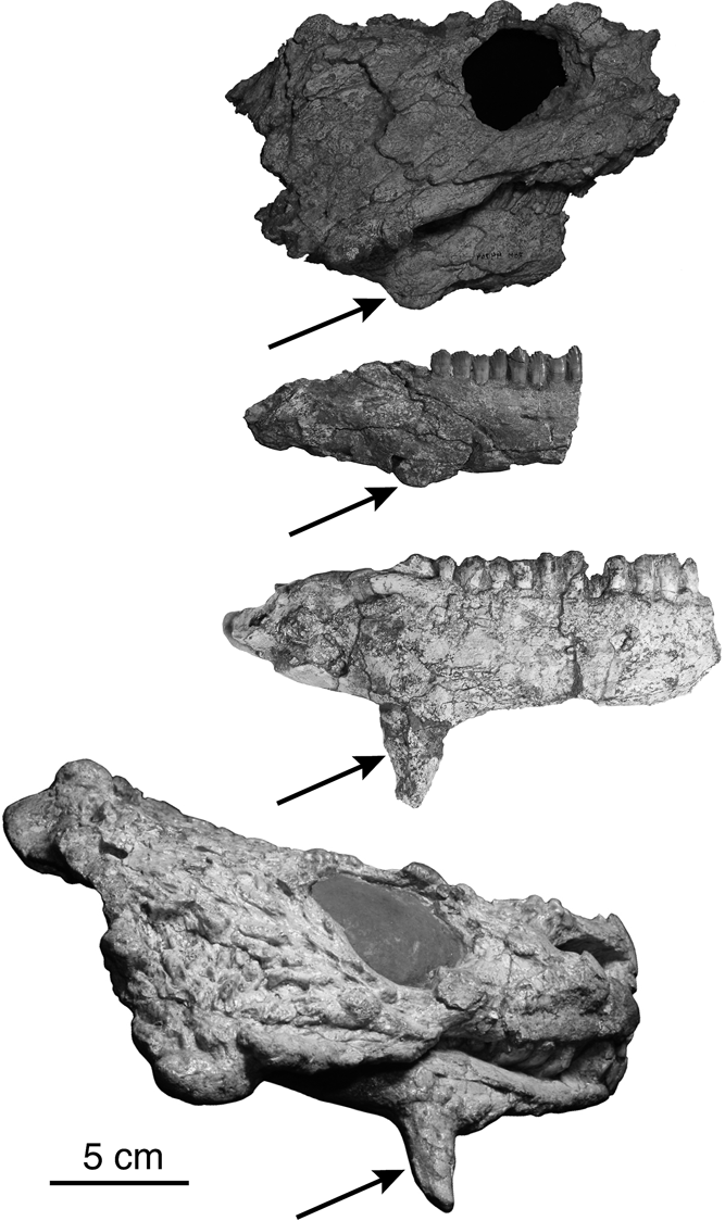
The angular (Figs 3, 4, 14, 20, 22) forms the ventralmost part of the mandible, and can be seen in both medial and lateral views. Medially, the majority of the dorsal border of the angular is formed by a suture with the prearticular, and it meets the splenial anteriorly and the articular posteriorly. Externally (laterally), the surangular forms the majority of its dorsal border, and it meets the dentary anterodorsally. One of the most distinctive synapomorphies of pareiasaurs is the prominent boss towards the anterior end of the angular, which often takes the form of an obvious horn. In the largest specimens of Deltavjatia, the boss is large and pointed, but still more gracile than in pareiasaurs such as Bradysaurus. In the smaller specimens of Deltavjatia, the boss is rather small, low, and laterally compressed (Figs 14, 20B). The dimensions and prominence of the angular boss increase with increasing size and, presumably, age of the individuals (Fig. 27; see also discussion below).
The growth and development of the angular boss in Deltavjatia rossicus (boss indicated by arrow). Specimens, from top to bottom: ROM 44764, PIN 2212/6, SMNS 58317, KPM 232.

The articular (Figs 3, 14B, 22B) has very little lateral or medial exposure and is overlapped by the prearticular internally and the angular and surangular externally. The majority of the visible part of the bone is exposed in dorsal aspect in the form of the glenoid fossa for the articulation with the quadrate of the skull. This articulation surface is concave, with a central raised and rounded anteroposteriorly-directed ridge dividing the articulation surface into two sections, the more medial of which is more concave. The articulation surface itself also tilts ventrally on its posterior end. The posterior and ventral edge of the articular forms the entirety of the retroarticular process. In Deltavjatia, and indeed in all pareiasaurs, this feature is very small, considerably smaller than in other parareptiles such as bolosaurids (Reisz et al. Reference Reisz, Müller, Tsuji and Scott2007), and bears a small slightly upturned flange (termed a ‘dorsal lump’ by Lee Reference Lee1995b) at its posteriormost end.
2.2.5. Hyoid
In general, the hyoid apparatus of parareptiles tends to be preferentially preserved (Carroll & Lindsay Reference Carroll and Lindsay1985; Reisz & Scott Reference Reisz and Scott2002; Tsuji Reference Tsuji2006). It is also known from other pareiasaurs (Lee et al. Reference Lee, Gow and Kitching1997); however, none are preserved in the specimens of Deltavjatia available for study.
2.2.6. Dentition
The teeth of Deltavjatia are leaf shaped; labio-lingually compressed, with a number of rounded cusps evenly distributed across the crown. There are 9–11 cusps in the teeth of the maxilla as well as the dentary. Contrary to what is described by Lee (Reference Lee, Benton, Shishkin, Unwin and Kurochkin2000), some of the teeth have a cingulum on the lingual surface of the tooth, the largest of which also bear small cusps (Fig. 15B). Lee has asserted that the unerupted teeth of pareiasaurs are simple and conical in shape (Lee Reference Lee1997a), but barely-erupted replacement teeth in specimens of Deltavjatia already bear a nearly complete set of cusps and their crowns are still expanded (Fig. 15) (see also Kordikova & Khlyupin Reference Kordikova and Khlyupin2001). The upper jaw has 12 alveoli (SMNS uncat), while the mandible has 11.
2.3. Postcranial
Postcranial material of Deltavjatia is not as numerous as skull material, but a considerable amount has been excavated and prepared in the last years. The specimens UMZC T1321, ROM 44764, TMP 95.44.10, SMNS 58317, PMU R1259, KPM uncat/P1, KPM 11/94 and KPM 232 contain significant amounts of postcranial material and, for the most part these have been prepared and/or mounted in situ. It is interesting to note that the majority of the skeletons that include postcranial material were prepared as in-situ mounts; most of these preserve the skeleton oriented upright as if mired. This style of preservation reflects the taphonomy observed in a number of other pareiasaur skeletons (Lee Reference Lee1997b). The postcranial reconstruction in lateral view (Fig. 26E) is based primarily on KPM 232, with additional information from PMU R1259, SMNS 58317, TMP 95.44.10 and UMZC T1321.
2.3.1. Vertebral column and ribs
The presacral vertebral count of basal pareiasaurs is lower than for those that are more derived, while the caudal vertebral count is generally higher in basal forms (Lee Reference Lee1997a). There are 17 presacral vertebrae in KPM 232, but there may be as many as 18 in UMZC T1321. There are four or five sacral vertebrae (see discussion below). The last part of the tail in KMP 232 has been grafted on from another specimen, so an exact number of caudal vertebrae cannot be determined, but it is obvious that the tail is not short. Although gastralia have been identified in Emeroleter (Tsuji et al. Reference Tsuji, Müller and Reisz2012), there is no evidence of these structures in specimens of Deltavjatia, nor in the nycteroleter Macroleter (Tsuji, pers. obs.).
Whereas they are obscured or absent in most specimens of Deltavjatia, the elements of the atlas–axis complex (Fig. 24) can be seen in UMCZ T1321 in dorsal and lateral views (Fig. 24). Although a proatlas was not found by Boonstra (Reference Boonstra1934a) in his study of Karoo pareiasaurs, an element here identified provisionally as a proatlas has drifted lateral to its natural position. The two more lateral elements are the paired atlas neural arches, which are almost square in outline when viewed in dorsal aspect. Their broad flat surfaces are tilted laterally, such that they form long, antero-posteriorly oriented ‘spines’. In lateral view, a large anterior element is identified as the atlantal intercentrum, but presumably most of the dorsal part of this structure belongs to the atlantal centrum, which is the condition present in most basal amniotes (Sumida et al. Reference Sumida, Lombard and Berman1992). The bone surface is badly preserved in this area, however, and the delineation between these two elements is not clear. Posterior to the atlantal intercentrum is a semi-lunate axial intercentrum, followed posteriorly by the axial (pleuro-) centrum, which appears to be fused with the neural arch of this element (Fig. 24B). The axial neural arch is a tall, prominent structure, whose dorsalmost tip appears to be slightly flattened, perhaps to accommodate the anteriormost osteoderms (Fig. 24A). The bone surface of these elements is not well preserved; therefore it is not possible to describe the nature of the rib articulations.
The remaining presacral vertebrae of Deltavjatia are typical for pareiasaurs. They have wide, swollen neural arches – more specifically, the dorsal edge of the postzygopophysis forms a tall, rounded arch that adjoins the neural spine. The typical dorsal vertebra has transverse processes that are approximately three times as wide as the centrum length, a condition that imparts a much broader appearance than that seen in most other basal amniotes (Sumida & Modesto 2001). The neural arches also possess a significant furrow on their posterior-facing surface, delimiting the high swollen arch from a surrounding flattened area, below which sits the flat, horizontally-oriented posterior zygopophysis for articulation with the anterior zygapophysis of the succeeding vertebra. The neural spines in Deltavjatia are short compared to other pareiasaurs such as Scutosaurus and Bradysaurus, which in well-preserved specimens extend dorsally to a height approaching that of the centrum and increase in height posteriorly (Boonstra Reference Boonstra1934b). In the articulated specimens of Deltavjatia, the neural spines are often broken. However, as evidenced by the presence of semi-articulated osteoderms, in some cases the spines are preserved in their entirety. It is clear that the neural spines never reached the height of the centra, increasing to a height of approximately 2 cm in KPM 232 as they approach the sacrum. Broken surfaces indicate that the neural spines were diamond in cross-section, and were considerably more robust posteriorly than anteriorly.
The centra are almost completely circular in outline, significantly antero-posteriorly shorter than wide, and amphicoelous. The transverse process for the articulation of the ribs consists of a thin, dorsally oriented flange that approaches the height of the centrum and is hosted by both the centrum and neural arch. The height of transverse processes increases considerably more posteriorly in the vertebral column as the size of the ribs increases (Fig. 6).
In pareiasaurs, the presacral (dorsal) ribs (Figs 6, 8, 9) form a continuous cage around the torso for the entire length of the presacral vertebral column, unlike the condition seen in the closely related nycteroleters, which do not bear ribs on their posterior dorsal vertebrae (Tsuji et al. Reference Tsuji, Müller and Reisz2012). In Deltavjatia, the presacral ribs are slightly flattened and curve forward slightly. The ribs are single-headed (holocephalus), and articulate with the extensive transverse processes (rib facets) of the vertebrae. The rib heads are very broad, but antero-posteriorly very restricted, reaching their peak size midway along the presacral vertebral column. The main body of the ribs is long and gently curved in the first two-thirds of the presacral vertebral column, and then decreases in length and curves more strongly ventrally and internally as they approach the sacrum. Overall, the ribs are slightly flattened and often possess a median groove on their posterior surface. The ribs of Deltavjatia are also widely separated, unlike the condition seen in Pumiliopareia, in which there is little separation between successive ribs (Lee Reference Lee1997b).
The sacral vertebrae (Fig. 6) in Deltavjatia have fused into a single structure. Judging from the number of sacral ribs involved in the articulation with the sacrum, this structure is likely composed of four vertebrae. The top of the neural spine expands significantly in the sacral vertebrae, providing a larger area for articulation for the osteoderms. Starting with first sacral vertebra, neural arches are greatly reduced, and the sacral ribs are fused to the body (Fig. 6).
The presence of three or more sacral ribs (Fig. 6) is a pareiasaurian synapomorphy, with some taxa in possession of as many as five. Deltavjatia appears to have four, with a fifth that, while it does not directly contact the innominate, is curved anteriorly and is fused to the rib of the vertebra immediately anterior (4th sacral vertebra), making it functionally supportive. The first two sacral ribs are the most robust, with the second slightly more so than the first. The third is relatively straight, while the fourth tapers at the midpoint, subsequent to which it flares broadly where it fuses to the sacrum (Fig. 6).
There are, unfortunately, no specimens of Deltavjatia available or prepared that preserve the complete set of caudal vertebrae (Fig. 6). Only KPM 232 shows what appears to be a long tail, but the majority of it belongs to another specimen (Fig. 6, grey). Despite this, some information can still be gleaned about the anatomy from this specimen (Fig. 6). The caudal vertebrae are considerably smaller than the presacral vertebrae. The swelling of the neural arches seen in the presacral vertebrae is absent. In the first few vertebrae posterior to the sacrum, the ribs are fused to the body of the vertebra and project laterally. These vertebrae also retain a flattened cap on the top of the neural spine for the articulation of osteoderms, though after the first five, the neural spine is much reduced. The more posterior caudal vertebrae have haemal arches, which articulate with the ventralmost portion of the centrum. The centra themselves appear to be more laterally compressed and slightly lengthened, such that the caudal vertebrae are taller than wide. There is a minimum of 26 caudal vertebrae preserved in KPM 232 (Fig. 6). Lee (Reference Lee, Benton, Shishkin, Unwin and Kurochkin2000) also mentions a long tail in the Deltavjatia. This count is lower than in Bradysaurus, but higher than in other, more derived pareiasaurs such as Scutosaurus or Anthodon (Lee Reference Lee1997b).
2.3.2. Pectoral girdle and forelimb
The scapulocoracoid (Fig. 6) of Deltavjatia is more gracile in appearance than that of the more basal pareiasaurs, but differs significantly from the condition seen the derived dwarf taxa (Boonstra Reference Boonstra1932; Lee Reference Lee1997a). The scapular blade is relatively thin, flaring only slightly at its dorsal end, and it is approximately two times as wide at the dorsal end as at the most constricted point. The blade is flattened throughout its length, however, unlike the cylindrical cross section of the blade seen among the more derived pareiasaurs such as Anthodon (Lee Reference Lee1997b). In Deltavjatia, the shape of scapular blade also contrasts with the relatively rectangular, unflaring outline of the more primitive pareiasaurs (Lee Reference Lee1997b). At the base of the scapular blade, a small flat anterolaterally-facing spur can be seen, identified as an ‘acromion-like’ process (Fig. 6), since it is not necessarily homologous with this structure in turtles (Lee Reference Lee1998). Only the dorsalmost portion of the glenoid can be seen in the specimens, with the majority of the coracoid portion remaining unprepared below the matrix of multiple specimens. In pareiasaurs, the coracoid plate of the posterior coracoid generally extends posteriorly well beyond the glenoid (Lee Reference Lee1997a).
Although the cleithrum is present in at least two pareiasaurs (Embrithosaurus schwarzi and Bradysaurus (Lee Reference Lee1997a, Reference Leeb)), it does not appear in any known specimen of Deltavjatia, and is assumed to be absent in this taxon.
The clavicles (Figs 6, 9) are relatively thin and blade-like and connect the scapulocoracoids on either side of the skeleton with the ventrally-positioned interclavicle. These elements are well preserved and in articulation in UMZC T1321 and KPM 232, and are very gracile (Fig. 6). The clavicle originates in a deep dorsally-facing facet of the interclavicle, and would have met its pair at the midline. The element then curves posterodorsally and laterally, twisting slightly where it is applied to the anterior edge of the scapular blade. The clavicle follows the edge of the scapular blade dorsally, before ending just ventral to where the latter structure increases in width – about halfway along its length. The clavicle remains about the same width throughout, tapering slightly at its dorsal end. The posterior face of the clavicle has a groove running along the length of the bone, indicating an area for the origin of the deltoid muscle.
The shape of the interclavicle (Figs 8, 23) of Deltavjatia clearly places this taxon within the Ankyramorpha – a group of more derived parareptiles (deBraga & Reisz Reference deBraga and Reisz1996). The element is vaguely anchor-shaped, with long thin laterally-projecting ‘arms’ that curve slightly dorsally at their ends (Fig. 23), and a thin stem, contrasting with the diamond-shaped interclavicle characteristic of lower tetrapods (deBraga & Reisz Reference deBraga and Reisz1996). The interclavicle of Deltavjatia would have been oriented primarly posteriorly in life, with the facets for the articulation of the interclavicles facing dorsally and slightly anteriorly, and with the relatively long stem running posteriorly. The stem also curves slightly dorsally (posteriorly) at its pointed tip. The tip of the stem in KPM 11/94 appears to be slightly forked (Fig. 8), a condition that might be considered a taphonomic artifact, were it not for a similar morphology present in a specimen of Macroleter poezicus (Tsuji pers. obs.). There is a slight ridge running along the ventral face of the stem of the interclavicle (Fig. 23A), likely related to the attachment of the pectoralis muscle. The bone does not possess any dermal sculpture, unlike the condition seen in seymouriamorphs (Romer Reference Romer1956).
In Deltavjatia the humerus (Fig. 6) retains a relatively basal form. Although the element is only exposed in dorsal view in the specimens in which it is present, certain morphologies can still be discerned. The humerus shares a fairly robust morphology with more basal pareiasaurs, but in contrast with the Tapinocephalus and Tropidostoma Assemblage Zone pareiasaurs, there is a definite, relatively gracile shaft (Fig. 6). The proximal end of the bone is flat and flared anteroposteriorly, bearing a broad surface for the articulation with the glenoid of the scapulocoracoid. The angle between the proximal and distal planes of expansion is slightly greater than 45 degrees, which is neither particularly basal nor derived. Bradysaurus seeleyi, a basal pareiasaur, has a humeral torsion of approximately 60 degrees, and the humeri of the most derived pareiasaurs such as Anthodon have an angle of around 20 degrees (Lee Reference Lee1997a). The distal end of the humerus in Deltavjatia does not flare as widely as the proximal end. A large entepicondyle foramen is present on the moderately developed entepicondyle. The end distal end of the humerus bears a broad trochlea for the reception of the large olecranon of the ulna. Although the humeri available for study are not ideally preserved, there is no evidence of an ectepicondylar foramen.
The radius (Fig. 6) and ulna are similar to those of other pareiasaurs, and indeed display a consistent morphology across most early amniote groups and are easily recognisable. The radius is slightly shorter than the ulna. It is primarily columnar, with slightly expanded proximal and distal ends. The proximal end of the bone has a slightly concave surface for articulation with the radial condyle of the humerus.
The ulna (Fig. 6) has a large expanded head and a tall and robust olecranon, but a relatively shallow sigmoidal notch for the reception of the trochlear condyle of the humerus. The head of the ulna is significantly larger than the distal end of the bone and, indeed, the radius has a larger distal end than the ulna.
The manus (Figs 6, 25) of Deltavjatia also does not differ greatly from the general pareiasaurian form. Although none of the specimens examined retain a complete manus, certain elements can be distinguished. Boonstra (Reference Boonstra1929a) describes the proximal carpus of pareiasaurs as composed of a radiale, ulnare, intermedium and centrale. In UMCZ T1321, a large, block-like element in close association with the radius can be identified as the radiale, and a slightly smaller element close to the ulna is identified as the ulnare (Fig. 25). An incomplete element located in between the radiale and ulnare is provisionally identified as an intermedium, which would also have articulated primarily with the ulna (Fig. 25). Boonstra (Reference Boonstra1929a) illustrates a centrale in the manus of Bradysaurus. A small element in between the intermedium and radiale of UMCZ T1321 is possibly a centrale, or it could also be a distal carpal that has drifted out of position. The set of distal carpals in UMCZ T1321 is incomplete, but three, or possibly four, distal tarsals are present (Fig. 25).
The metacarpals are much wider than they are long, a typically pareiasaurian feature. The terminal phalanx is robust, and takes the form of a slightly recurved claw. The phalangeal formula of the manus appears to be 2, 3, 3, 3, 2, which is typical for pareiasaurs (Lee Reference Lee1997a).
2.3.3. Pelvic girdle and hind limb
No isolated postcranial elements of Deltavjatia were available at the time of this study, as most of the skeletons including postcranial material have been prepared or mounted in situ. The pelvic girdle and portions of the hindlimb of Deltavjatia are preserved in three specimens. However, as they are mounted in situ, of the pelvic girdle only the anatomy of the ilium can be described in detail.
The ‘neck’ of the ilium (Fig. 6) of Deltavjatia is tilted anteriorly, at an angle of approximately 60 degrees to the plane of the body of the pelvic girdle and the blade is flared and slightly everted. The anterior end of the iliac blade is longer and more pointed than the posterior end. The posterior margin of the iliac blade is oriented almost vertically. The forward slant of the ‘neck’ of the ilium is typical of the more derived pareiasaurs, with a more upright blade typifying the more basal members of the clade (Lee Reference Lee1997a). It has been suggested that the anterior and dorsal expansion of the iliac blade is related to a shift in limb posture indicating a more upright stance of the hind limb, a trend which is paralleled by the contemporary synapsids (Romer Reference Romer1956).
The ischium and pubis (Fig. 6), though present in the mounted skeleton (KPM 232) are still embedded in matrix and thus features of the acetabulum and puboischiadic plate, including the obturator foramen, cannot be described for this taxon. The lateral portion of the ischium can be seen in dorsal view in KPM 232, as can the posterior edge of the pubis, which forms a relatively straight posterior margin.
The femur (Fig. 6) of Deltavjatia is relatively robust (Fig. 6). The proximal end (or head) of the bone is tilted anteriorly. It is also quite broad, with a visible and relatively large internal trochanter and corresponding adductor ridge for the attachment of the adductor musculature, that is slightly separated from the main body of the bone. The shaft of the femur is only minimally constricted, and is a flattened oval in cross-section, rather than being circular. On the distal end of the femur, the articular condyles for the tibia and fibula are well defined and separated by a significant intercondylar fossa (Fig. 6).
The tibia (Fig. 6) of Deltavjatia is not particularly distinct from the pareiasaurian and, indeed, the early amniote condition. The proximal end of the bone is expanded such that it is subtriangular in cross-section, and is divided into two articular surfaces that articulate with the femoral condyles. There is an evident cnemial crest on the dorsal side of the proximal end of the bone, bordered anteriorly by a significant furrow (Fig. 6).
The fibula (Fig. 6) is also a primitive-looking bone that is only slightly shorter, but is significantly more gracile than the tibia. The distal end is slightly expanded and slightly flattened where it articulates with the astragalocalcaneum. The shaft of the fibula curves lightly away from the tibia (Fig. 6).
The tarsus (Figs 6, 7) of pareiasaurs is distinctive. In adult animals, a single element comprises the proximal tarsus – the astragalocalcaneum – a structure resulting from the complete fusion of the astragalus and the calcaneum. This condition is also present in Macroleter (Tsuji Reference Tsuji2006), Emeroleter (Tsuji et al. Reference Tsuji, Müller and Reisz2012), and also in some procolophonoids (Ketchum & Barrett Reference Ketchum and Barrett2004). The astragalocalcaneum is only preserved in two known specimens of Deltavjatia, KPM 232 and TMP 95.95.44.10. The outline of the element can be described as sub-triangular or house-shaped, with a relatively flat posterior side and a pointed anterior end. This apex is displaced slightly laterally, such that the fibular articulation surface is shorter than the tibial. A single foramen for the perforating artery can be seen entering the element slightly posterior to the centre of the bone in KPM 232. The foramen does not run straight through the bone, entering close to the ventral (posterior) margin on the dorsal surface, but emerging closer to the centre on the ventral surface (Fig. 7A, B). The dorsal surface of the element consists of two planes that meet at a raised ridge, running down from the point dividing the articulating surfaces for the tibia and the fibula on the anterior side. On the ventral surface, there is a prominent set of grooves forming an inverted ‘y’ (Fig. 7B).
The distal tarsal elements have been described in other pareiasaurs (Broom Reference Broom1913), but are rarely preserved and prepared in situ (Boonstra Reference Boonstra1929b), making it difficult to determine the constituent elements. It is often the case that the distal tarsals are missing from specimens, and this also appears to be the case in specimens of Deltavjatia that preserve this area. Only the large astragalocalcaneum complex remains (KPM 232, TMP 95.44.10) (Fig. 6).
The metatarsals of Deltavjatia are wide and flat, approximately 1.5 times longer than wide. The phalanges of Deltavjatia, like those of all parieasaurians, are very robust, wider than long, and each digit terminates in a robust and recurved claw (Boonstra Reference Boonstra1929b). The phalangeal formula in the pes appears to be 2, 3, 4?, 4, 3.
2.3.4. Dermal armour
The large number of articulated specimens of Deltavjatia that include postcranial material and span multiple size classes permits a unique opportunity to examine the dermal armour of a pareiasaur. Deltavjatia has very little dermal armour, even in the largest specimens, approaching the condition seen in the more basal and oldest members of the clade such as Bradysaurus (Boonstra Reference Boonstra1934b). Although four of the specimens examined during this study had significant amounts of articulated postcranial material, few osteoderms were found in direct association with the vertebral column. Oddly, scutes appear to be more closely associated with each other and more likely to be found in place in the smaller, juvenile specimens of Deltavjatia (see discussion below).
The general form of a single scute in a larger individual is intermediate between the condition seen in the oldest Tapinocephalus Assemblage Zone pareiasaurs and the younger, more derived forms. Whereas the osteoderms of the basalmost (sensu Lee Reference Lee1997a) pareiasaurs such as Bradysaurus are relatively smooth and convex ovoid masses (Boonstra Reference Boonstra1934b), the osteoderms of the more derived forms such as Pareiasaurus and Nanoparia possess a raised central boss, sometimes elaborated into a horn or spike, with an extensive, flattened surrounding area bearing ridges radiating outwards from the boss (Boonstra Reference Boonstra1934b; Maxwell Reference Maxwell1991). In the case of Deltavjatia, the central boss is rounded and spherical, not approaching the horn-like form seen in the more derived pareiasaurs such as Scutosaurus and Elginia (Lee Reference Lee1997a). However, the larger osteoderms do possess a small, flat shelf surrounding the boss that is relatively featureless, with few, or only faint, ridges radiating outwards and a slightly crenulated edge in some cases (Fig. 28C). The osteoderms associated with the posterior dorsal vertebrae in a couple of specimens consist of small, almost spherical structures with a smooth surface and no associated flattened area.
Ontogenetic series of dermal osteoderms with anterior dorsal vertebrae in Deltavjatia rossicus. Based on specimens (top to bottom): KPM 11/94, KPM uncat/P1, KPM 232.

Where scutes are preserved, they are usually in relatively close association with the neural spines of the vertebrae. None are found in close association with the ribs or limbs. This pattern of the dermal armour in Deltavjatia is similar to that of the less-armoured forms such as Bradysaurus and Embrithosaurus, which show only a single or paired row of osteoderms associated with the neural spines (Boonstra Reference Boonstra1934b). The osteoderms are more closely associated with each other more anteriorly, beginning their close association with the neural spine of the vertebra immediately following the axis neural arch. They are particularly closely associated with each other over the cervical region of the vertebral column, even forming interdigitating sutures between them in the smaller of the specimens. They are less closely associated farther posteriorly in the vertebral column. More derived forms such as Pumiliopareia and Anthodon have a pattern of scutes that approximates a full dorsal carapace, with rows of articulated osteoderms following the ribs and bony studs also over the limbs and limb girdles (Lee Reference Lee1996, Reference Lee1997b).
In Deltavjatia the osteoderms are positioned in an offset single or double row over the neural arches of the vertebrae. A pair of osteoderms articulates with the lateral edges of the expanded and flattened dorsal surface of each neural spine, but in larger individuals, these osteoderms do not meet each other at the midline. There is also a medial row of osteoderms that is attached to the anterior side of the flattened neural spine, bridging the gap between consecutive spines in smaller individuals, similar to the configuration illustrated by Brink (Reference Brink1955) for Nanoparia pricei. The osteoderms are more closely associated to each other and to the neural spines, and are also larger, more anteriorly.
3. Qualitative observations of ontogenetic change in Deltavjatia
In addition to allowing a more detailed description of the anatomy of Deltavjatia, the abundance of well-preserved material of multiple size classes permits the observation of ontogenetic changes in the taxon. Because this material often includes both skull and postcranial elements, changes that occur in multiple areas of the skeleton can be described.
The dermal sculpture of the skull in presumably juvenile individuals of Deltavjatia is less ornate than that of the adults. In the largest specimens, including PIN 2212/8, the bosses on dermal skull roof elements such as the nasal are especially globular and show a high relief from the rest of the skull roof (Fig. 2). Such marked cranial sculpture allometry is present in many other taxa and has been demonstrated in another pareiasaur, Elginia mirabilis (Spencer & Lee Reference Spencer and Lee2000). In addition, in concordance with an increased rugosity of sculpturing, the bosses on the ventral edge of the cheek flange (quadratojugal) also increase in size and prominence in the larger individuals. It is also clear that the ossification of the skull elements increases with increasing size. The braincase of smaller individuals does not appear to be firmly fused to the palate as it is in larger individuals, and in PIN 2212/6, the individual braincase elements themselves are unfused. Elements of the skull are also disarticulated in small specimens (Fig. 8).
Spencer & Lee (Reference Spencer and Lee2000) suggest that the ‘supernumerary elements’ (tabulars) in Elginia are not present in the smallest specimen, and instead were incorporated into the skull roof later in ontogeny. However, even in the smallest specimens of Deltavjatia, the supernumerary elements are present and fully integrated into the skull roof, suggesting that one of these conditions is taxon rather than clade-specific. Given the presence of the tabulars in successive outgroups to pareiasaurs, and the identification of this element as a tabular in pareiasaurs (Tsuji Reference Tsuji2006), it seems most reasonable to interpret the condition seen in Elginia as either incorrectly interpreted or unique to this taxon. This assertion is also likely because the sutures of the posterior skull table in the juvenile specimen of Elginia cannot be discerned, and so the absence of the ‘supernumerary elements’ was only implied and not definitive (Spencer & Lee Reference Spencer and Lee2000).
In addition to the cranial ornament, there is a marked change in the size and shape of the angular boss of the mandible that is related to ontogenetic stage (Fig. 27). In the smallest specimen, PIN 2212/6, the angular forms only a small lump of low relief, whereas in larger specimens the boss is extended into a horn (Fig. 27). The function of the angular boss in pareiasaurs is not clear. An analysis of the functional morphology of the skull of pareiasaurs has suggested that the angular boss had very little functional import for oral processing, and instead suggested that this feature served as protection for the vulnerable neck area (Lee et al. Reference Lee, Gow and Kitching1997). According to the currently understood phylogenetic relationships of pareiasaur taxa, the angular boss was significantly reduced in some of the most derived ‘dwarf’ forms (Lee Reference Lee1997a), implying that these taxa retain juvenile features as adults. However, this suggestion must be interpreted with caution, as these dwarf pareiasaurs could potentially simply be juveniles of other, larger taxa. A more comprehensive understanding of pareiasaurian anatomy and ontogeny could help to determine whether features interpreted as a retention of juvenile features in the adults of new taxa are in fact simply indications that they are juvenile specimens of known taxa.
Because the available specimens of Deltavjatia often include articulated postcranial material, it is also possible to study osteoderm morphology and placement in multiple size classes in Deltavjatia, something that has only been recorded to a far more limited extent in Elginia mirabilis (Spencer & Lee Reference Spencer and Lee2000) (Fig. 28). It is of note that the scutes preserved and described below are all associated with the anterior dorsal vertebrae. Indeed, the preservation of osteoderms is not as prevalent further posteriorly in any of the observed specimens. In the smallest, and presumably juvenile, specimens of Deltavjatia, the scutes are closely associated with each other, even forming interdigitating sutures between them, whereas in the adult specimens, the osteoderms are widely separated and much more loosely associated with the neural spines of the vertebrae (Fig. 28). It appears that during growth, the osteoderms stay associated with the flat terminal process of the neural spine. As the vertebrae increase in size and the distance between neural spines likewise increases, the osteoderms separate from each other while remaining associated with the neural spine. In addition, the appearance of the osteoderms themselves changes considerably from juvenile to adult form (Fig. 28). The osteoderms in the smallest specimen have a very well defined central boss, which almost appears to be a separate ossification from the flat surrounding portion. The central boss is also much more amplified in height in the smallest individual (KPM 11/94), and the apices of the bosses are pointed rather than rounded. The sides of this central boss are also ornamented in the form of faint ridges running down from the apex (Fig. 28). In the larger specimens, the boss is much more rounded and spherical, with barely any indication of a separate ossification of the raised boss from the surrounding flat section. There are still slight pits and ridges on the flat area closest to the boss (Fig. 28). On the largest specimen, however, the flat area is almost featureless and smooth, and the boss itself is very rounded and fully continuous with the flat area. This condition appears to contrast that seen in Elginia, where it is assumed that the osteoderms are less ossified or absent in juveniles (Spencer & Lee Reference Spencer and Lee2000). This difference could be explained by preservational factors, or possibly that the juvenile specimen of Elginia is of an even earlier ontogenetic stage than that of the smallest specimen of Deltavjatia. The differences in suture patterns between the osteoderms indicate that features that were seen as diagnostic for some pareiasaur taxa may in fact be ontogenetic.
These changes in Deltavjatia elucidate a number of ontogenetic trends within pareiasaurs. The qualitative description of such changes makes a direct comparison between taxa possible, but the addition of quantitative methods can augment these results. A more in-depth and quantitative analysis of allometric changes in the skull of Deltavjatia is undertaken below.
4. Geometric morphometric analysis of cranial ontogeny in Deltavjatia rossicus
The use of geometric morphometric methods for analysing ontogenetic patterns in Permo-Triassic vertebrates has recently become more prevalent (see Stayton & Ruta Reference Stayton and Ruta2006; Witzmann et al. Reference Witzmann, Scholz and Ruta2009; Campione & Evans Reference Campione and Evans2011). These methods have the potential to address numerous questions about growth and evolution in early amniote groups. As is the case for many other pareiasaurian features, the ontogeny of these animals is very poorly known, particularly the quantitative aspects. This situation is in part due to the relatively small number of specimens of each taxon (indeed, at least three taxa are known from fewer than three specimens) in addition to the extreme ossification and unfavourable preservation and preparation of the majority of the specimens. Juveniles of a number of known parieasaur taxa have been reported, including Elginia (Spencer & Lee Reference Spencer and Lee2000) and Bradysaurus, and a neonate Deltavjatia consisting of a partial palate has been described (Kordikova & Khlyupin Reference Kordikova and Khlyupin2001), but multiple individuals of widely differing size classes within a single taxon is rare. It is thus extremely advantageous to have access to multiple well-preserved specimens of Deltavjatia of differing ontogenetic stages, as this allows a preliminary analysis of changes in skull proportions during growth. An understanding of the role of ontogenetic stage in relation to skull size and/or shape in Deltavjatia can serve as the basis for the study of ontogenetic change in pareiasaurs. In particular, this data could help to determine whether one or more of the ‘dwarf’ pareiasaurs from the South African Karoo are in fact juvenile members of other, ‘larger’ taxa, an issue introduced above.
As in all methods that use statistics and involve the comparison of multiple individuals, it is ideal to use as many specimens as possible to try to mitigate the influence of individual variation in addition to providing statistical support. As it is quite obvious, however, geometric morphometric analysis of fossils is limited by the availability of fossil material. While the number of well-preserved specimens of Deltavjatia and their size range is relatively high for pareiasaurs as a whole, the absolute number is very small. Thus it must be noted that patterns observed here can only be considered preliminary, and are unlikely to be strongly supported statistically.
4.1. Materials
The geometric morphometric analysis of the skull table of Deltavjatia was based on five skulls of presumably differing ontogenetic stages, due to the obvious difference in size (ROM 44764, PIN 2212/6, PIN 2212/2, KPM 232, PIN 2212/8). While there is evidence that at least one pareiasaur, Scutosaurus karpinskii, displays some degree of sexual dimorphism (Lee Reference Lee1995a), there is no extraordinary variation in the known skulls of Deltavjatia, and sexual dimorphism is difficult to prove in the fossil record (see Sullivan et al. Reference Sullivan, Reisz and Smith2003 for a well-supported case in the Permian). As such, the variation seen in these skulls is presumed to result from processes related to allometry.
4.2. Methods
4.2.1. Specimen selection and reconstruction
Whereas undistorted material is the ideal candidate for analysis, it is rare that undistorted fossils are available, particularly in fossils as old as the Permian. Most of the specimens of Deltavjatia examined for this study are relatively well preserved and appear to show only minimal distortion. While any distortion of a fossil will almost certainly introduce error into the analysis, it can only be assumed that a reconstruction of a distorted fossil will likely have the additional bias of an imprecise method of 2-dimensional reconstruction implemented by the researcher. Experimental tests of the effect of tectonic deformation and the various processes of retrodeformation have demonstrated that the ontogentic signal is likely to remain, although the variance does increase, when deformation has occurred, and that retrodeformation – which would here be applied as an attempt at reconstruction – does not significantly improve the results (Angielczyk & Sheets Reference Angielczyk and Sheets2007). As such, for this analysis only well preserved – and ostensibly only minimally distorted – specimens were used. In addition, only the dorsal view of the skull, consisting primarily of the skull table, has been analysed here.
In geometric morphometric analysis using two-dimensional data, only points within the same plane are used, as three-dimensional data cannot be properly assessed using this method (Zelditch et al. Reference Zelditch, Swiderski, Sheets and Fink2004). The fossils used in this analysis are relatively undistorted, but the skulls are relatively deep, with a flat skull table, but with a steeply angled cheek area. In pareiasaurs, the flaring cheek flanges are especially vulnerable to distortion, as they are free on their ventral and posterior edges. It is thus almost impossible to reconstruct the original orientation of the cheek flanges. Pareiasaurs are sometimes preserved upright, in life position, as if mired (Lee Reference Lee1997b), and this is often the case in specimens of Deltavjatia from the Kotelnich locality (Hartmann-Weinberg Reference Hartmann-Weinberg1937; Coffa Reference Coffa2007). Thus, the skulls tend to be dorso-ventrally flattened as opposed to being laterally compressed. This, for the most part, appears to leave the skull table relatively undistorted.
4.2.2. Landmark selection and digitisation
A set of 20 landmarks that lie in the same plane on the dermal skull roof were selected (Fig. 29). These landmarks were indicated on one side of the skull only. In most of the cases, one side of the skull was better preserved than the other. It is acknowledged that the left and right halves of the skull are rarely exact mirror images of each other due to variation within the individual (Stayton & Ruta Reference Stayton and Ruta2006), and in the particular case of fossils, variation is also due in part to distortion or missing data associated with preservational processes. In the case where a landmark could not be located on one side of the skull, the image was reflected across the axis and the corresponding point on the other side was used. When the position of landmarks varied widely between the two sides on one specimen, the average position was taken as the landmark, unless one side showed considerable distortion compared to the other. The selection of the landmarks was made with the intention that it will capture information about ontogenetic change in specific areas of the skull, and the landmarks were chosen to try to encapsulate as much information about shape as possible.
Location of landmarks used in the geometric morphometric analysis of allometric patterns in the skull table of Deltavjatia rossicus.

The majority of the landmarks correspond to Type 1 (sensu Bookstein Reference Bookstein1991; Zelditch et al. Reference Zelditch, Swiderski, Sheets and Fink2004). They are thus locally defined, meaning that they lie at “discrete juxtapositions of tissues”, in this case at suture junctions. Two of the landmarks used in this analysis are classified as Type 2; landmarks 17 and 20 are points that lie at local minima or maxima of curvature (Fig. 29).
Outlines of the skull tables of the specimens specified above were made from photographs, and the landmarks were digitised, using tpsDig2 (Rohlf Reference Rohlf2006). MorphoJ (Klingenberg Reference Klingenberg2008) was used to examine the data for outliers, and any specimens that were significantly different from the others were excluded from the analysis. The landmarks were then aligned in MorphoJ using a Procrustes superimposition, which serves to eliminate variation not related to shape (i.e., scale, rotation, position) from the dataset (see Zelditch et al. Reference Zelditch, Swiderski, Sheets and Fink2004).
MorphoJ was then used to perform a principal components analysis of the procrustes coordinates generated by the superimposition, and the resulting lollipop graphs and transformation grids were prepared with the same program. These diagrams illustrate the shape changes along the principal coordinate axes and within morphospace, with the dot representing average location of the landmark between the samples and the line representing the direction and magnitude of change with increasing values along the principal component (Zelditch et al. Reference Zelditch, Swiderski, Sheets and Fink2004). The relationship between size and shape was investigated by regressing shape (the individual principal components) on size (represented by the log-transformed centroid size), also using MorphoJ.
4.3. Results
Specimen PIN 2212/6 was found to be an outlier to the rest of the data set, differing greatly from the average shape. The deviation of the landmarks from the average shape shows that the entire skull table is significantly narrower than the rest of the specimens, an indication that the skull has been laterally compressed. Therefore, this specimen has been excluded from the remainder of the analysis.
The Eigenvalues resulting from the principal components analysis of the remaining four specimens show that the first principal component (hereafter PC1) explains 54% of the shape variation (Fig. 30A), the second principal component (PC2) explains 34% (Fig. 30B), and the third principal component (PC3) is responsible for 12% of the variation in shape (Fig. 30C). The results are illustrated in the form of a transformation grid for the three PC axes (Fig. 30A, B, C). The regression of the principal components (shape vectors) on size revealed that only PC1 is correlated with size, with increasing size correlated with increasing PC1 scores. Thus only changes in PC1 will be discussed here, as the morphological changes along the other two principal component axes are not correlated with size. It must be noted, however, that the significance of this result is P=0.084, indicating some doubt about the significance of this correlation, but given the low number of specimens, this result should only be taken as preliminary.
Results of the principal components analysis of the procrustes coordinates, from the skull of Deltavjatia: (A) PC1; (B) PC2; (C) PC3.

As shown in PC1 (Fig. 30A), with increasing size, the snout length increases, as does the length of the postorbital region, resulting from a corresponding reduction in the posterior extent of the orbit. The width of the posterior skull table is also reduced with increasing size, with a decreasing importance or width of the tabular. There is apparently a small decrease in the relative length of the parietal foramen, accompanied by a slight posterior displacement of this structure. With increasing size, the anterior extent of the medial embayment of the posterior border of the skull table also seems to increase slightly, but the overall size of the postparietal decreases as a proportion of the total length of the skull, in length as well as width.
4.4. Discussion
It is clear that growth is not isometric in Deltavjatia, with changes in the relative proportions of certain areas of the skull and the displacement of features with increasing size. The decrease in the relative size of the orbit with increasing size is not unexpected, as the same trend can be seen in other reptiles and indeed most animals (e.g. Jones Reference Jones2008). Also, the lengthening of the snout with size appears to be a trend in other reptiles (ie rhynchosaurs – Benton & Kirkpatric Reference Benton and Kirkpatric1989; general patterns – Emerson & Bramble Reference Emerson, Bramble, Hanken and Hall1993; Triceratops – Horner & Goodwin Reference Horner and Goodwin2006). The increase in the proportion of the postorbital region of the skull to total skull length with increased size is a trend opposite to that seen in rhynchosaurs (Benton & Kirkpatric Reference Benton and Kirkpatric1989), though this could be a functional change in the case of rhynchosaurs. The typically short length of this area in the nycteroleters (outgroup to pareiasaurs) is similar to the pattern seen in the smaller specimens of Deltavjatia. It has been suggested that the postparietal and tabular of pareiasaurs has been ‘dragged up’ onto the skull roof from the occiput (Tsuji Reference Tsuji2006), and an increased length of the postorbital region of the skull compared to nycteroleters appears to support this assertion. The close relationship of these groups suggests ontogenetic scaling (in this case hypermorphosis), though this is a very simplistic treatment (see Emerson & Bramble Reference Emerson, Bramble, Hanken and Hall1993).
These ontogenetic trends have particular relevance to the identity of the ‘dwarf pareiasaurs’, Nanoparia luckhoffi and Pumiliopariea pricei. Because the growth patterns of pareiasaurs are not well known, the differing proportions and morphology displayed by these small taxa have been cited to justify their identification as new species. Better knowledge of the allometric trends within a single pareiasaur taxon could help to understand ontogenetic change in pareiasaurs as a whole, and this information can be applied to questions of paedomorphosis versus juvenility in small pareiasaur specimens. The highly reduced angular boss of these dwarf taxa already suggests that they are either immature individuals, or possibly neotenic (see above). However, more detailed knowledge of the changes that occur during skull growth has the potential to test these hypotheses. Both of these dwarf taxa must also be better described in order to complete such an analysis, but it can be presumed that if they fall within an ontogenetic trajectory of another contemporaneous pareiasaur taxon, they could be declared a junior synonym of this taxon; a line of reasoning used for other Permo-Triassic taxa (i.e., Abdala & Giannini Reference Abdala and Giannini2000).
The current geometric morphometric analysis is simplistic in its scope, only seeking to explore the changes in allometry in one species of pareiasaur represented by a limited number of specimens, but the results lead to many additional avenues of inquiry. Although the results of this analysis are not particularly striking, they are of considerable interest and relevance to observed patterns within the pareiasauromorphs. An interesting future study would be to test if the observed allometric changes seen in these specimens of Deltavjatia are consistent with those of other pareiasaurs, and also to compare these patterns with other closely related taxa such as the nycteroleters. There are other pareiasaur taxa with individuals that range across multiple size classes, including some of the Tapinocephalus Assemblage Zone taxa and Scutosaurus, the other Russian pareiasaur that is represented by a large number of specimens (Lee Reference Lee, Benton, Shishkin, Unwin and Kurochkin2000), and there is also a growth series of Macroleter (Tsuji pers. obs.). It must be noted, however, that most of the pareiasaur material is not as well preserved as the Deltavjatia specimens analysed herein, with the ability to identify skull sutures thus severely limited. It is, therefore, very difficult to identify Type 1 landmarks (sensu Bookstein Reference Bookstein1991) in many pareiasaur specimens.
5. Pareiasaurian phylogeny
5.1. Background
Early speculations about the relationships of pareiasaurs recognised that the taxa found in the Tapinocephalus and Pristerognathus assemblage zones of the South African Karoo appeared to be more ‘primitive’ than those found in the younger Karoo strata and the faunas of Russia and Europe (Haughton & Boonstra Reference Haughton and Boonstra1929; Boonstra Reference Boonstra1932; Hartmann-Weinberg Reference Hartmann-Weinberg1933). These hypotheses were based on qualitative observations such as tooth cusp number and skull shape and proportion, as well as the extent of osteodermal coverage in the postcranium. After a review of pareiasaur taxa in 1969 (Kuhn Reference Kuhn and Kuhn1969), however, no attempts at a complete classification or analysis of the ingroup relationships were published for nearly 30 years, due in large part to a taxonomic mess involving a clearly inflated number of taxa. Before a modern phylogenetic analysis could be undertaken, an alpha-taxonomic review of pareiasaurs was needed. After completing one of the first analyses of the relationships among parareptiles (Lee Reference Lee1995b), Lee revised the alpha taxonomy of pareiasaurs (Lee Reference Lee1997b), and subsequently performed the first phylogenetic analysis to determine relationships within Pareiasauria (Lee Reference Lee1997a). This analysis has served as the basis for all subsequent investigations, with all papers discussing pareiasaurian phylogeny adding to or only slightly modifying this initial matrix (Jalil & Janvier Reference Jalil and Janvier2005; Tsuji & Müller Reference Tsuji and Müller2008). In the succeeding decade since its publication, however, there have been considerable advances in parareptilian anatomy, phylogeny, and systematics, which taken together justify a reevaluation pareiasaur relationships.
This phylogenetic analysis aims to incorporate morphological information from the detailed study of Deltavjatia and other examined pareiasaurs and their close relatives, the ‘nycteroleters’ (Tsuji Reference Tsuji2006; Müller & Tsuji Reference Müller and Tsuji2007; Tsuji et al. Reference Tsuji, Müller and Reisz2012), secondarily assessing if the absence of turtles and characters related to turtle evolution have an effect on the overall topology. New characters have been selected with the intention of describing the variation within pareiasaurs, yet the analysis is still based largely on the work done by Lee (Reference Lee1995a, Reference Leeb, Reference Lee1997a) and a modification of his basic matrix by Jalil & Janvier (Reference Jalil and Janvier2005).
5.2. Excluded taxa
The anatomy and phylogenetic position of many of the outgroup taxa used by Lee (Reference Lee1995b, Reference Lee1997a) have been considerably revised since his analysis. Certain taxa have been excluded from the present study, and are listed, along with justification for their exclusion, below.
5.2.1. Sclerosaurus armatus
Sclerosaurus was found to be the sister taxon to Pareiasauria by Lee (Reference Lee1995b, Reference Lee1997a) but a recent revision revealed that it is in fact nested well within the leptopleurinine procolophonids (Sues & Reisz Reference Sues and Reisz2008), a position that is stratigraphically consistent with this taxon's Early Triassic age. Sclerosaurus has been excluded from the analysis because other closely related procolophonids – the Leptopleurinae – are not included.
5.2.2. Lanthanosuchus watsoni
Lanthanosuchus has an elaborately sculptured and highly dorso-ventrally compressed skull, with an altogether aberrant morphology. The taxon was found by Lee to have close affinities to pareiasaurs, based primarily on characters relating to the palate and braincase (Lee Reference Lee1995b, Reference Lee1997a). However, a revision of the taxon and further phylogenetic analysis showed that Lanthanosuchus was in fact most closely related to the Early Permian Oklahoman parareptile Acleistorhinus pteroticus (deBraga & Reisz Reference deBraga and Reisz1996), with this group nesting more basally within Parareptilia (deBraga & Reisz Reference deBraga and Reisz1996; Tsuji et al. Reference Tsuji, Müller and Reisz2012). Thus, as it is in fact many nodes removed from this latter group, it appears that Lanthanosuchus shares a number of convergent characters with pareiasaurs. Since the analysis in this study only involves pareiasaurs and their closest outgroups, Lanthanosuchus watsoni was not included and was eliminated as an outgroup taxon.
5.2.3. Turtles
In another major change from Lee (Reference Lee1995b, Reference Lee1997a), this analysis excludes turtles as a terminal taxon. A general consensus regarding the phylogenetic placement of turtles has emerged in the last few years (eg. deBraga & Rieppel Reference deBraga and Rieppel1997; Hedges & Poling Reference Hedges and Poling1999; Rieppel & Reisz Reference Rieppel and Reisz1999; Zardoya & Meyer Reference Zardoya and Meyer2001; Meyer & Zardoya Reference Meyer and Zardoya2003; Müller Reference Müller, Arratia, Wilson and Cloutier2004; Hill Reference Hill2005), which places them well within Eureptilia as archosauromorphs, although some still argue for their inclusion within Parareptilia (Lyson et al. Reference Lyson, Bever, Bhullar, Joyce and Gauthier2010) and even Lepidosauromorpha (Lyson et al. Reference Lyson, Sperling, Heimberg, Gauthier, King and Petersen2012). Including turtles in this analysis without other purportedly related groups would not be an adequate assessment of their position within early amniotes. The elimination of turtles resulted in a number of characters becoming phylogenetically uninformative, because some characters were solely synapomorphies uniting Anthodon with turtles, or became autapomorphic for turtles upon the exclusion of other taxa.
5.3. Additional included taxa
Additional taxa were also included in the analysis. As determined by Tsuji et al. (Reference Tsuji, Müller and Reisz2012), in addition to other recent studies (Tsuji Reference Tsuji2006; Müller & Tsuji Reference Müller and Tsuji2007; Tsuji & Müller Reference Tsuji and Müller2009; Tsuji et al. Reference Tsuji, Müller and Reisz2010), the nycteroleters are now considered to be the closest outgroup taxa to the Pareiasauria and may even be paraphyletic with respect to pareiasaurs, a position that could have an effect on character evolution within the clade. It is reasonable to include these taxa in order to help to determine the identification and polarity of characters within Pareiasauria, as well as within the nycteroleters themselves, so Macroleter poezicus, Emeroleter levis, Rhipaeosaurus tricuspidens, Bashkyroleter mesensis, Bashkyroleter bashkyricus and Nycteroleter ineptus were also included in the matrix. Millerettidae and Owenetta were also included as more distant outgroup taxa for the analysis, and were rescored based on new data (Reisz & Scott Reference Reisz and Scott2002; Cisneros et al. Reference Cisneros, Rubidge, Mason and Dube2008).
5.4. Pareiasaur selection
The selection of pareiasaurs for inclusion in the ingroup has also been carefully considered. As noted by Lee (Reference Lee1997a) and Jalil & Janvier (Reference Jalil and Janvier2005), considerable uncertainty (in the form of unresolved nodes) is found in the topologies resulting from the analysis, in particular associated with a small number of poorly known taxa. Thus, only named and currently accepted pareiasaurs were considered in the analysis. As a result, three pareiasaurs have been excluded; the ‘Welgevonden pareiasaur’, Argana 1 and Argana 2. The first of these, the ‘Wegevonden pareiasaur’ from the Karoo Basin of South Africa, consists of a single partial skeleton, and in previous analyses (Lee Reference Lee1997a; Jalil & Janvier Reference Jalil and Janvier2005) has had a large number of most parsimonious placements on the tree. It has been excluded along with the two unnamed Argana pareiasaurs from Morocco, which consist only of partial postcrania.
5.5. Analysis of relationships and phylogenetic methods
Previous analyses of pareiasaurian relationships (Lee Reference Lee1997a; Jalil & Janvier Reference Jalil and Janvier2005) have used the initial Lee (Reference Lee1997a) character matrix as their basis, and have all used a parsimony algorithm to analyse the data. In addition to the new information about pareiasaur anatomy described in this work, information from recent studies on close pareiasaurian relatives, along with the interrelationships between these and pareiasaurs, have the potential to further knowledge of the phylogenetic relationships within this clade of derived parareptiles. In addition, the increased use and acceptance of Bayesian inference using morphological characters provides a novel method of producing a phylogeny; not an alternative, but rather as a complement or contrast to parsimony. A comparison of the methods and results of the parsimony and Bayesian analyses is made and the results discussed below.
5.6. Parsimony
The use of a parsimony algorithm has been the traditional method of phylogenetic inference using morphological characters. All previous analyses of pareiasaur relationships have used parsimony, as have the majority of analyses of parareptilian relationships. The analysis uses an algorithm to find the most parsimonious topology from a given data matrix (the topology that results from the least number of steps), with each transition in character state counting as a step (Wiley et al. Reference Wiley, Siegel-Causey, Brooks and Funk1991). Using different types of character ordering and weighting, depending on the philosophy of the researcher, can recover slightly different results.
5.6.1. Methods
The character matrix was constructed using Mesquite 2.72 (Maddison & Maddison Reference Maddison and Maddison2009) and consisted of 125 informative characters (character list – , character matrix – ; see Supplementary Material), and included the following 29 taxa; Millerettidae (outgroup), Owenetta, Bashkyroleter bashkyricus, ‘Bashkyroleter’ mesensis, Rhipaeosaurus tricuspidens, Nycteroleter ineptus, Emeroleter levis, Macroleter poezicus, Bradysaurus baini, ‘Bradysaurus’ seeleyi, Nochelesaurus alexanderi, Embrithosaurus schwarzi, Deltavjatia rossicus, Shansisaurus xuecunensis, Shihtienfenia permica, Pareiasuchus peringueyi, Pareiasuchus nasicornis, Pareisaurus serridens, Scutosaurus karpinskii, Sanchuansaurus pygmaeus, Parasaurus geinitzi, Elginia mirabilis, Provelosaurus americanus, Anthodon serrarius, Pumiliopareia pricei, Nanoparia luckhoffi, Bunostegos akokanensis, Arganaceras vacanti and Obirkovia gladiator. The definitions for these taxa can be found in Lee (Reference Lee1997b), Sidor et al. (Reference Sidor, Blackburn and Gado2003), Bulanov & Yashina (Reference Bulanov and Yashina2005), Cisneros et al. (Reference Cisneros, Abdala and Malabarba2005), Jalil & Janvier (Reference Jalil and Janvier2005), Tsuji & Müller (Reference Tsuji and Müller2008) and Tsuji et al. (Reference Tsuji, Müller and Reisz2012).
The analysis was run using TNT (Goloboff et al. Reference Goloboff, Farris and Nixon2008), with all characters weighted equally and left unordered. Each analysis was run using the traditional search TBR (tree bisection reconnection) algorithm with 50000 addition sequences and ten trees per replication. A bootstrap resampling consisting of 5000 replicates was also run, and the Bremer decay index for each of the nodes was calculated (both with TNT) to assess support.
5.6.2. Results
In the analysis, ten most parsimonious topologies were recovered, each consisting of 206 steps, a strict consensus of which can be found in Figure 31. The Consistency Index (CI) is 0.77, the Retention Index (RI) is 0.89 and the Rescaled Consistency Index (RC) is 0.68. The topology described below refers to the strict consensus tree. As in previous assessments, there is no support for the monophyly of the genus Bradysaurus, or for the monophyly of Bashkyroleter. Also, as is seen in the Bayesian analysis of the nycteroleters from Tsuji et al. (Reference Tsuji, Müller and Reisz2012), there is no support for a monophyletic nycteroleter clade, with these taxa rather grading into pareiasaurs, and Macroleter the closest sister taxon to the clade. The large Middle Permian Tapinocephalus and Pristerognathus Assemblage Zone pareiasaurs fall out basally, with ‘Bradysaurus’ seeleyi the basalmost pareiasaur, followed by a polytomy of Bradysaurus baini and Nochelesaurus, and then Embrithosaurus in successive steps crownward. Parasaurus, Deltavjatia and Bunostegos follow, grouping in an unresolved polytomy. The clade of the more derived pareiasaurs consists of three major groups, one involving Nanoparia, Provelosaurus, Pumiliopareia and Anthodon, with this clade sister to a group that is broken down into two additional clades; one with the Chinese pareiasaurs Shansisaurus and Shihtienfenia and the two Pareiasuchus species, and the other consisting of a polytomy of the remaining six taxa (Obirkovia, Arganaceras, Elginia, Sanchuansaurus, Scutosaurus and Pareiasaurus).
Strict consensus of the ten most parsimonious topologies recovered from parsimony analysis of pareiasauromorph data. Support values listed on branch leading to the node; Bremer decay indices to the left and bootstrap percentages to the right, listed when values are one or more and over 50%, respectively.

Although the tree itself is relatively well resolved, very few of the nodes in this cladogram are well supported. Twelve internal nodes in the cladogram are supported by unambiguous synapomorphies (characters that have only one character state optimisation). Characters correspond to (see Supplementary Material) and nodes to cladogram in Figure 31:

Three nodes are retained in over 50% of bootstrap replicates (nodes E, K and O), with only the monophyly of pareiasaurs showing strong support of 85% (Fig. 31). Two nodes – Node F (that supporting the grouping of Macroleter with pareiasaurs) and Node G (Pareiasauria) – are supported by a Bremer support of more than two (Fig. 31), meaning that the majority of the nodes collapse when the tree length is increased by one.
5.6.3. Parsimony – discussion of results
The addition of morphological information from Deltavjatia rossicus, the addition of the nycteroleters, and the elimination of outgroup taxa that have since been revised has had a definite influence on the final topology under a parsimony algorithm. This analysis strove to include as many taxa as possible and still maintain a reasonable number of most parsimonious solutions, rather than eliminating somewhat problematic taxa. The integration of the new morphological information from the nycteroleters resulted in them becoming the closest sister taxa to Pareiasauria, which differs from other analyses in which the procolophonomorphs hold this position (outside of the two other outgroups eliminated for the reasons stated above) (Lee Reference Lee1997a; Jalil & Janvier Reference Jalil and Janvier2005). The genus ‘Bradysaurus’ remains paraphyletic, as it has in all other analyses, with ‘Bradysaurus’ seeleyi retaining the basalmost position among the pareiasaurs. Indeed, the topology recovered in this analysis is broadly similar to that recovered by Lee (Reference Lee1997a) and Jalil & Janvier (Reference Jalil and Janvier2005), a fact not surprising, given the fact that the basis of the three analyses is the same.
The main differences within Pareiasauria between the current topology and those published previously include the more basal position of Parasaurus geinitzi (referred to in previous analyses as “the Kupferschiefer pareiasaur”). Following Tsuji & Müller (Reference Tsuji and Müller2008), additional morphological information and the reinterpretation of the bones of the skull roof from the state implied by the original skull description by Wild (Reference Wild1985) indicate a less derived morphology, although this position is equivocal, with placements among the more derived pareiasaurs also being among the most parsimonious topologies. More complete specimens, especially those with well-preserved cranial material, would help to further support one of these positions. The phylogenetic position of Deltavjatia rossicus, on the other hand, does not appear to be considerably altered. It remains a relatively basal taxon amongst those pareiasaurs not present in the Tapinocephalus and Pristerognathus assemblage zones, as would be expected from it possessing a mix of basal and derived characters.
The parsimony analysis has resulted in a poorly resolved cladogram, with relatively poor support for the majority of the nodes, including those within Pareiasauria. This state is similar to other analyses of pareiasaurian relationships (Lee Reference Lee1997a; Jalil & Janvier Reference Jalil and Janvier2005), suggesting that more information is needed for many of the taxa. In particular there is poor resolution amongst the basal members of the clade, and these Tapinocephalus and Pristerognathus assemblage zone taxa require more taxonomic as well as anatomical study, though due to the high degree of ossification and poor preservation and preparation of the fossils, their anatomy is notoriously difficult to discern. Whereas there are some informative postcranial characters and more information can be gleaned from these elements should they be adequately preserved, there is more potential to find new and informative characters in the skull, when and if the sutures can be determined.
One difference between the current analysis and that of Lee (Reference Lee1997a) is the exclusion in the current character matrix of ordered characters, a practice that imposes additional cost for transitions between character states that are not consecutive. The ordering of characters can in some cases be justified; certain continuous morphological characters (those that show quantitative variation along a single axis) are delimited, based on the fact that the similarity between the states is informative (Wiens et al. Reference Wiens, Fetzner, Parkinson and Reeder2005), and not ordering these types of characters implies that the character itself is not informative and thus should otherwise be excluded from the analysis. While this is true for these types of characters, for most other discrete characters the path of evolution is not as clear. What constitutes “a clear morphocline” is also not always the same to every observer, particularly when the justification is based on changes in character states that are, directly or indirectly, based on presumed phylogenetic trajectory (and hence circular reasoning), and thus the more conservative practice is to use the ordered characters only minimally (Hauser & Presch Reference Hauser and Presch1991; Slowinski Reference Slowinski1993). Lee (Reference Lee1997a) also uses an all-zero ancestor (a combination of eureptiles and millerettids were used to infer the ‘primitive’ state of the ingroup) as the outgroup, a practice which presumes the ‘basal’ condition ‘a priori’. It is the opinion of the present author that the determination of these characters is better implied by the states possessed by an outgroup taxon.
Despite the elimination of character ordering and the use of a real terminal taxon as the outgroup in this analysis, these changes have not significantly affected the topology recovered, nor have they had a significant effect on the CI or RI that result from them. Apart from the removal from the character matrix of a number of characters shared by only turtles and Pumiliopareia, there was little effect on the analysis that resulted from the removal of turtles. A proper test of the relationships of turtles requires the inclusion of a wider range of terminal taxa, in particular those of suspected eureptilian relatives, a task outside the scope of this analysis.
5.7. Bayesian inference
Initially used only for analysis of molecular (sequence) data, the Bayesian method of maximum likelihood has recently been adopted for use in analyses involving morphological characters (Lewis Reference Lewis2001). The use of Bayesian analysis for exclusively fossil taxa has become increasingly common (Snively et al. Reference Snively, Russel and Powell2004; Müller & Reisz Reference Müller and Reisz2006; Tsuji et al. Reference Tsuji, Müller and Reisz2010, Reference Tsuji, Müller and Reisz2012; Lee & Worthy Reference Lee and Worthy2011) as an alternative, or contrast to, parsimony. Very basically, the method uses one of a choice of models of character evolution and the Markov chain Monte Carlo procedure to sample the posterior probabilities of the range of possible topologies and find the most probable topologies consistent with the model in an efficient manner (Holder & Lewis Reference Holder and Lewis2003).
Bayesian inference of phylogenies has only recently been applied to the analysis of parareptilian relationships (Tsuji et al. Reference Tsuji, Müller and Reisz2010, Reference Tsuji, Müller and Reisz2012) and, in general, the behaviour of Bayesian inference with strictly discrete morphological characters is not completely understood (Müller & Reisz Reference Müller and Reisz2006). The parsimony analysis of pareiasaurian relationships above recovers a topology that is not particularly well supported, and it is an interesting exercise to test whether the application of an alternative method of evaluation produces an obviously different topology, or whether it largely conforms to that generated by parsimony.
Bayesian analysis also allows for the inclusion of autapomorphies of terminal taxa, whereas the same are uninformative in a parsimony analysis, which takes into account only shared derived characters. In Bayesian inference, autapomorphies influence branch lengths and evolutionary rates, which in turn may have an influence on the posterior probability of a topology.
5.7.1. Methods
All analyses were run using MrBayes 3.2 (see Huelsenbeck & Ronquist Reference Huelsenbeck and Ronquist2001; Ronquist & Huelsenbeck Reference Ronquist and Huelsenbeck2003) and based on the character matrix used in the parsimony analysis above (125 characters, ; see Supplementary Material), which was constructed using Mesquite (Maddison & Maddison Reference Maddison and Maddison2009) and modified for input in MrBayes using a text-editing program. The Mk model, whereby coding was set to variable, was implemented as recommended by Lewis (Reference Lewis2001) for use with morphological characters. Each Bayesian run consisted of 2,000,000 generations, sampled every 100th generation, with a burn-in of 2000 sampled trees (confirmed as adequate via a plot of generation vs log probability generated in MrBayes 3.2, showing that the analysis had reached stationarity), four chains and four runs, and a heating temperature of 0.2. Millerettidae was specified as the outgroup taxon.
In total, four different Bayesian analyses of Pareiasauromorpha were run. Two different data sets were analysed, one that used the character matrix from the parsimony analysis (; see Supplementary Material), and a second matrix that also incorporated 72 known autapomorphies for the included taxa (list of autapomorphies, ; added to character matrix, ; see Supplementary Material). In order to test the effect of application of different rate parameters, each analysis was run twice, once implementing a gamma-shaped parameter allowing rates to vary, and once without this application where character rates are equal, for a total of four separate analyses.
The runs implementing the differing models were assessed for fit to the data by calculating the Bayes factor (a means of calculating the posterior probability that one of two theories is preferred given the data) (Kass & Rafferty Reference Kass and Rafferty1995). The Bayes factor was determined using the marginal likelihood estimation procedure, which compares the estimation of the harmonic means (determined with MrBayes 3.2) of the analyses. Two times the difference between the log of the harmonic means of the analyses is then compared to a table (found in Kass & Rafferty Reference Kass and Rafferty1995) that assesses the probability of the data providing evidence against the null hypothesis (that no one method better explains the data than the other) (Kass & Rafferty Reference Kass and Rafferty1995).
5.7.2. Results
There was no difference in topology between the two analyses that included autapomorphies (Fig. 32A). The log harmonic mean of the analysis with a gamma-shaped parameter implemented was −1223.52, and the one with equal rates was −1231.86, resulting in a Bayes factor (two times the difference between the two) of 16.68. This implies that the run implementing gamma was strongly preferred over the analysis using equal rates. There were only slight differences in the posterior probabilities of certain nodes, which might be due to stochastic sampling (Fig. 32A). There is also no topological difference between the analyses that did not include autapomorphies (Fig. 32B); the log harmonic mean of the analysis that implemented gamma was −824.02, and the run without a gamma-shaped parameter was −822.62, resulting in a Bayes factor of 2.8. This is a relatively low value, implying that neither parameter was preferred over the other. When discussing posterior probabilities of the respective topologies, the values from the analysis that was preferred (including gamma for the analysis with autapomorphies and without gamma in the analysis without autapomorphies), however slightly, are used. The difference in magnitude of the posterior probabilities between the implementation of gamma or not on any given node was never more than 0.03.
Cladogram of pareiasauromorph relationships resulting from Bayesian analysis including autapomorphic characters (A) and without autapomorphic characters (B); both with and without the application of a gamma parameter. Posterior probabilities listed on the branch leading to the node; values the same for both parameters; when they differ, value from analysis with gamma to the left of the branch, value without gamma to the right.

There is, however, a significant difference in topology between the two sets of analyses that used autapomorphies and the two that did not, though admittedly the differences lie in clades that are not strongly supported. In the analysis that did not include autapomorphies (Fig 32B), Rhipaeosaurus was the sister taxon to all other pareiasauromorphs, and the rest of the ‘nycteroleter’ taxa, with the exception of Macroleter, formed a monophyletic clade, with Macroleter constituting the first outgroup to Pareiasauria. Interestingly, this analysis also shows a monophyletic Bradysaurus, with the two species grouped in a small clade with Nochelesaurus. This topology is unique, in that all previous analyses have placed ‘Bradysaurus’ seeleyi as the basalmost pareiasaur, followed by a paraphyletic Bradysaurus baini. There is a fairly strongly supported clade within pareiasaurs consisting of the more derived, all non-Tapinocephalus and Pristerognathus Assemblage Zone taxa (posterior probability, hereafter pp=0.82). Bunostegos, Parasaurus and Deltavjatia were succeeding outgroups to a clade of more derived pareiasaurs (pp = 0.77). A well-supported group consisting of Provelosaurus, Nanoparia, Anthodon and Pumiliopareia was produced (pp=0.90). The relationships among the more derived pareiasaurs were otherwise poorly supported.
In the analysis that did include autapomorphies (Fig 32A), Rhipaeosaurus is again the most basal taxon within Pareiasauromorpha, and Macroleter is the taxon most closely related to Pareiasauria. Pareiasauria, is well supported (pp=1.00), as is a clade of the non-Tapinocephalus and Pristerognathus Assemblage Zone pareiasaurs (pp=0.97). Velosauria is also relatively well supported (0.83). The Provelosaurus–Anthodon–Nanoparia–Pumiliopareia clade is again strongly supported (pp=0.88). There is poor support for the nodes among the more derived pareiasaurs; however, one clade appears that groups Shansisaurus with a monophyletic Pareiasuchus, and the two other Chinese pareiasaurs amongst a clade of the more derived pareiasaurs, including Elginia, Arganaceras and Obirkovia.
The branch lengths, representing the expected number of changes per character across the branch (Lewis Reference Lewis2001), vary significantly between the trees. The topologies containing autapomorphies tend to have longer terminal branch lengths and shorter internal lengths, with the opposite situation for the topologies without (Fig. 33). The topologies resulting from the analyses that included gamma tend to have a slightly higher branch length for nodes consistent between the different topologies than those that did not implement the gamma parameter.
Topologies resulting from Bayesian analysis of pareiasauromorph relationships showing branch lengths of the respective analyses: (A) without autapomorphies and no model (without gamma); (B) with autapomorphies and no model (without gamma); (C) without autapomorphies and with gamma; (D) with autapomorphies and with gamma.

5.7.3. Bayesian – discussion of results
Although the basic phylogenetic signal remained the same, the considerable difference found between the different Bayesian analyses – that including autapomorphies and that without – indicates that autapomorphies have a definite effect on the analysis under a Bayesian framework. In its traditional use in molecular estimations of phylogeny, autapomorphies are naturally included in the data set, along with constant characters. However, in an analysis of morphological characters using a parsimony algorithm, these characters are traditionally excluded, because only shared derived characters are informative in determining the most parsimonious topology. Using maximum likelihood methods, however, autapomorphies are phylogenetically informative, because they influence branch lengths of terminal taxa, and estimates of the mean and variance of evolutionary rates across characters, and therefore affect the likelihood of a given tree, and thus should be included in a Bayesian analysis when possible (Lewis Reference Lewis2001). It is interesting to note that autapomorphies made up a high proportion of the total number of characters used in the analysis – 72 out of 197 characters (36.5%). It is also interesting to note that the analysis that included autapomorphies in many ways more closely approximates the parsimony tree than that without. This result suggests that the inclusion of autapomorphies does indeed have a significant, and possibly positive, effect on the topology recovered in a Bayesian analysis. However, there is no standardised technique for the identification of autapomorphies in fossil taxa, and readily available autapomorphy lists exist only for taxa that have been defined using autapomorphies, a practice that is relatively new, and by no means exhaustive. Many of the taxa that lack autapomorphies in this analysis are poorly known, or have not been reexamined in many years.
Branch lengths, as was found by Müller & Reisz (Reference Müller and Reisz2006), are not directly correlated with the posterior probability of the branch. Differences in branch lengths between the models were minimal, while between the different data sets, the internal branch lengths of the analysis that did not include autapomorphies were often noticeably longer than the same branches in the analysis with autapomorphies. This is a considerable change from the findings of Müller & Reisz (Reference Müller and Reisz2006), perhaps relating to the greater proportion of autapomorphic characters in the present analysis (0.58 vs. 0.47).
In the current analysis, the Bayes factor indicates that in the analysis including autapomorphies, the run implementing gamma was preferred over that with no model, and yet in the analysis without autapmorphies, there is little to choose between excluding and including the gamma parameters. This situation is interesting, because in other cases, including Tsuji et al. (Reference Tsuji, Müller and Reisz2012), and Müller & Reisz (Reference Müller and Reisz2006), there was a significant and obvious preference for the implementation of the gamma parameter.
Currently, Bayesian analysis using morphological characters uses the most simplistic form of character change – the Mk model – which assumes that a character can change its state at any time with equal probability for all instantaneous time intervals along the branch (Lewis Reference Lewis2001). Whether more sophisticated models of character rate substitution would have a significant effect on topologies recovered in Bayesian analysis remains to be seen. In addition, an interesting question is whether the type or definition of morphological characters created for use in parsimony analysis are also the most appropriate way of defining characters for Bayesian methods. This model of character evolution, although the most simplistic one available, was designed to model the changes in molecular sequences – those related to base substitutions. The transitions between discrete morphological characters such as those involving presence/absence can be assumed to proceed differently from continuous characters such as vertebral number, which are related to changes in serial homology. Thus, using the same type of model to represent them may not model their changes realistically. However, the use of this simplistic model seems to give relatively similar results to parsimony, so these speculations are at this time only academic in nature.
5.8. Comparison of parsimony and Bayesian
There is basic agreement between the Bayesian and parsimony topologies. The differences observed appear to be on a similar scale to those seen in other analyses, where the two methods were compared (ie Müller & Reisz Reference Müller and Reisz2006; Tsuji et al. Reference Tsuji, Müller and Reisz2010). These differences may in part be due to the fact that the data set itself is not particularly clear on the issue. None of the recovered topologies contain a large number of nodes that are particularly well supported. Those that are well supported in one analysis are also present and are relatively well supported in another, whether Bayesian or parsimony, appearing to indicate that nodes supported by many characters are reproduced by either method. The most problematic taxa in terms of the congruence between the two types of analysis are Rhipaeosaurus and Shihtienfenia. Rhipaeosaurus is part of an unresolved polytomy of ‘nycteroleters’ in the parsimony analysis, whereas in both Bayesian analyses it is the basalmost. Shihtienfenia is closely related to Shansisaurus and Pareiasuchus in the parsimony analysis and the Bayesian analysis ignoring autapomorphies, while in the Bayesian analysis involving autapomorphies, it is among the most derived pareiasaurs. These two taxa in particular are afflicted by a lack of data – Rhipaeosaurus can be scored for almost no cranial characters and is only 30% complete and Shihtienfenia can only be scored for 17.5% of the characters. This suggests that in these specific cases the missing data is significant, but it must be noted that other taxa missing significant amounts of data are not as variable within or between analyses (Shansisaurus, Obirkovia), and also that some taxa that are much more complete show variability of placement (B. baini). This result reflects, in part, the fact that incomplete taxa (those with many missing data) are not necessarily to blame for uncertainty (Wiens Reference Wiens2003; Wiens & Moen Reference Wiens and Moen2008). In parallel, being well known does not necessarily mean that the position of a taxon position is well resolved. Bradysaurus baini, for example, possesses a mix of homoplastic characters that make its position variable.
6. Conclusion
This comprehensive redescription of Deltavjatia rossicus represents one of the most complete descriptions of a pareiasaur. The relatively unadorned skull and the morphology and placement of the osteoderms suggest that Deltavjatia retains some primitive features of the more basal Tapinocephalus and Pristerognathus Assemblage Zone pareiasaurs, yet shares derived features of the ilium and humerus with the more derived members of the clade. A geometric morphometric analysis of the skull roof of the taxon demonstrates that growth is not isometric, instead showing a reduction in the width of the posterior skull table and a reduction in the proportional length of the snout, coupled with a broadening of the snout in the larger specimens. The potential for additional research using this technique, perhaps with an eye on variation within Pareiasauromorpha, should be considered.
Deltavjatia has been shown to share features with basal pareiasaurs as well as with more derived taxa. The taxon is not a highly autapomorphic form, suggesting that some of the observed features are useful for examining clade-wide patterns. This quality, along with the availability of abundant and well-preserved material, makes Deltavjatia an ideal taxon for studying patterns within Pareiasauria. An analysis of relationships of known pareiasauromorphs reveals a topology that contains relatively few well-supported clades. Slight differences were found between the topologies recovered using parsimony and Bayesian methods, and differences between the Bayesian analyses that included autapomorphies and those that did not were also found.
Annual fieldwork is carried out by the Kotel'nich Museum of Paleontology in localities along the Vjatka River, and more skeletons of Deltavjatia are found and collected each year, with many awaiting preparation at the museum. Additional information from well-preserved new specimens could add further statistical support to the current data, or potentially reveal new patterns.
7. Acknowledgements
I am grateful to M. Lowe, J. Clack and R. Asher (University Museum of Zoology, Cambridge), A. Khlyupin (Kotel'nich Paleontological Museum, Kotel'nich), V. Bulanov, Y. Gubin and V. Golubev (Paleontological Institute, Moscow), and J. Gardner (Royal Tyrrell Museum, Drumheller), who provided access to collections. R. Schoch (Staatliches Museum für Naturkunde, Stuttgart) and K. Seymour and D. Evans (Royal Ontario Museum, Toronto) kindly allowed me to borrow specimens under their care. J. Ebbestad (Paleontological collections of the Museum of Evolution, Uppsala) sent photographs of the Uppsala specimen of Deltavjatia. Carole Radtke (Museum für Naturkunde, Berlin) took the photos of SMNS 58317, and H. Stöhr prepared a number of the specimens studied in this project. I am grateful to T. Liebrecht for bringing the nomenclatural confusion surrounding this taxon to my attention. I thank C. Bickelmann, F. Witzmann, C. Sidor, A. Huttenlocker, and particularly J. Müller, for helpful discussions. Thanks also to R. Reisz and M. Lee, who provided thoughtful reviews that greatly improved quality of the manuscript, M. Benton for editing the work, and V. Hammond for assistance with submission and formatting. Funding for this project was provided by a grant of the Deutsche Forschungsgemeinschaft (DFG Mu 176-/2-33), and also a grant from the Welles Fund, University of California Museum of Paleontology, Berkeley.
8. Supplementary Material
(List of characters used in the phylogenetic analysis), (Character matrix for phylogenetic analysis) and (List of autapomorphic characters for taxa involved in phylogenetic analysis) are provided as Supplementary Material with the online version of this paper. This is hosted by the Cambridge Journals Online service and can be viewed at http://journals.cambridge.org/tre.