Case report
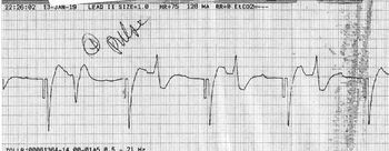
An 8-year-old boy (30 kg) was transferred to our cardiac ICU from an outside emergency room for further management after a witnessed sudden cardiac arrest at home. As per the mother, he had not wiped his buttocks properly after using the toilet and was being reprimanded for the same when he abruptly collapsed. The mother did not feel any pulses or a heartbeat upon palpation; therefore, emergency medical services were called and cardiopulmonary resuscitation was immediately initiated. Cardiopulmonary resuscitation was continued while he was being transported to the emergency room (adult facility) for stabilisation and he was defibrillated a few times during transport without return of pulses. In the emergency room, ventricular fibrillation was documented and he was defibrillated twice (ventricular fibrillation terminated but restarted immediately), intubated and ventilated, and also administered 150 mg of amiodarone intravenously. After 15 minutes of cardiopulmonary resuscitation and several additional defibrillation attempts, transcutaneous pacing was initiated with intermittent capture and alternating episodes of a slow ventricular-paced rhythm with premature ventricular contractions (Fig 1) and severe sinus bradycardia (heart rate 30–40 bpm) during which weak femoral and carotid pulses could be palpated. At this time, transport to our facility was initiated. A chest radiograph was also obtained and showed appropriate endotracheal tube position, normal-sized cardiac silhouette, and clear lung fields. Unfortunately, he lost pulses en route with severe sinus bradycardia (heart rate 20 bpm) due to complete loss-of-capture and required prolonged cardiopulmonary resuscitation which was continued in our cardiac ICU. Transcutaneous pacing was attempted again without successful capture. Due to prolonged cardiopulmonary arrest time, a decision was made to not initiate extracorporeal membrane oxygenation (ECMO) after extensive discussion with the family. Genetic testing (Catecholaminergic polymorphic ventricular tachycardia panel, Ambry Genetics, Aliso Viejo, CA, USA) was obtained and revealed a likely pathogenic mutation in RYR2 coding exon 8 [c.506G > T (p.R169L)]. In addition, p.G1339 (c.4015G > T) variant of unknown significance in ABCC9 which has not been associated with any abnormality and p.R52H (c.155G > A) variant of unknown significance in KCNJ5 which has been associated with sporadic primary hyperaldosteronism were detected.Reference Murthy, Xu and Massimo1 The child’s past history was significant for two episodes of emotion-triggered syncope in the month prior to the fatal event – the first episode occurred immediately after falling down from a swing at home and the second during a visit with his dentist when a “scary tool” was brought out to explore his teeth. During both the episodes, he passed out abruptly for a brief period of time (30–45 seconds) but immediately reverted back to his usual self upon regaining consciousness. He was not connected to a cardiac monitor at the dentist’s office. Unfortunately, none of these two episodes were further investigated. He had otherwise been completely asymptomatic from a cardiovascular standpoint and the parents denied a history exercise-triggered syncope or hypertension. The family history was significant for sudden death in the maternal great grandmother in her early 30s. His father, mother, and younger brother are negative for the RYR2 mutation suggesting that the mutation had arisen de novo. The father who has been asymptomatic is positive for both the variants of unknown significance, while the brother who has also been asymptomatic and has a normal Holter, echocardiogram, and exercise stress test is only positive for the KCNJ5 variants of unknown significance. The mother who denies any cardiovascular symptoms is negative for both the variants of unknown significances.
A slow ventricular-paced rhythm followed by premature ventricular contractions is noted following successful defibrillation.

An autopsy was performed and revealed left ventricular hypertrophy [weight 128 g, mean normal 71.44 g (52.84–98.64 g), Z score 2.9;Reference Foster, Mackie, Mitsnefes, Ali, Mamber and Colan2 left ventricular thickness measured as follows: anterior wall 1.4 cm, lateral wall 1.0 cm, posterior wall 1.2 cm, interventricular septum 1.2 cm]. Fibromuscular bands were also noted in the right atrium. An echocardiogram was not performed as our focus was on restoring effective circulation without interrupting the cardiopulmonary resuscitation in this critically sick child who was being considered for ECMO support.
Of note, the patient was tested for 30 genes (ACTC1, ACTN2, ALPK3, ANKRD1, CSRP3, FHL1, FLNC, GLA, JPH2, LAMP2, MYBPC3, MYH6, MYH7, MYL2, MYL3, MYPN, NEXN, PLN, PRKAG2, PTPN11, RAF1, RIT1, SOS1, TCAP, TNNC1, TNNI3, TNNT2, TPM1, TTR, VCL) associated with hypertrophic cardiomyopathy and other cardiomyopathy associated genes such as MYOZ2 and TTN as a part of broad arrhythmia and cardiomyopathy panel which includes variants in 92 genes but did not carry any variant which has been associated with hypertrophic or any other cardiomyopathy. In addition, the parents and the brother have been investigated and have structurally normal hearts.
Discussion
Our patient carried a de novo mutation [c.506G > T (p.R169L)] in exon 8 of RYR2 and had left ventricular hypertrophy on autopsy, a combination which has not been reported before. He presented at 8 years of age with two episodes of emotion-triggered syncope which were followed by emotion-triggered sudden death. Given his acute fatal presentation, exercise or pharmacological stress testing could not be carried out; however, the family denied a past history of exercise-triggered syncope, dizziness, or seizures.
Catecholaminergic polymorphic ventricular tachycardia is a rare (prevalence: 1/10,000) channelopathy characterised by premature ventricular contractions, and monomorphic, bidirectional, or polymorphic ventricular tachycardia.Reference Kohli, Aziz, Beaser and Nayak3 In addition to exercise, as seen in our patient, arrhythmias in catecholaminergic polymorphic ventricular tachycardia patients are often triggered by acute emotion or stress.
While genotype–phenotype correlation in catecholaminergic polymorphic ventricular tachycardia patients is overall lacking, certain associations do exist. Mutations in the C-terminal channel-forming RYR2 domain have been associated with an increased risk of non-sustained ventricular tachycardia than the N-terminal domain mutations;Reference van der Werf, Nederend and Hofman4 in contrast, variants found at the interface between neighbouring subunits in the RYR2 N-terminal domain, in the S5 and S6 helices, and the S4–S5 linker of the RYR2 C-terminus have been associated with more severe phenotypic features such as cardiac arrest.Reference Roston, Yuchi and Kannankeril5 In addition, a large deletion in RYR2 exon 3 has been associated with dilated and non-compaction cardiomyopathies and a severe and at times nadolol and/or flecainide refractory catecholaminergic polymorphic ventricular tachycardia phenotype.Reference Kohli, Aziz, Beaser and Nayak3 In contrast to RYR2 exon 3 deletions, RYR2 mutations are not thought to be associated with structural cardiac alterations. However, several recent reports have challenged this paradigm.
Fujino et al screened 187 sarcomere mutation negative probands with hypertrophic cardiomyopathy for RYR2 mutations and identified a novel missense mutation, Thr1107Met (p.A1107M), in a proband who had severe left ventricular outflow tract obstruction (pressure gradient: 80 mmHg) and ventricular fibrillation. Familial evaluation of the proband revealed four other mutation carriers (aged 41.0 ± 13.6 years) all of whom showed asymmetric septal hypertrophy (septal thickness 21.0 ± 5.9 mm) and a normal left ventricular posterior wall thickness (11.3 ± 2.3 mm). Left ventricular end-diastolic dimension, percentage fractional shortening, and right ventricular size and function were normal in all of these probands. DNA analysis of the family members revealed that the RYR2 p.A1107M mutation was inherited in this pedigree and co-segregated with the affected status suggesting that this mutation caused the disease. Furthermore, this mutation was absent in the other 299 probands or 200 normal controls.Reference Fujino, Ino and Hayashi6 The cellular effects of this mutation were subsequently elucidated in vitro where it was found to increase the threshold for CaReference Foster, Mackie, Mitsnefes, Ali, Mamber and Colan2 release termination and decrease the fractional Ca2 release in HEK293 cell lines.Reference Tang, Tian, Wang, Fill and Chen7
Only one other patient with RYR2 p.R169L mutation has been reported till date.Reference Ohno, Hasegawa and Horie8 The mutation had arisen de novo in a young girl who had catecholaminergic polymorphic ventricular tachycardia and presented with a syncopal episode at 9 years of age. The echocardiographic findings in this patient were not reported.
An alternate amino acid substitution, p.R169Q, at this position has been reported in six patients with catecholaminergic polymorphic ventricular tachycardia. Hsueh et al reported an 18-year-old female who presented with sudden collapse during exercise and was found to have episodes of bidirectional ventricular tachycardia during exercise stress testing. This patient had a structurally normal heart and responded well to β-blockers.Reference Hsueh, Weng and Chen9 Ohno et al reported two girls, aged 5 and 9 years, with the same mutation, both of whom presented with cardiopulmonary arrest.Reference Ohno, Hasegawa and Horie8 This alternative mutation had also arisen de novo in these three reported patients.Reference Ohno, Hasegawa and Horie8,Reference Hsueh, Weng and Chen9 Nozaki et al recently reported three unrelated female carriers of RYR2 p.R169Q mutation who had onset of exercise-induced syncope between 5 and 7 years of age. The mutation had arisen de novo in two of these patients; familial evaluation could not be carried out for the other patient. More importantly, all of these three patients had left ventricular non-compaction cardiomyopathy and two of them initially presented with sudden cardiac arrest.Reference Nozaki, Kato and Uike10 It is noteworthy that there is a considerable overlap between left ventricular non-compaction and hypertrophic cardiomyopathies and the same mutations have been associated with expression of either of these two phenotypes.Reference Oechslin and Jenni11 These three patients were screened for other mutations in cardiomyopathy-associated genes but were negative for the same. In vitro functional analysis of the RYR2 p.R169Q has shown decreased thresholds for overload-induced Ca2+ release and increased fractional Ca2+ release from the sarcoplasmic reticulum.Reference Nozaki, Kato and Uike10
The average age at diagnosis of catecholaminergic polymorphic ventricular tachycardia has been reported to be between 11 and 27 years in unselected cohorts of RYR2 mutation carriersReference van der Werf, Nederend and Hofman4,Reference Roston, Yuchi and Kannankeril5 and only 50–65% of unselected RYR2 mutation carriers identified by familial screening of probands express catecholaminergic polymorphic ventricular tachycardia phenotype.Reference Roston, Yuchi and Kannankeril5,Reference Bauce, Rampazzo and Basso12 An acute event rate of 58% has been reported in a cohort which consisted of untreated asymptomatic and symptomatic RYR2 mutation carriers over 8 years of follow-up.Reference Hayashi, Denjoy and Extramiana13 Severe symptoms such as sudden cardiac death or aborted sudden cardiac death were only seen in 15% of unselected RYR2 mutation carriers in the same cohort.Reference Hayashi, Denjoy and Extramiana13 In contrast, though the overall numbers are small, more than 70% of the reported patients with a loss of arginine at 169th position of RYR2 have presented with severe symptoms such as sudden cardiac arrest or aborted sudden cardiac arrest. Moreover, an asymptomatic carrier of a mutation which leads to replacement of arginine at 169th position of RYR2 has not been reported till date.
Arginine at the 169th position of RYR2, which is known to be highly conserved among several vertebrate species and is located within one of the mutation hot spots of RYR2,Reference Ohno, Hasegawa and Horie8,Reference Hsueh, Weng and Chen9 plays an important role is functioning of RYR2 as suggested by nuclear magnetic spectroscopic evaluation of the structural effects of R169Q mutation in vitro. These include a lack of visibility in the electron density which is consistent with a higher degree of flexibility of the α-helix and a moderate conformational effect on the hot-spot loop and the core RYR2 structure. These findings suggest that R169Q causes local structural perturbations within or near the hot-spot loop of RYR2 which then lead to functional effects.Reference Amador, Kimlicka and Stathopulos14 In addition, RYR2 p. R169Q leads to diminished size of the side chains and reduced positive charge and stacking interaction of the RYR2 protein. These alterations possibly affect allosteric regulation without induction of a conformational change or structural instability. It is noteworthy that the same mechanism, impaired allosteric regulation, has been proposed in patients with RYR2 exon 3 deletions, which are known to be associated with cardiomyopathies and a more severe catecholaminergic polymorphic ventricular tachycardia phenotype.Reference Kohli, Aziz, Beaser and Nayak3,Reference Nozaki, Kato and Uike10,Reference Amador, Kimlicka and Stathopulos15,Reference Borko, Bauerová-Hlinková and Hostinová16
Though less likely, it is possible that the patient also carried a yet unidentified hypertrophic cardiomyopathy-associated mutation which had arisen de novo. The chance of simultaneous expression of catecholaminergic polymorphic ventricular tachycardia and hypertrophic cardiomyopathy phenotypes at 8 years of age is however less than 1 in 5 million.
Haemodynamically unstable brady-arrhythmia was a potential contributor to mortality in this patient. Sinus node dysfunction is a well-established finding in patients with catecholaminergic polymorphic ventricular tachycardia due to RYR2 mutations and exon 3 deletions;Reference Kohli, Aziz, Beaser and Nayak3,Reference Kohli, Aziz, Beaser and Nayak17 however, its contribution to hemodynamic instability during acute arrhythmic crises in catecholaminergic polymorphic ventricular tachycardia patients had not been systematically evaluated. The sinus bradycardia could have been further exacerbated by prolonged cardiopulmonary resuscitation which did not succeed in restoring effective spontaneous circulation and intravenous amiodarone which perhaps should have been avoided in this patient.
To conclude, our patient presented with emotion-triggered sudden death and was found to have RYR2 p.R169L mutation and left ventricular hypertrophy. Another mutation at the same residue RYR2 (p.Q169L) has been associated with catecholaminergic polymorphic ventricular tachycardia and left ventricular non-compaction cardiomyopathy in three unrelated patients. In addition, a mutation (p.A1107M) at a different locus of RYR2 has also been associated with hypertrophic cardiomyopathy in five members of a family. These findings suggest that some RYR2 mutations, including mutations that lead to loss of arginine at 169th position of RYR2, could be associated with an atypical catecholaminergic polymorphic ventricular tachycardia phenotype which includes structural alterations such as hypertrophic and left ventricular non-compaction cardiomyopathies. The precise mechanisms that underlie structural alterations in carriers of these RYR2 mutations have not been elucidated. Though the numbers are too small to come to any conclusion, loss of arginine at 169th position of RYR2 could also be associated with a more severe catecholaminergic polymorphic ventricular tachycardia phenotype. There however is an overall dearth of genotype–phenotype correlation studies in catecholaminergic polymorphic ventricular tachycardia patients and in vitro and in vivo effects of simultaneous expression of catecholaminergic polymorphic ventricular tachycardia and cardiomyopathy phenotypes have been poorly characterised. These studies are important to identify catecholaminergic polymorphic ventricular tachycardia patients who might be at a higher risk of adverse events including sudden cardiac death and therefore could benefit from a more aggressive therapeutic approach.
Acknowledgements
None.
Financial support
This research received no specific grant from any funding agency, commercial, or not-for-profit sectors.
Conflicts of interest
None.