In lichens with a competitive ecological strategy, reproduction, dispersal and maintenance of the habitat are considered equally important (Ott Reference Ott1987). While wind, heavy rain or large animals may detach whole thalli or thallus pieces, small herbivores such as snails, slugs, mites or insects may cause physical damage by grazing systematically on whole lobes or particular thallus parts. In this way, herbivores can decimate preferred species or limit their thallus growth (Lawrey Reference Lawrey1983; Fröberg et al. Reference Fröberg, Baur and Baur1993, Reference Fröberg, Baur and Baur2006; Baur et al. Reference Baur, Baur and Fröberg1994, Reference Baur, Fröberg and Baur1995, Reference Baur, Fröberg, Baur, Guggenheim and Haase2000; Benesperi & Tretiach Reference Benesperi and Tretiach2004; Asplund & Gauslaa Reference Asplund and Gauslaa2008; Asplund et al. Reference Asplund, Larsson, Vatne and Gauslaa2010; Asplund Reference Asplund2011). In the case of Lobaria pulmonaria (L.) Hoffm., it seems that grazing invertebrates may reduce populations of recently established young thalli (Asplund & Gauslaa Reference Asplund and Gauslaa2008), but grazing damage does not necessarily change growth rates of this species if growth zones are not affected (Gauslaa et al. Reference Gauslaa, Holien, Ohlson and Solhøy2006). There has been considerable research on regeneration of structures developed after artificially caused damage (Henssen & Jahns Reference Henssen and Jahns1974; Armstrong Reference Armstrong1979, Reference Armstrong2010; Honegger Reference Honegger1996a ; Honegger et al. Reference Honegger, Conconi and Kutasi1996). Moreover, studies assessing wounds caused by grazing snails have revealed that different lichen species respond with the same regeneration process; in both cases they activate lobe primordia along wound margins and start with the formation of secondary lobes (Baur et al. Reference Baur, Fröberg, Baur, Guggenheim and Haase2000; Fröberg et al. Reference Fröberg, Baur and Baur2006). A few studies suggest that regenerative abilities of lichens might have the potential to restructure new thalli from fragments that have survived the gut passage of feeding invertebrates (Fröberg et al. Reference Fröberg, Björn, Baur and Baur2001; Meier et al. Reference Meier, Scherrer and Honegger2002; Boch et al. Reference Boch, Prati, Werth, Rüetschi and Fischer2011). However, evidence for successful regeneration under field conditions is still lacking.
During fieldwork, we regularly observe thalli with feeding damage or dying lobes that had formed regeneration lobes. All previous studies, however, have focused on regeneration structures and too little attention has been paid to repair mechanisms in wound healing. In this study, we report on morphological reactions to physical damage in field specimens of L. pulmonaria and Lobaria amplissima (Scop.) Forss. that were wounded through natural processes including herbivory and mechanical stress.
The study had to be limited to a small number of specimens, because L. pulmonaria and L. amplissima are threatened species in Switzerland (Scheidegger et al. Reference Scheidegger, Clerc, Dietrich, Frei, Groner, Keller, Roth, Stofer and Vust2002; www.swisslichens.ch). Mature thalli of L. pulmonaria and L. amplissima (seven specimens of each) growing on bark of Acer pseudoplatanus L. were collected in an upper mountain mixed forest in the Wägital valley, canton of Schwyz, Switzerland, (47°04′70″ N, 8°56′40″ E). The collection site was 900 m above sea level. All specimens were air dried and subsequently processed for microscopic analysis within a few days after collection, following the protocols described by Cornejo & Scheidegger (Reference Cornejo and Scheidegger2013). We studied grazing damage for both species, but dying lobes were only studied for L. pulmonaria.
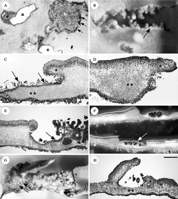
All thalli collected exhibited damage restricted to small areas of the thallus caused by slugs or snails that showed all the same wound characteristics. Concordant with previous studies, we noted that feeding herbivores have particular food preferences because the upper cortex and the algal layer were regularly grazed but not the medulla or cephalodia containing cyanobacteria (Fig. 1A, C, E & F). Some wounds were restricted to the upper cortex only, leaving most algal cells intact (Fig. 1A, asterisks). Freshly grazed parts left an exposed, white medulla and on wound margins the visible algal layer was a vivid green colour (Fig. 1A, ‘a’ & ‘c’). Other wounds, however, were brownish and exhibited, in semi-thin sections, a secondary cortex with a paraplectenchymatous stratum: thick-walled cells with small lumina that sealed the open medulla (Fig. 1A, C, E & F). In many cases, the secondary cortex was covered by a whitish tomentum; single hyphae that protruded from the secondary cortex (Fig. 1A, at ‘b’ & 1C, arrows). We observed that in all cross-sections the secondary cortex was thinner and the medulla below it was formed by tighter interwoven hyphae than in the medulla of undamaged thallus parts (Fig. 1C, E & F). Lobaria amplissima formed regeneration lobes along wounds in peripheral or central thallus parts or on wounds that slit the thallus (Fig. 1A, C & E), but in L. pulmonaria new lobes were only observed along vertically damaged parts of the thallus (Fig. 1B). Additionally, in L. pulmonaria we found few soredia and mainly globular to cylindrical isidia-like regeneration lobes covered with a cortex but not stratified internally (Fig. 1B, arrow & 1D, asterisk), whereas L. amplissima formed flattened lobes stratified in the upper and lower cortex, algal layer and medulla (Fig. 1C, asterisk & 1E). While wounds above cephalodia of L. pulmonaria seemed not to affect their position within the thallus or their morphology (Fig. 1F), we regularly found within wounds of L. amplissima the coralloid, external type of cephalodia, referred to as Dendriscocaulon umhausense (Auersw.) Degel. (Fig. 1E, asterisk).
Grazed wounds on thallus surface and dying lobes. A, four grazing marks on a thallus of L. amplissima. On wounds ‘a’ & ‘c’, the white medulla is visible and parts of the upper cortex are grazed but not the algal layer (asterisks), around the wound ‘b’, new regeneration lobes are formed (arrows) and, within the wound, the brown-pigmented secondary cortex and its whitish tomentum can be seen; B, a wound on L. pulmonaria that crosses the thallus vertically, on the thallus border new cylindrical lobules are formed (arrow); C & D, semi-thin sections of wounds shown on (A) and (B), in both species, the medulla below the secondary cortex appears formed by tightly interwoven hyphae (double asterisks) and a secondary cortex seals the wound; C, this lobule of L. amplissima is dorsi-ventrally stratified (asterisk) and the secondary cortex is covered by single hyphae that protrude from the secondary cortex (arrows); D, this marginal lobe of L. pulmonaria is internally nonstratified (asterisk); E & F, grazed lobes containing cephalodia (asterisks) and a secondary cortex (arrows); E, semi-thin section of a coralloid cephalodium of L. amplissima growing out of a wound; F, hand-cut sections of wounds above internal cephalodia of L. pulmonaria; G & H, dying lobes of L. pulmonaria with regeneration structures; G, narrow lobe where the algal layer is reduced to small groups (black arrows), if present at all, and the upper cortex is disintegrated, the most developed lobe (asterisk) grows upright on the parent thallus and keeps contact with its base, soredia were also found on dying lobes (white arrow); H, semi-thin section of two regeneration lobes; the smaller one is not stratified (asterisk), the upper cortex and algal layer are partially missing and the parent thallus does not develop a secondary cortex or tightly interwoven medulla (double asterisks). Scale bar in H (for all figures): A=388 µm; B=137 µm; C=381 µm; D=245 µm; E=548 µm; F=143 µm; G=326 µm; H=443 µm.

We also examined dying lobes of L. pulmonaria (Fig. 1G & H), which produced new lobes within the affected areas. In semi-thin sections, dying lobes could be clearly distinguished because, not only the upper cortex and algal layer were missing, but also a secondary cortex (Fig 1G & H). It is notable that dying thalli formed regeneration lobes that were tightly arranged (Fig. 1G). In semi-thin sections, these lobes were identical to lobes formed along wound margins (Fig. 1H). New lobes grew upright from the former thallus and kept contact with the old lobe at their base (Fig. 1G & H), except for those growing at the thallus margin that grew horizontally (not shown).
Lobaria pulmonaria and L. amplissima revealed two different and species-specific responses to damage, either by mechanical stress or grazing, namely repair and regeneration. Both species studied displayed a repair mechanism during wound healing processes that resulted in the sealing of open wounds with a secondary cortex. We have not observed a repair of the algal layer below the secondary cortex, but Gauslaa et al. (Reference Gauslaa, Holien, Ohlson and Solhøy2006) noted in L. pulmonaria old grazing marks, “in which green algal cells had already re-colonized the algal deficient patches”. A secondary cortex also repairs wounds above cephalodia. In L. pulmonaria, cephalodia do not change their morphology and our observations do not confirm Jordan's (Reference Jordan1970) conclusion that internal cephalodia would grow to the upper or lower cortex depending on least mechanical resistance. Additionally, internal cephalodia of L. amplissima seem to develop into the coralloid form of this species. It is difficult to establish if these cephalodia would break through anyway or whether the new anatomical situation after feeding triggers formation of the coralloid type. It is also conceivable that the coralloid cephalodia observed were formed before grazing and herbivores have avoided feeding on them. Experimental studies are required to address this question. The formations shown here demonstrate that regeneration occurs depending on the position of injuries on the thallus. In L. pulmonaria, new lobes develop only on wounds that slit the thallus, while in L. amplissima new lobes grow in any part of the thallus. Dying lobes of L. pulmonaria form a high number of new lobes where green algae are still available. In most cases we found globular to cylindrical lobes that had a cortex, but also soredia were observed, indicating that lobes in dying thalli might also begin their development as soredia. We could not discover why these lobes of L. pulmonaria were declining; however, we know that herbivores may graze the algal layer of whole lobes and induce a necrosis. It is also well known that senescent thalline areas disintegrate regularly and are overgrown (Ott et al. Reference Ott, Meier and Jahns1995; Honegger Reference Honegger and Nash1996b ).
The empirical findings in this study have demonstrated the ability of lichenized fungi to repair injured lobes by sealing open wounds. Additionally, the two species studied react with the formation of new lobes that regenerate lost parts. In this investigation, repair and regeneration were observed in two rather closely related species and they are, nonetheless, highly distinct at the species level. It can therefore be assumed that repair and regeneration are genetically controlled mechanisms resulting from a divergent evolutionary history. Along with dispersal, growth and regeneration, repair is an integral element of coherent strategies for successfully gaining and maintaining a presence in a habitat. But wound repair in particular, and its consequences for life-history traits of lichens, is still poorly understood. More experimental research on the effects of wound repair on growth and reproductive output is needed.
We thank the anonymous reviewer and especially Yngvar Gauslaa for constructive comments on the manuscript.