Introduction
Nasopharyngeal carcinoma (NPC) is unique among head and neck cancers because of its epidemiology – it is relatively rare worldwide and is predominantly found in men.Reference Chan, Plich, Kuo, Barnes, Eveson, Reichart and Sidransky1 However, in China the age-adjusted incidence of NPC ranges from 25 to 50 per 100 000.Reference Ayan, Kaytan and Ayan2 Although NPC is markedly radiosensitive, the curative effect is far from perfect in later-stage patients due to the tumour's invasiveness and metastatic behaviour. These factors, along with the resistance of NPC cells to chemotherapeutic drugs, are major problems in NPC treatment.
Our previous observations indicated that overexpression of epidermal growth factor and insulin-like growth factor 1 receptors is readily detectable in approximately 65.3 and 56 per cent of NPC cases, respectively. However, most normal tissues and benign tumours do not show evidence of such overexpression.Reference Yulin, Xuhong, Jian, Xiaoping, Jun, Linfeng, Xiaoping and Dong3 Therefore, these receptor molecules could be attractive anti-cancer treatment targets. Inhibitors of the epidermal growth factor receptor (ErbB1) and of human epidermal growth factor receptor (Her2) (ErbB2) have already been shown to have clinical application in lung and breast cancer.Reference Hortobagyi4
The insulin-like growth factor 1 receptor is a heterotetrameric tyrosine kinase receptor with close homology to the insulin receptor. Insulin-like growth factor 1 receptor is synthesised as a proreceptor, which undergoes cleavage into alpha and beta subunits before assembly into α2β2 tetramers and insertion into the plasma membrane.Reference Ward, Garrett, McKern, Lou, Cosgrove and Sparrow5 Insulin-like growth factor 1 receptor is becoming increasingly recognised as an attractive anti-cancer treatment target; it is frequently overexpressed in tumours, often as a result of tumour suppressor gene loss, and it mediates tumour cell proliferation, survival, and various properties required for invasion and metastasis.Reference Werner and Roberts6 These functions are predominantly mediated through insulin-like growth factor induced activation of the phosphoinositide 3 kinase–AKT, AKT–RAS–RAF and p38 mitogen-activated protein kinase signalling cascades.Reference Vincent and Feldman7
Our previous research also showed that dual epidermal growth factor receptor and insulin-like growth factor 1 receptor gene silencing could reduce further expression of these receptors in the CNE2 NPC cell line.Reference Yuan, Zhou, Song, Qiu, Li and Yed8 In CNE2 cells, ribonucleic acid (RNA) interference is triggered by the presence of double-stranded RNA, and results in rapid destruction of messenger RNA that contains an identical or near-identical sequence to the double-stranded RNA.Reference Hannon9, Reference Liu, Ma, Liu, Liu and Sehon10 In other words, a post-transcriptional mechanism is involved. Ribonucleic acid interference is thought to have evolved as a defence mechanism to suppress viral replication and transposon mobilisation.Reference Montgomery11 Since its discovery in 1998, this mechanism has become one of the most widely applied technologies in molecular and cellular research.Reference Fire, Xu, Montgomery, Kostas, Driver and Mello12 Ribonucleic acid interference has been shown potently to ablate targeted messenger RNA within a variety of species.Reference Paddison, Caudy, Bernstein, Hannon and Conklin13 The use of deoxyribonucleic acid (DNA) vectors constructed to mediate RNA interference by expressing short hairpin RNA from U6 promoters is a newly established technique which can produce long-term, stable and highly specific gene silencing.Reference Brummelkamp, Bernards and Agami14
In our previous work, we found that treatment with short hairpin RNA expression vectors induced a significant decrease both in epidermal growth factor and insulin-like growth factor 1 receptor messenger RNA expression and in the levels of these receptor proteins, within the CNE2 NPC cell line.Reference Yulin, Xuhong, Jian, Xiaoping, Jun, Linfeng, Xiaoping and Dong3, Reference Yuan, Zhou, Song, Qiu, Li and Yed8 In the present study, we investigated the influence of such short hairpin RNA expression vectors on the in vivo growth of human NPC in an animal model, in order to test the therapeutic efficacy of this treatment.
Materials and methods
Materials
The following were commercially obtained: rabbit polyclonal anti-insulin-like growth factor one receptor β chain (1:1000; C20, Santa Cruz, California, USA); goat anti-mouse primary polyclonal antibody to epidermal growth factor receptor (Santa Cruz Biotechnology, Santa Cruz, California, USA); secondary antibody immunoglobulin (Ig) G (Pierce, Rockford, Illinois, USA); Trizol reagent (Gibco, Carlsbad, California, USA); deoxynucleoside triphosphates (dNTP) (Invitrogen, Carlsbad, USA); ribonuclease inhibitor (RNasein) (Invitrogen); RTase (Gibco); and Taq DNA polymerase (Promega, Madison, Wisconsin, USA). The CNE2 nasopharyngeal carcinoma (NPC) cell line was purchased from Type Culture Conservation (Wuhan, PR China). The primer and probe were designed by Primer 5.0 software (RPMI-1640 culture medium) and synthesised by Invitrogen.
Cell culture
The human NPC CNE2 cell line was cultured with 5 per cent CO2 in RPMI-1640 medium (Gibco), with 10 per cent heat-inactivated fetal calf serum (Hyclone, Logan, Utah, USA), 100 U/ml penicillin and 100 mg/ml streptomycin.
Construction of short hairpin ribonucleic acid expression plasmids
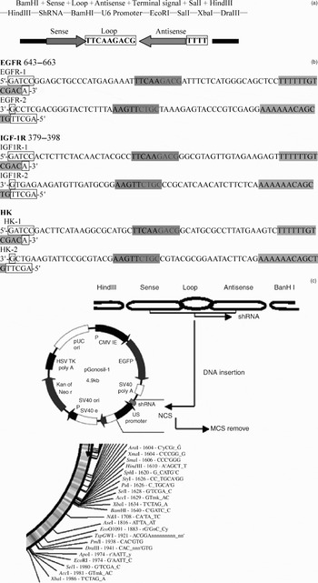
Short hairpin RNA segments targeting plasmids for epidermal growth factor receptor and insulin-like growth factor 1 receptor were designed, according to the complementary DNA sequence of the relevant receptor: GenBank accession number NM_201283 for epidermal growth factor receptor and GenBank accession number NM_000875 for insulin-like growth factor 1 receptor. A short hairpin RNA segment targeting the HK plasmid, which did not target any specific human gene, was also designed as a control. Short hairpin RNA segments encoding a DNA template were designed as follows: a 19-nucleotide target sequence (as a sense strand), followed by a spacer and complementary antisense strand, and then four continuous thymines as a terminate signal (Figure 1a and 1b). The short hairpin RNA segments were subcloned into the HD5a (Figure 1c), with human U6 promoter, between the BamHI and HindIII restriction sites. A short hairpin RNA expression plasmid, which carried an enhanced green fluorescence protein gene (constructed by JingSai Biotechnology, Wuhan, PR China) was used. All of the constructs used in this study were verified by DNA sequencing.
The structure of short hairpin ribonucleic acid (shRNA) segments and their vector. (a) Predicted structure of shRNA segments. (b) Design of shRNA template for epidermal growth factor receptor (EGFR), insulin-like growth factor 1 receptor (IGF-1R) and (HK). (c) Diagram of the vector. An shRNA encoding template was inserted between BamHI and HindIII restriction sites downstream of the U6 promoter. Transcripts of the template (see part (b)) will form a 19-nucleotide, double-stranded stem with a 9-nucleotide loop hairpin that targets either mouse EGFR or IGF-1R transcriptase messenger RNA (mRNA) or no human gene mRNA. T = thymine; C = cytosine; G = guanine; A = adenine; CMV IE = cytomegalovirus; EGFP = enhanced green fluorescent protein; HSV TK = thymidine kinase; DNA = deoxyribonucleic acid; NCS = neocarzinostatin; MCS = multiple cloning sites

Forty female Balb/c nude mice aged between five and six weeks (body weight 16–18 g) were purchased from the Experimental Animal Centre of Hubei province. All mice were housed in a cage, under laminar airflows and in pathogen-free conditions. The mice were maintained at a constant temperature (18–22°C) and relative humidity (50–80 per cent), with 12-hour dark–light cycles. They were fed on a standard diet and given water ad libitum. All experiments were performed with aseptic technique under laminar airflow. The animals were inspected daily and any sign of discomfort was recorded.
The animals were divided into five groups of eight mice each, according to whether they received one of the following injections: short hairpin RNA epidermal growth factor receptor plasmid; short hairpin RNA insulin-like growth factor 1 receptor plasmid; short hairpin RNA epidermal growth factor receptor plasmid and short hairpin RNA insulin-like growth factor 1 receptor plasmid (1:1 combination); short hairpin RNA HK plasmid; or saline (i.e. control).
Tumour method and analysis
Approximately 1 × 10⁷ NPC cells in 0.2 ml of serum-free media were inoculated subcutaneously into the right flank. Tumour growth was monitored using a caliper every two or three days. Tumour volume (V) was calculated by the formula (L × WReference Ayan, Kaytan and Ayan2)/2, where L = length (mm) and W = width (mm). Tumour treatment was commenced when the maximum tumour diameter reached 5–7 mm. Twenty micrograms of short hairpin RNA epidermal growth factor receptor plasmid or short hairpin RNA insulin-like growth factor 1 receptor plasmid, or both, or short hairpin RNA HK plasmid, dissolved in 300 µl of serum-free medium with 30 µl transfection reagent (Metafectene, Biontex, Munich, Germany), was directly injected into the tumour once every two days for a total of seven times. Saline injection was used as a control. Inhibition ratio was calculated by the formula (V − V1)/V, where V = volume before the treatment (mm3) and V1 = volume after the treatment (mm3).
Mice were sacrificed by depleting, seven days after the final treatment. The tumours were then removed and weighed. Part of the tumour tissue was frozen instantly, cryosectioned at 20 µm and observed for fluorescence under a confocal laser scanning fluorescent microscope. Of the remaining tumour tissue, half was analysed by quantitative real-time polymerase chain reaction, and the other half was fixed in 4 per cent paraformaldehyde in phosphate-buffered saline overnight, embedded in paraffin wax and then sectioned at 5 µm. Peripheral blood was also collected for haematological and biochemical analysis.
Cell staining and apoptosis detection
Tumour sections were stained with haematoxylin and eosin for morphological observation. In order to study how the short hairpin RNA had inhibited tumour growth, apoptotic tumour cell death was examined. Apoptotic cells were identified using the modified end-labelling technique originally described by Zhang et al. Reference Zhang, Zhang, Yin and Zhao15 All procedures were performed following the manufacturer's instructions (in situ cell apoptosis detection kit, Wuhan Boster Biological Technology, Wuhan, PR China).Reference Zhang, Zhang, Yin and Zhao15 Positive cells were visualised using diaminobenzidine tetrahydrochloride staining and methyl green counterstaining. Apoptotic cells were quantified according to morphological criteria and positive reactivity. Cells undergoing apoptosis had a shrunken cell body, a pyknotic nucleusReference Zhang, Zhang, Yin and Zhao15 and brown particles in the nucleus on labelling. The numbers of apoptotic cells and total cells within a microscope viewing field were counted at ×400 magnification. The cells counted in five viewing fields selected randomly by computer were used to calculate the apoptotic index. The apoptotic index was obtained by dividing the number of apoptotic cells by the total number of tumour cells, multiplied by 100, i.e. apoptotic index = (apoptotic cells/total cells) × 100.
Polymerase chain reaction analysis
Total RNA was extracted from 0.5 mg of tumour tissue using Trizol reagent. Complementary deoxynucleic acid (cDNA) was synthesised from 1 µg total RNA using 1.0 µl Oligo (dT) primer (50 µg/ml). The mixture was heated at 70°C for 5 minutes and chilled on ice for 2 minutes. The following were then added: 5× buffer 4.0 µl, 10 mM dNTP 2.0 µl; 20 units of ribonuclease inhibitor (RNasein); and 200 units of reverse transcriptase (RTase). The resultant mixture was incubated at 37°C for 60 minutes, heated at 95°C for 5 minutes and then stored at −20°C.
The RNA was then analysed for the integrity of the 28S, 18S and 5S ribosomal RNA bands by Northern hybridisation, using formaldehyde-containing agarose gels. During polymerase chain reaction, 1 µl of cDNA production was amplified with gene specific primers, in a total volume of 50 µl. This mixture contained: 1 µl copy DNA; 10 × buffer 50; 7 µl of 25 mM Mgcl2); 1 µl of 10 mM dNTP; 0.8 µl of 20 pmol/μl insulin-like growth factor 1 receptor or epidermal growth factor receptor or β-actin upstream primer; 0.8 µl of 20 pmol/μl epidermal growth factor receptor or insulin-like growth factor 1 receptor or β-actin downstream primer; 0.4 µl of 20 pmol/μl epidermal growth factor receptor or insulin-like growth factor 1 receptor or β-actin probe; 0.5 µl of 5 U/μl Taq DNA polymerase; and H2O added to make up 50 µl.
The upstream primer of insulin-like growth factor 1 receptor was 5′-AACGCTTCAGTTCCTTCCATTC-3′ (where A = adenine, C = cytosine, G = guanine and T = thymine). The downstream primer was 5′-CTCTTCCGGGTCTGTGATATTGT-3′.
The upstream primer of epidermal growth factor receptor was 5′-CCAAGGCACGAGTAACAAGC-3′, and the downstream primer was 5′-CCAAATTCCCAAGGACCACC-3′.
The TaqMAN probe sequence was 5′-FAM-ACGCAGTTGGGCACTTTTGAAGATC-TAMARA-3′.
The reacting conditions were as follows: pre-degenerate at 94°C for 5 minutes, then degenerate at 94°C for 30 seconds, anneal at 52°C for 30 seconds and extend at 72°C for 30 seconds. There were 50 cycles in total.
With β-actin as a native reference, the upstream primer was 5′-GAACGGTGAAGGTGACAG-3′ and the downstream primer was 5′-TAGAGAGAAGTGGGGTGG-3′. The TaqMAN probe sequence was 5′-FAM-GACTTTGATTGCACATTGTTGTT- TAMARA-3′.
The reacting conditions were as follows: pre-degenerate at 94°C for 5 minutes, degenerate at 94°C for 30 seconds, anneal at 50°C for 30 seconds and extend at 72°C for 30 seconds. There were 50 cycles in total.
Negative contrast excluding cDNA was used. The results were analysed using the FTC-2000 real-time polymerase chain reaction system (Funglyn Biotech, Shanghai, China) to obtain the cycle threshold value. The experiment was repeated three times. The polymerase chain reaction quantitative measurement equation was as follows: the relative value of the tested group was equal to 2(ΔCt test group _ ΔCt β-actin) ×100.
Receptor protein expression
To detect expression of the epidermal growth factor receptor and insulin-like growth factor 1 receptor proteins, tumour sections were immunostained with an anti-epidermal growth factor receptor antibody and an anti-insulin-like growth factor 1 receptor antibody.
Briefly, paraffin sections were routinely deparaffinised and incubated in 3 per cent H2O2 for 10 minutes to block endogenous peroxidase. To prevent nonspecific antibody binding, the sections were pre-incubated in normal goat serum for 15 minutes, then incubated with rabbit anti-human insulin-like growth factor 1 receptor or goat anti-human epidermal growth factor receptor IgG (Santa Cruz Biotechnology) at 4°C overnight. After rinsing with distilled water, the sections were incubated with biotin-labelled anti-rabbit or anti-goat IgG at 37°C for 10 minutes, then treated with streptavidin–horseradish peroxidase complex at 37°C for 10 minutes. The labelled cells were visualised using 0.05 per cent 3;3'-diaminobenzidine as the chromagen. Finally, the sections were counterstained with Meyer's haematoxylin and dehydrated through serial ethanols. Negative control sections were prepared by substituting phosphate-buffered saline for anti-epidermal growth factor receptor or insulin-like growth factor 1 receptor antibody. Positive cells were stained brown.
The method for scoring epidermal growth factor receptor and insulin-like growth factor 1 receptor expression was modified from that described by Zhou et al. Reference Zhou, Yu, Liu, Luo, Chen and Chen16 Positive tumour cells were quantified by two independent observers. The mean percentage of positive tumour cells was determined from at least five random microscope viewing fields (at a magnification of ×400) and assigned to one of five categories: zero (5 per cent); one (5–25 per cent); two (25–50 per cent); three (50–75 per cent); or four (>75 per cent). The intensity of receptor protein immunostaining was scored as: one (weak), two (moderate) or three (intense). For tumours showing heterogeneous staining, the predominant pattern determined the scoring. Cases with weighted scores of less than two were defined as negative; otherwise, they were defined as positive.
Statistical analysis
Values were expressed as the mean ± standard deviation of multiple experiments. Analysis of covariance using the Dunnett or Tukey–Kramer post-tests was employed for multiple groups, and Fisher's test was used for comparison of the five groups, using the Statistical Package for the Social Sciences version 11.5 software (SPSS Inc, Chicago, Illinois, USA).
Results
During the experiments, the general condition of all the treated animals was good and did not differ from that of the controls.
Study data are shown in Tables I to IV.
Effect of shRNA plasmid treatment on tumour volumes and inhibition ratios in nude mice

Data obtained after 20 days' treatment; eight animals in each treatment group. *p < 0.01, compared with saline control and short hairpin ribonucleic acid (shRNA) HK plasmid groups. †p < 0.01, compared with saline control and shRNA HK plasmid groups. ‡p < 0.05, compared with shRNA epidermal growth factor receptor (EGFR) plasmid alone or shRNA insulin-like growth factor 1 receptor (IGF-1R) plasmid alone. SD = standard deviation
Relative value of EGFR and IGF-1R MRNA in the treatment groups

Data are presented as (mean ± SD) × 100. Epidermal growth factor receptor (EGFR) and insulin-like growth factor 1 receptor (IGF-1R) messenger ribonucleic acid (mRNA) expression were detected by quantitative real-time polymerase chain reaction. In the statistical results, the same letter indicates that no significant difference existed between the two groups of data, while different letters indicate significant differences between the groups of data. shRNA = short hairpin RNA; HK
Effect of shRNA plasmid treatment on peripheral blood in nude mice

Data obtained after 20 days' treatment, presented as mean ± standard deviation. *p > 0.05, compared with short hairpin ribonucleic acid (shRNA) epidermal growth factor receptor (EGFR) plasmid alone, shRNA insulin-like growth factor 1 receptor (IGF-1R) plasmid alone, shRNA (HK) plasmid and saline control group. RBC = red blood cells; Hb = haemoglobin; PLT = platelets; WBC = white blood cells
Effect of shRNA plasmid treatment on biochemical parameters in nude mice

Data obtained after 20 days' treatment, presented as mean ± standard deviation. *p>0.05, compared with short hairpin ribonucleic acid (shRNA) epidermal growth factor receptor (EGFR) plasmid alone, shRNA insulin-like growth factor 1 receptor (IGF-1R) plasmid alone, shRNA HK plasmid and saline control group. ALT = alanine transaminase; AST = glutamic oxalacetic transaminase; ALP = alkaline phosphatase; TP = total protein; ALB = albumin; BUN = blood urea nitrogen; TB = total bilirubin; DB = direct bilirubin; GLU = glucose
Tumour growth inhibited by dual plasmid treatment
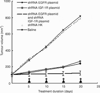
As shown in Table I and Figure 2, the growth of the CNE2 subcutaneous nasopharyngeal carcinoma tumours was significantly suppressed following treatment with both short hairpin RNA epidermal growth factor receptor plasmid and short hairpin RNA insulin-like growth factor 1 receptor plasmid, compared with that of tumours treated with short hairpin RNA epidermal growth factor receptor plasmid alone, short hairpin RNA insulin-like growth factor 1 receptor plasmid alone (p < 0.05), saline and short hairpin RNA HK plasmid (p < 0.01), respectively.
Subcutaneous tumour growth. Tumour growth was greatly suppressed following treatment with short hairpin ribonucleic acid (shRNA) epidermal growth factor receptor (EGFR) and shRNA insulin-like growth factor 1 receptor (IGF-1R) plasmids together, compared with treatment with shRNA EGFR plasmid alone or shRNA IGF-1R plasmid alone (*p < 0.05), and also compared with treatment with shRNA (HK) plasmid or saline (▴p < 0.01). Whiskers show outliers.

In animals treated with both short hairpin RNA epidermal growth factor receptor plasmid and short hairpin RNA insulin-like growth factor 1 receptor plasmid for 20 days, the mean tumour volume was 124.55 ± 11.04 mm3. In contrast, animals receiving other treatments had the following mean tumour volumes: short hairpin RNA epidermal growth factor receptor alone, 242.34 ± 18.98 mm3; short hairpin RNA insulin-like growth factor 1 receptor plasmid alone, 268.33 ± 17.08 mm3; short hairpin RNA HK plasmid, 818.25 ± 117.35 mm3; and saline, 786.23 ± 116.45 mm3.
There was no statistically significant difference between the mean tumour volumes of mice injected with short hairpin RNA epidermal growth factor receptor plasmid alone and short hairpin RNA insulin-like growth factor 1 receptor plasmid alone (p > 0.05), suggesting that injection with either of these two plasmids had a similar inhibitory effect on tumour growth in nude mice. However, injection of both plasmids together had a relatively strong inhibitory effect on the tumour.
Expression of short hairpin RNA plasmid in the tumour
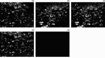
Green fluorescence was found in the majority of tumour cells harvested from mice injected with short hairpin RNA epidermal growth factor receptor plasmid alone, short hairpin RNA insulin-like growth factor 1 receptor plasmid alone, both of these plasmids together, and short hairpin RNA HK plasmid. This suggests that all these tumour cells were successfully transfected with the various plasmids. As expected, no green fluorescence was detected in tumours injected with saline (as shown in Figure 3).
Fluorescent protein expression in the tumours. Many tumour cells with green fluorescence were present in mice transfected with (a) short hairpin ribonucleic acid (shRNA) epidermal growth factor receptor (EGFR) plasmid alone, (b) shRNA insulin-like growth factor 1 receptor (IGF-1R) plasmid alone, (c) both shRNA EGFR and shRNA IGF-1R plasmids together, and (c) shRNA HK plasmid. No fluorescence was found in tumours from mice transfected with saline (e).

More tumour cell death following dual vs single plasmid transfection
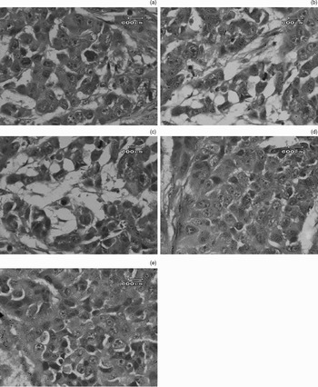
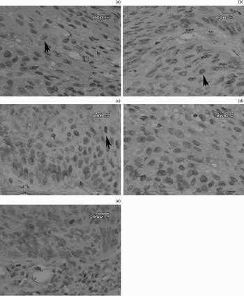
Many necrotic tumour cells were present in tumour tissue from mice treated with both short hairpin RNA epidermal growth factor receptor plasmid and short hairpin RNA insulin-like growth factor 1 receptor plasmid. However, tumour cells grew well in mice treated with short hairpin RNA HK plasmid or saline (as shown in Figure 4).
Histopathological structure of the subcutaneous tumours. Necrotic tumour cells were present in mice transfected with: (a) short hairpin ribonucleic acid (shRNA) epidermal growth factor receptor (EGFR) plasmid alone (moderate amount of necrosis); (b) shRNA insulin-like growth factor 1 receptor (IGF-1R) plasmid alone (moderate amount); (c) both shRNA EGFR and shRNA IGF-1R plasmids (large amount); (d) shRNA HK plasmid (small amount); and (e) saline (small amount) (H&E; ×1000).

More apoptotic cells were found in tumours transfected with both short hairpin RNA epidermal growth factor receptor plasmid and short hairpin RNA insulin-like growth factor 1 receptor plasmid. However, a few apoptotic cells were also detected in tumours transfected with short hairpin RNA epidermal growth factor receptor plasmid alone or short hairpin RNA insulin-like growth factor 1 receptor plasmid alone. The apoptotic index of tumours treated with both short hairpin RNA epidermal growth factor receptor plasmid and short hairpin RNA insulin-like growth factor 1 receptor plasmid also higher than that of tumours treated with short hairpin RNA HK plasmid (2.73 ± 1.35 per cent) or saline (2.98 ± 1.75 per cent) (p < 0.01). These findings are illustrated by Figure 5.
Photomicrographs showing apoptotic cells within the subcutaneous tumours, seen as scattered, dark-staining cells (arrowheads) with numerous nuclear particles. Only a few apoptotic cells were present in tumours from mice transfected with (a) short hairpin ribonucleic acid (shRNA) epidermal growth factor receptor (EGFR) plasmid alone or (b) shRNA insulin-like growth factor 1 receptor (IGF-1R) plasmid alone; these tumours stained light. More apoptotic cells were present in tumours from mice transfected with (c) both shRNA EGFR and shRNA IGF-1R plasmids. Apoptotic cells were not easy to see in tumours from mice transfected with (d) shRNA HK plasmid or (e) saline (terminal deoxynucleotidyl transferase-mediated dUTP-biotin nick end labeling (TUNEL); ×400).

Dual receptor silencing leads to significant down-regulation of receptor protein expression
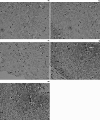
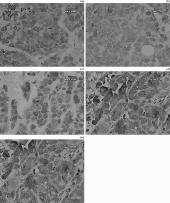
Only a few cells expressing epidermal growth factor receptor and insulin-like growth factor 1 receptor were observed in tumours treated with short hairpin RNA epidermal growth factor receptor plasmid alone or short hairpin RNA insulin-like growth factor 1 receptor plasmid alone. However, even fewer such cells were observed in tumours treated with both short hairpin RNA epidermal growth factor receptor plasmid and short hairpin RNA insulin-like growth factor 1 receptor plasmid. In contrast, epidermal growth factor receptor and insulin-like growth factor 1 receptor protein expression was detected in all tumours treated with short hairpin RNA HK plasmid or saline (seen by dark staining, as shown in Figures 6 and 7). This suggests that the expression of epidermal growth factor receptor and insulin-like growth factor 1 receptor proteins was significantly down-regulated by treatment with short hairpin RNA epidermal growth factor receptor plasmid alone, short hairpin RNA insulin-like growth factor 1 receptor plasmid alone, and both short hairpin RNA epidermal growth factor receptor plasmid and short hairpin RNA insulin-like growth factor 1 receptor plasmid.
Photomicrographs showing epidermal growth factor receptor (EGFR) protein expression in the subcutaneous tumours. Cells with EGFR protein expression were present in tumours from mice transfected with: (a) short hairpin ribonucleic acid (shRNA) EGFR plasmid alone (moderate staining); (b) shRNA insulin-like growth factor 1 receptor (IGF-1R) plasmid alone (moderate staining); (c) both shRNA EGFR and shRNA IGF-1R plasmids (light staining); (d) shRNA HK plasmid (dark staining); and (e) saline (dark staining) (Streptavidin-horseradish peroxidase; ×1000).

Photomicrographs showing insulin-like growth factor 1 receptor (IGF-1R) expression in the subcutaneous tumours. Cells with IGF-1R receptor protein expression were present in mice transfected with: (a) short hairpin ribonucleic acid (shRNA) epidermal growth factor receptor (EGFR) plasmid alone (moderate staining); (b) shRNA IGF-1R plasmid alone (moderate staining); (c) both shRNA EGFR and shRNA IGF-1R plasmids (light staining); (d) shRNA HK plasmid (dark staining); and (e) saline (dark staining) (Streptavidin-horseradish peroxidase; ×1000).

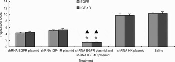
Protein expression scores for epidermal growth factor receptor (EGFR) and insulin-like growth factor 1 receptor (IGF-1R) expression within tumour samples, following the five treatments. The score following treatment with both shRNA EGFR and shRNA IGF-1R plasmids was much lower than that following treatment with shRNA EGFR plasmid alone, shRNA IGF-1R plasmid alone, shRNA HK plasmid or saline. ▴p < 0.01, compared with treatment with shRNA HK plasmid or saline; *p < 0.05, compared with treatment with shRNA EGFR plasmid alone or shRNA IGF-1R plasmid alone. Whiskers show outliers.

Weighted protein expression scores were calculated for epidermal growth factor receptor and insulin-like growth factor 1 receptor. In tumours treated with both short hairpin RNA epidermal growth factor receptor plasmid and short hairpin RNA insulin-like growth factor 1 receptor plasmid, protein expression scores were in the range zero to two (being 1.4 ± 0.56 for epidermal growth factor receptor and 1.45 ± 0.35 for insulin-like growth factor 1 receptor). In cases treated with one or the other plasmid, protein expression scores were in the range three to six following treatment with short hairpin RNA epidermal growth factor receptor plasmid alone (being 4.3 ± 0.59 for epidermal growth factor receptor and 4.4 ± 0.55 for insulin-like growth factor 1 receptor) and in the range four to seven following treatment with short hairpin RNA insulin-like growth factor 1 receptor plasmid alone (being 5.0 ± 0.6 for epidermal growth factor receptor and 5.6 ± 0.58 for insulin-like growth factor 1 receptor). In tumours treated with short hairpin RNA HK plasmid, protein expression scores were in the range six to 12 (being 9.67 ± 1.45 for epidermal growth factor receptor and 9.70 ± 1.76 for insulin-like growth factor 1 receptor). In tumours treated with saline, the protein expression scores were in the range eight to 12 (being 10.26 ± 1.7 for epidermal growth factor receptor and 10.30 ± 1.25 for insulin-like growth factor 1 receptor). The protein expression score for tumours treated with both short hairpin RNA epidermal growth factor receptor plasmid and short hairpin RNA insulin-like growth factor 1 receptor plasmid was much lower than the protein expression scores for tumours treated with either plasmid alone (p < 0.05). The protein expression scores for tumours treated with short hairpin RNA HK plasmid or saline were the highest of all the treatment groups; this difference was statistically significant (p < 0.01).
Effect on relative values of receptor messenger ribonucleic acid
Following dual silencing of epidermal growth factor and insulin-like growth factor 1 receptors, the relative value of epidermal growth factor receptor and insulin-like growth factor 1 receptor messenger RNA expression of different samples was conveyed with epidermal growth factor receptor and insulin-like growth factor one receptor β-actin mRNA percentage.
The PCR quantitative measurement equation is as follows: the relative value of the tested group is equal to 2(Δct test group-Δct β-actin)×100, where ΔctΔct negative control- Δct test group, ct negative control is 50.
Effect of plasmids on blood, liver and kidney
All the mice survived the whole experiment. There were no significant differences in peripheral blood haematological and biochemical parameters, comparing the following treatment groups: both short hairpin RNA epidermal growth factor receptor plasmid and short hairpin RNA insulin-like growth factor 1 receptor plasmid; short hairpin RNA epidermal growth factor receptor plasmid alone; short hairpin RNA insulin-like growth factor 1 receptor plasmid alone; shRNA HK plasmid and saline. This suggests that the short hairpin RNA plasmid treatment did not affect haematological, hepatic or renal functions (p > 0.05) (Tables III and IV).
Discussion
Ribonucleic acid interference has the potential to ‘knock down’ oncogenes in cancer. Some studies have shown that the percentage of small interference ribonucleic acid (siRNA) targeting of insuline like growth factor-1 receptor (IGF1R) or epidermal growth factor receptor (EGFR) repressed EGFR and IGF-1R expression to reach as high as 74–90 per cent.Reference Bai, Zhu, Chen, Gao, Zhang and Wang17, Reference Pardridge18
Our previous studies showed that epidermal growth factor receptor and insulin-like growth factor 1 receptor play an important role in cancer cell biology. Firstly, they are overexpressed in most malignant tumour cells but not in normal cells, suggesting that such overexpression may be involved in cancer development.Reference Yulin, Xuhong, Jian, Xiaoping, Jun, Linfeng, Xiaoping and Dong3 Secondly, in most cancers, levels of such overexpression generally correlate with tumour cell proliferation, recurrence, five-year mortality rate and drug resistance.Reference Yuan, Zhou, Song, Qiu, Li and Yed8 Our previous investigations also indicated that inhibition of epidermal growth factor and insulin-like growth factor 1 receptor function may alter the growth of malignant cells.Reference Yuan, Zhou, Song, Qiu, Li and Yed8
In the present study, after the tumours were transfected with short hairpin RNA plasmids, many green fluorescent cells were observed under confocal microscopy. This result indicates that the constructed plasmid effectively transfected the tumour cells in vivo. The more marked reduction in tumour volume found in the dual plasmid treatment group (84.78 per cent), compared with the single plasmid treatment groups (70.38 per cent for epidermal growth factor receptor plasmid and 67.21 per cent for insulin-like growth factor 1 receptor plasmid), demonstrates that dual silencing of epidermal growth factor and insulin-like growth factor 1 receptors can more significantly inhibit tumour growth. We speculate that our short hairpin RNA epidermal growth factor receptor plasmid and short hairpin RNA insulin-like growth factor 1 receptor plasmid expressed specific short hairpin RNA segments and selectively degraded the epidermal growth factor receptor and insulin-like growth factor 1 receptor messenger RNA, respectively. Loss or down-regulation of epidermal growth factor receptor and insulin-like growth factor 1 receptor expression can lead to limitation of cancer cell growth. Finally, these cells undergo cell death through necrosis or apoptosis. This is consistent with our observation that dual silencing of epidermal growth factor and insulin-like growth factor 1 receptors could result in a low level of protein expression in transplanted tumours, as shown by immunohistochemical analysis.
In our study, we observed no significant differences between the expression of messenger RNA and protein, comparing the negative HK control plasmid and saline groups, and comparing the short hairpin RNA epidermal growth factor receptor plasmid group and the short hairpin RNA insulin-like growth factor 1 receptor plasmid group. Our results indicated no inhibitory effect when mismatched short hairpin RNA was used. Furthermore, our results showed that the short hairpin RNA epidermal growth factor receptor plasmid alone and the short hairpin RNA insulin-like growth factor 1 receptor plasmid alone (with the same transfection reagent) had the same suppressive effect on the CNE2 cells. Our results indicate that dual silencing of epidermal growth factor receptor and insulin-like growth factor 1 receptor has a relatively strong inhibitory effect. The repressive effect of the short hairpin RNA plasmids on the target receptor genes and proteins was more specific when using dual gene plasmids (targeting both epidermal growth factor receptor and insulin-like growth factor 1 receptor), compared with using single gene plasmids (i.e. for epidermal growth factor receptor alone or insulin-like growth factor 1 receptor alone).
These results provide a basis for developing multigene RNA interference. In our study, the efficacy of repression of dual gene short hairpin RNA plasmids on epidermal growth factor receptor and insulin-like growth factor 1 receptor expression did not equate to the sum of the repressive effects of the two single gene short hairpin RNA plasmids. Furthermore, dual gene short hairpin RNA plasmids could not completely knock out the gene. Further studies on this topic are required. It is probable that incomplete inhibition of epidermal growth factor receptor and insulin-like growth factor 1 receptor expression is sufficient to abolish growth factor-mediated signalling.
Our results indicate that treatment both with a null, mismatched HK short hairpin RNA plasmid vector and with saline has no effect on apoptosis. We also found that RNA interference targeting epidermal growth factor receptor and insulin-like growth factor 1 receptor can effectively induce apoptosis. Furthermore, we found that dual gene short hairpin RNA plasmids can much more effectively induce cell apoptosis than single gene plasmids; while epidermal growth factor receptor and insulin-like growth factor 1 receptor expression decreases, apoptosis increases.
The above results show that epidermal growth factor receptor and insulin-like growth factor 1 receptor play a very important role in tumour cell apoptosis. Other authors have stated that epidermal growth factor receptor and insulin-like growth factor 1 receptor both down-regulate apoptosis, through insulin-like growth factor mediated down-regulation of Bcl-xS expression, via the mitogen-activated human non-small cell lung cancer (A549 and SPC-A1 cells) protein pathway (findings similar to those of the present study).Reference Bai, Zhu, Chen, Gao, Zhang and Wang17 This indicates that the same short hairpin RNA segment will have a similar effect on different kinds of tumour cells, making it possible for an active short hairpin RNA sequence to be used as an anti-tumour remedy. In our study of nude mice, the combined use of two short hairpin RNA gene plasmids resulted in a tumour inhibition ratio of 84.78 per cent. There are significant differences between dual gene and single gene interference kinase (mitogen-activated protein kinase).Reference Oldak and Malejczyk19 A decrease in apoptosis is the main characteristic of malignant tumour cells. Thus, accelerating tumour cell apoptosis rates by targeting repression of epidermal growth factor receptor and insulin-like growth factor 1 receptor may represent an anti-tumour mechanism. Treatment which suppresses epidermal growth factor and insulin-like growth factor 1 receptors, with the aim of promoting apoptosis, has been demonstrated to be effective in tumour therapy.Reference Harari and Huang20
• Nasopharyngeal carcinoma (NPC) is unique among head and neck cancers because of its epidemiology; it is relatively rare worldwide and is predominantly found in males
• This study examined the effects of dual silencing of epidermal growth factor and insulin-like growth factor 1 receptors on the growth of NPC in nude mice; it also assessed potential side effects in these animals
• Results indicated that dual silencing of both epidermal growth factor and insulin-like growth factor 1 receptors induced more apoptosis and more growth suppression of NPC cells, compared with silencing of either epidermal growth factor receptor alone or insulin-like growth factor 1 receptor alone, without significant side effects in the experimental animals
• This study suggests new therapeutic modalities for the treatment of human NPC; the findings may represent a promising strategy for such treatment
It must be noted that a short hairpin RNA expression vector which targeted either epidermal growth factor or insulin-like growth factor 1 messenger RNA significantly inhibited the growth of nasopharyngeal carcinoma (NPC) in nude mice, with no significant side effects. The main advantages of short hairpin RNA treatment are efficient tumour growth inhibition and systemic safety. This suggests that RNA interference has a potential application in treating NPC. The mice injected with short hairpin RNA remained healthy; the treatment did not affect their haematological, hepatic or renal function. This indicates that short hairpin RNA treatment may possibly be safer than chemotherapy. Our results suggest that RNA interference has a potential application in treating human NPC.
We believe that molecular targeting therapy represents the future for tumour therapy in the twenty-first century. Via targeting of epidermal growth factor receptor and insulin-like growth factor 1 receptor, RNA interference can inhibit growth and induce apoptosis of NPC in nude mice, especially when dual gene short hairpin RNA plasmids are used. These study results lay a solid foundation for multigene therapy. Our results suggest new therapeutic modalities in the treatment of human NPC, which may represent promising strategies for the treatment of this cancer.
Acknowledgement
This work was supported by grants from the National Natural Science Foundation of China (number 30471873, Beijing, PR China).