



Introduction
Infertility affects approximately 17.5% of couples during their lifetime (Cox et al., Reference Cox, Thoma, Tchangalova, Mburu, Bornstein, Johnson and Kiarie2022), with various aetiologies contributing to the failure to establish a pregnancy. The introduction of assisted reproductive technologies (ART) marked a breakthrough in the treatment of infertility, resulting in the birth of millions of babies (Editorial, 2023). However, a small percentage of couples remain unable to achieve pregnancy despite undergoing multiple ART cycles (Bashiri et al., Reference Bashiri, Halper and Orvieto2018).
One of the prerequisites for successful ART treatment is the presence of a mature oocyte with normal morphology (Keefe et al., Reference Keefe, Kumar and Kalmbach2015). Human oocytes are formed during fetal development and arrested at the germinal vesicle (GV) stage. Oocyte maturation is induced by an endogenous luteinizing hormone (LH) surge or by human chorionic gonadotropin (hCG) injection during stimulated cycles (Hartshorne et al., Reference Hartshorne, Lyrakou, Hamoda, Oloto and Ghafari2009). As a result, the oocyte progresses to the metaphase II (MII) stage, during which a small polar body (PB) containing sister chromatids and minimal cytoplasm is extruded (Jones, Reference Jones2008). This maturation process is regulated by a complex network of molecular mechanisms (Pei et al., Reference Pei, Deng, Xu and Zhang2023), and any abnormalities in these events can result in oocytes that are incapable of fertilization or normal embryonic development (Pei et al., Reference Pei, Deng, Xu and Zhang2023).
Current literature has identified numerous genes involved in oocyte maturation and fertilization (Baldini et al., Reference Baldini, Ferri, Malvasi, Laganà, Vimercati, Dellino, Baldini and Trojano2024). We previously reported that pathogenic variants in TLE6, PADI6, PATL2 and ASTL are associated with fertilization failure, oocyte maturation defects, embryonic arrest and polyspermy (Alazami et al., Reference Alazami, Awad, Coskun, Al-Hassan, Hijazi, Abdulwahab, Poizat and Alkuraya2015; Maddirevula et al., Reference Maddirevula, Coskun, Awartani, Alsaif, Abdulwahab and Alkuraya2017a; Reference Maddirevula, Coskun, Alhassan, Elnour, Alsaif, Ibrahim, Abdulwahab, Arold and Alkuraya2017b; Reference Maddirevula, Coskun, Al-Qahtani, Aboyousef, Alhassan, Aldeery and Alkuraya2022). Additionally, many other genes have been implicated in oocyte maturation. Among these, TUBB8 has been shown to cause oocyte maturation arrest at the metaphase I (MI) stage (Feng et al., Reference Feng, Sang, Kuang, Sun, Yan, Zhang, Shi, Tian, Luchniak, Fukuda, Li, Yu, Chen, Xu, Guo, Qu, Wang, Sun, Liu, Shi, Wang, Feng, Shao, Chai, Li, Xing, Zhang, Nogales, Jin, He, Gupta, Cowan and Wang2016). Mutations in TUBB8 are associated with a range of phenotypes beyond oocyte arrest, including embryonic developmental arrest, fertilization failure and enlarged PBs (Zhao et al., Reference Zhao, Guan, Wang, Chen, Xu, Wu, Yan, Li, Fu, Shi, Shi, Du, Li, Zhang, Mu, Zhou, Dong, Jin, He, Sun, Kuang, Wang and Sang2020).
More recently, variants in the MOS gene have been reported to result in oocytes with large PBs or early embryonic arrest in humans (Wang and Sang, Reference Wang and Sang2021; Jiao et al., Reference Jiao, Lian, Xing, Chen, Du and Liu2022; Zhang et al., Reference Zhang, Zheng, Ren, Jin, Hu, Liu, Fan, Gong, Lu, Lin, Zhang and Tong2022; Zeng et al., Reference Zeng, Shi, Xu, Shi, Wu, Li, Xue, Zhu, Chen, Sang and Wang2022). The function of MOS had previously been well characterized in mouse oocytes, where it was found to regulate the size and degradation of the first PB (Choi et al., Reference Choi, Fukasawa, Zhou, Tessarollo, Borror, Resau and Vande Woude1996).
Here, we report a case of an infertile woman with a MOS gene variant, presenting with the phenotype of enlarged PBs and parthenogenetic activation of oocytes.
Materials and methods
A 30-year-old woman with a 12-year history of infertility was referred for evaluation and treatment. She reported regular menstrual cycles, no history of oral contraceptive use and had previously undergone gastric sleeve surgery for weight loss and laparoscopic surgery for tubal reopening. The couple had undergone several unsuccessful infertility treatments, including Clomid and in vitro fertilization (IVF) cycles, but they could not recall specific details beyond negative pregnancy tests. The patient also had a sister with infertility; however, due to confidentiality concerns, no genetic screening was performed on family members. The husband had a normal semen analysis (including volume, count, motility and morphology), and he has a child from another marriage. The patient consented for IVF treatment, clinical exome sequencing (ES) and publication of the case with anonymized personal medical information.
The patient underwent two ovarian stimulation cycles using a gonadotropin-releasing hormone (GnRH) antagonist protocol, with daily administration of 0.25 mg Cetrorelix (Merck Europe B.V., The Netherlands), starting on the seventh day of stimulation. In the first cycle, 225 I.U. of urinary gonadotropin (Menopur, Ferring, Switzerland) was administered daily for 10 days. In the second cycle, 225 I.U. of recombinant FSH (Gonal-F, Merck, Germany) was administered daily for 12 days. In both cycles, ovulation was triggered with 5000 I.U. of (hCG, Pregnyl, Organon, U.S.A.) once the leading follicle reached at least 18 mm.
Follicular aspiration was performed 37 h after hCG injection via ultrasound-guided transvaginal aspiration. Cumulus-oocyte complexes were collected using needle suction, washed in handling medium (Cooper Surgical, U.S.A.) and cultured for one hour prior to denudation. Denudation was performed via mechanical pipetting in an 80 I.U./ml hyaluronidase solution (Cooper Surgical, U.S.A.), followed by intracytoplasmic sperm injection (ICSI).
Given the patient’s clinical presentation and family history (a sister with infertility), clinical ES was performed at the core laboratory of the Precision Medicine Laboratory Department, Genomic Medicine Center of Excellence (GMCoE), King Faisal Specialist Hospital and Research Center. The protein structure (AF-P00540-F1-model_v6.pdb) was downloaded from AlphaPDB (Cambridgeshire, UK). The protein structures have been viewed in PyMol (San Diego, CA, USA). The mutagenesis has been performed and bonds have also been interpreted using PyMol.
In the second cycle, oocytes were collected after 22 h for nuclear staining. Oocytes were first incubated in 4% paraformaldehyde for 10 min for fixation, then mounted on glass microscopic slides. The nuclei were stained with VECTASHIELD® antifade mounting medium containing 4′,6-diamidino-2-phenylindole (DAPI) for 5 min. Fluorescence imaging was conducted using an inverted microscope (Olympus IX71, Olympus), Tokyo, Japan connected to an epifluorescence unit (Olympus U-RFL-T, TH4-200, Olympus) and a digital camera (C13440, Hamamatsu, Saitama, Japan).
Results
The couple underwent two treatment cycles at our centre. In the first cycle, seven oocytes were retrieved. Of these, three were at the MII stage and were injected with the husband’s sperm; three were at the MI stage and one was abnormal. All MII oocytes displayed large polar bodies (PBs) (Figure 1D). Of the three injected MII oocytes, two developed more than four pronuclei (PN) (Figure 1E), suggesting abnormal fertilization or parthenogenetic activation. The cycle was subsequently cancelled. Two of the abnormally fertilized oocytes proceeded to cleavage (Figure 1F).
Control oocytes with normal size PB (A), and its progression during time-lapse monitoring showing 2 PN (arrows) at 18 h (B) and 4-cell embryo at 41 h (C) following ICSI. An oocyte from the patient with a MOS variant with a large PB (D) displaying 4 PN (arrows) at 17 h (E) and six blastomeres at 47 h (F) following ICSI. An MI oocyte (G) released its PB around 2.8 h (H) and spontaneous activation was evident with the presence of three PN at 21 h (I, arrows). Appearance of PN started around 10 h. Another MI oocyte (J) activated with the presence of multiple smaller PN (K, arrows) and asymmetrically cleaved (L). MI oocytes (G and J) were not injected with sperm.

Figure 1 Long description
The image consists of twelve microscopic images arranged in a grid, each showing different stages of oocyte maturation and fertilization. The first row shows a control oocyte with a normal-sized polar body (PB) at various stages: initial state (A), formation of two pronuclei (PN) at 18 hours (B), and a four-cell embryo at 41 hours (C) following intracytoplasmic sperm injection (ICSI). The second row depicts an oocyte from a patient with a MOS variant, characterized by a large PB, showing the initial state (D), formation of four PN at 17 hours (E), and six blastomeres at 47 hours (F) following ICSI. The third row illustrates an MI oocyte that releases its PB around 2.8 hours (H) and shows spontaneous activation with three PN at 21 hours (I). The final row shows another MI oocyte that activates with multiple smaller PN and asymmetrically cleaves. The images highlight the differences in maturation and fertilization processes between normal and abnormal oocytes.
The three MI oocytes were cultured in an embryoscope. Two of them showed spontaneous activation: one with a large PB and the other without any PB (Supplementary Video 1). Multiple PN were observed in both, and cleavage occurred.
In the second cycle, 11 oocytes were retrieved: four MII (all with large PBs), six MI and one at the GV stage. Of the four MII oocytes, three were injected with the husband’s sperm; the fourth displayed a significant abnormality and was excluded. One injected oocyte developed three PN, while the others remained at the 0 PN stage. The six MI oocytes were monitored in the embryoscope for 24 h. One extruded a large PB at 2.8 h (Figure 1G–H) and exhibited three PN at approximately 17 h of culture (Figure 1I, Supplementary Video 2). Another MI oocyte (Figure 1J) activated without PB extrusion, formed several small PN around 15 h (Figure 1K) and cleaved asymmetrically around 21 h (Figure 1L). The remaining MI oocytes did not undergo any changes. Spontaneous parthenogenetic activation was evident, with the presence of multiple PN and cleavage occurring without sperm injection.
ES revealed a novel homozygous variant in MOS (NM_005372.1:c.573C > A; p.(Phe191Leu)). According to the American College of Medical Genetics and Genomics (ACMG) guidelines, this variant is considered likely pathogenic (PM1_Supporting, PM2_Supporting, PP3_Strong). Replacing phenylalanine with leucine via p.(Phe191Leu) introduces a structural mismatch, despite both residues being hydrophobic. This is significant within the context of a kinase’s hydrophobic core, where specific residue interactions are required to keep the protein stable and functional (Figure 2).
Structural modelling (A-wildtype; B-mutant) indicates that the p. Phe191Leu substitution interferes with the correct packing of the MOS kinase domain as shown by the red disk in the mutant model. The protein structure (AF-P00540-F1-model_v6.pdb) has been downloaded from AlphaPDB. The protein structures have been viewed in PyMol. The mutagenesis has been performed and bonds have been interpreted using PyMol.
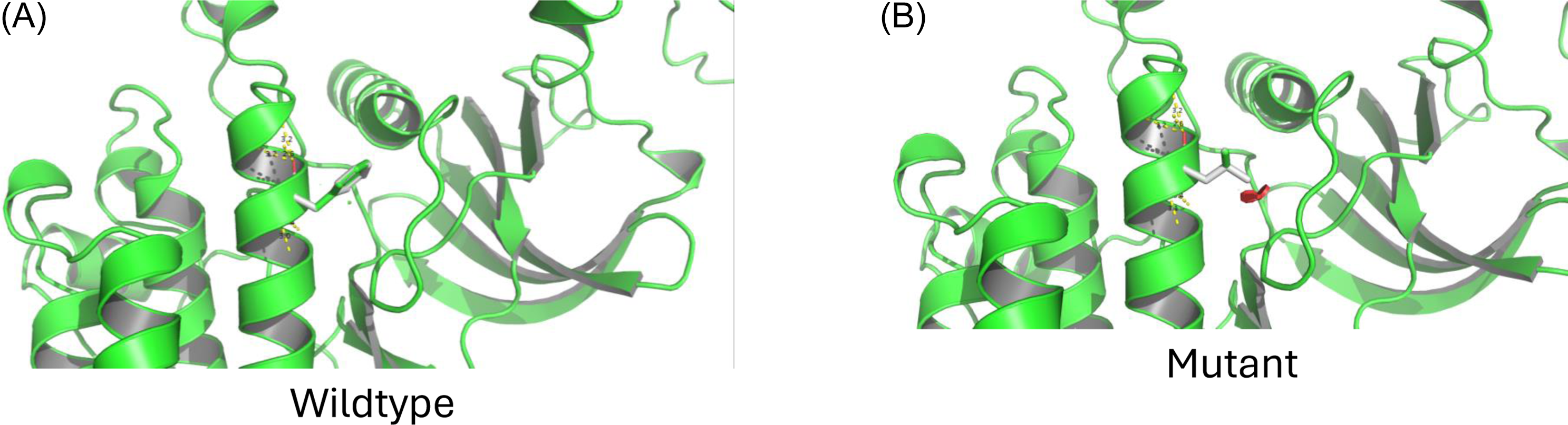
Figure 2 Long description
The image presents structural models comparing the wildtype and mutant forms of the MOS kinase domain. In the wildtype model, the protein structure is correctly packed. In contrast, the mutant model shows a red disk indicating that the p. Phe191Leu substitution interferes with the correct packing of the MOS kinase domain. The protein structure has been downloaded from AlphaPDB and viewed in PyMol. The mutagenesis and bond interpretations have also been performed using PyMol.
In the second cycle, after confirming the failure of normal fertilization, oocytes’ culture was terminated after 22 h for nuclear staining. Bright-field imaging of an MI oocyte that became MII and presented self-activation with three PN and a PB (Figure 3A) and nuclear staining of the same oocyte (Figure 3B) showing one nucleus inside the PB and the staining of two PN in oocyte, while the third pronucleus did not have staining.
Bright-field and fluorescent imaging of an oocyte showing nuclear materials inside the oocyte and the PB. An MI oocyte divided asymmetrically extruding a large PB (A) with nuclear materials in the PB (B, lower arrow) and two nuclear staining inside the oocyte (upper two arrows).

Figure 3 Long description
The left image shows an oocyte under bright-field imaging, highlighting its structure and the extrusion of a large polar body. The right image uses fluorescent imaging to reveal nuclear materials within the oocyte and the polar body. Arrows point to specific areas of interest: two nuclear stainings inside the oocyte and one in the polar body.
Discussion
In this study, we describe a novel homozygous missense variant in MOS, which resulted in large PB formation and parthenogenetic activation of oocytes. MOS has long been recognized as essential for oocyte maturation in animal models (Gebauer and Richter, Reference Gebauer and Richter1997). While male Mos knockout mice are fertile, female mice exhibit reduced fertility (Colledge et al., Reference Colledge, Carlton, Udy and Evans1994). Recently, several publications have demonstrated that MOS variants can also lead to female infertility in humans. Two major phenotypes have been described: early embryonic arrest (Zeng et al., Reference Zeng, Shi, Xu, Shi, Wu, Li, Xue, Zhu, Chen, Sang and Wang2022; Zhang et al., Reference Zhang, Zheng, Ren, Hu, Tong, Zhang, Li, Wang, Jiang, Jin, Yang, Cao, He, Ma, Zhang, Gu, Hu, Luo, Gong, Lu, Lin, Fan and Zhang2021) and large PB formation (Jiao et al., Reference Jiao, Lian, Xing, Chen, Du and Liu2022; Zhang et al., Reference Zhang, Zheng, Ren, Jin, Hu, Liu, Fan, Gong, Lu, Lin, Zhang and Tong2022). Our patient exhibited the latter phenotype.
This phenotype was previously observed in Mos knockout mice. Choi et al. (Reference Choi, Fukasawa, Zhou, Tessarollo, Borror, Resau and Vande Woude1996) reported that some mouse oocytes displayed large PBs resembling the first cleavage during in vitro maturation. This phenotype is consistent with what we observed in our patient. It has been suggested that MOS plays a role in positioning the metaphase spindle during meiosis. Normally, following GV breakdown at MI, the spindle is centrally located and later migrates to the periphery to facilitate PB extrusion. In mice, large PB formation was associated with a failure of this migration (Choi et al., Reference Choi, Fukasawa, Zhou, Tessarollo, Borror, Resau and Vande Woude1996; Verlhac et al., Reference Verlhac, Lefebvre, Guillaud, Rassinier and Maro2000).
During ART, oocytes with enlarged PBs are rarely seen, but they are associated with reduced fertilization, cleavage and good-quality embryo formation compared to oocytes with normal PB morphology (Navarro et al., Reference Navarro, de Araújo, de Araújo, Rocha, dos Reis and Martins2009). Such abnormal PB morphology has also been linked to variants in PATL2 (Cao et al., Reference Cao, Zhao, Wang, Cai, Xia, Zhang, Han, Xu, Zhang, Ling, Ma and Huo2021) and TUBB8 (Zhao et al., Reference Zhao, Guan, Wang, Chen, Xu, Wu, Yan, Li, Fu, Shi, Shi, Du, Li, Zhang, Mu, Zhou, Dong, Jin, He, Sun, Kuang, Wang and Sang2020). PATL2 is known to regulate MOS translation during oocyte meiosis, suggesting potential phenotypic overlap between PATL2 and MOS variants. Zhao et al. (Reference Zhao, Guan, Wang, Chen, Xu, Wu, Yan, Li, Fu, Shi, Shi, Du, Li, Zhang, Mu, Zhou, Dong, Jin, He, Sun, Kuang, Wang and Sang2020) described 34 TUBB8 variants with a wide range of phenotypes, including a subset of patients showing both normal and abnormal PB morphology. In our case, all mature oocytes exhibited large PBs, indicating a consistent phenotypic effect of this specific MOS variant.
A novel finding in our case is the presence of parthenogenetic activation caused by the MOS variant, a phenomenon previously demonstrated in mice (Choi et al., Reference Choi, Fukasawa, Zhou, Tessarollo, Borror, Resau and Vande Woude1996). MI oocytes from our patient developed multiple PN and cleaved in culture without sperm injection, closely resembling murine observations. Zhang et al. (Reference Zhang, Zheng, Ren, Jin, Hu, Liu, Fan, Gong, Lu, Lin, Zhang and Tong2022) reported cleavage-stage embryos from 0 PN oocytes and speculated that these may have resulted from either delayed fertilization or parthenogenetic activation which they cannot confirm since their oocytes were not monitored with time-lapse imaging. Our findings confirm their hypothesis by directly observing multiple PN and cleavage in MI oocytes via time-lapse monitoring. The nuclear materials inside the PB as shown in Figure 3 suggest that it is a real PB rather than a pseudo-cleavage.
One of the limitations of this report is the lack of pedigree data, which was not done due to the confidentiality issues. However, the protein structural modelling indicated that the p. Phe191Leu substitution interferes with the correct packing of the MOS kinase domain and creates a steric clash. This conflict likely destabilizes the protein core, abolishing kinase function and resulting in biological defects, probably leading to female infertility. Predicted protein structure, along with the well establish role of MOS in human and mice, this mutation can be considered probable cause of infertility in this couple.
In conclusion, our data further confirm that MOS is critical for proper oocyte maturation. The c.573C > A variant leads to the formation of large PBs and parthenogenetic activation. Patients with MOS variants should be counselled regarding their fertility prognosis, as no successful pregnancies have been reported in such cases to date.
Supplementary material
The supplementary material for this article can be found at https://doi.org/10.1017/S0967199426100409.
Competing interests
None of the co-authors has any competing interests, nor have they received or are due to receive any payment for writing this article.
Funding
This research did not receive any specific grant from funding agencies in the public, commercial or not-for-profit sectors.
Ethical standards
The authors assert that all procedures contributing to this work comply with the ethical standards of the relevant national and institutional committees on human experimentation and with the Helsinki Declaration of 1975, as revised in 2008.



