I. INTRODUCTION
Perfect crystalline diamond, a dense polymorph of carbon, has an indirect band gap of 5.5 eV and is transparent in the visible part of the light spectrum. However, most gem diamonds contain appm level of defects such as vacancies, interstitials, and substitutional impurity atoms. The presence of these defects can affect the associated energy levels within the band gap, resulting in changes in optical properties. The color change of a diamond arises because of the modification of the absorption and/or photoluminescence characteristics, which is dependent on the type and the concentration of the defects, including impurity atoms and irradiation-induced defects.
Black diamonds are relatively rare in nature, but for their beauty, they have been introduced as gemstone since late 1990s, both in natural and in artificial forms. A colored diamond has been produced by neutron irradiation in a nuclear reactor, and such diamond has shown deep blue to green color. Recently, a black diamond has been produced by high-temperature treatment of poor quality polycrystalline diamonds.Reference Willems, De Corte and Van Tendeloo1 In this treatment, the color of the diamond changed to black on the surface because of graphitization, often showing crack-like defects through the surface. It was reported that an introduction of nitrogen atoms into substitutional lattice sites of diamond produced a defect energy level at ∼1.7 eV below the conduction band,Reference Mita2 which resulted in yellow-brown color because of the characteristic absorption at the defects, so often called color centers.
This article reports that from a commercial grade single-crystalline diamond, a black diamond is produced by 70 keV N-ion implantation. X-ray photoelectron spectroscopy (XPS), Fourier transform infrared (FTIR), and Raman analyses indicate that the effect of N-ion implantation followed by an annealing is similar to that of a conventional doping on one hand, but irradiation causes atomic rearrangement producing a new type of carbon structure.
II. EXPERIMENTS
N-ions were irradiated onto transparent, pale yellow, commercial grade gem diamonds, which weighed about ∼0.02g and had been cut and polished to 57 facets. For irradiation, 70 keV N-ions were used at two fluence levels, 5 × 10¹⁶ and 1 × 10¹⁷/cm2. The base chamber pressure was 2 × 10⁻⁶ Torr, and the operational pressure during ion implantation was 2 × 10⁻⁵ Torr. The ion-beam irradiation was done on the table side and then culet side of the diamond to cover the whole surface area with N-ion beams. Postirradiation heat treatment was carried out at 600 °C for 2 h in vacuum (∼10−5 torr), under Ar gas atmosphere, and in air. On one occasion, the process was repeated for three times by irradiating to a fluence of 1 × 10¹⁷/cm2 followed by postimplantation annealing at 600 °C for 2 h for each cycle, to make the total fluence of 3 × 10¹⁷/cm2 with total 6 h of annealing. The intermittent annealing steps were necessary to repair the irradiation-induced damages and avoid blistering. The color change was examined with an optical microscope equipped with a halogen lamp. Fifty N-ion-implanted diamonds (about 2.7 mg each) were weighed with a microbalance before and after annealing to investigate the weight loss by oxidation. FTIR spectroscopy was carried out to examine the binding states of the implanted nitrogen, and XPS analysis was conducted for N1s in the N-ion-implanted diamonds after removing the surface by about 30 nm by milling with 500 eV Ar ions, both for annealed and unannealed specimens. UV–Vis–NIR spectroscopy was carried out for light transmittance in the range of 200 nm to 850 nm wavelength, using a deuterium lamp for shorter wavelength (200–350 nm) and a tungsten lamp for longer wavelength (350–850 nm). For Raman analysis, a 532 nm diode LASER (Verdi 5.5V, Coherent Inc., Santa Clara, CA) was used with 10 s exposure. Scattered light from the sample was dispersed through a triple-monochromator using 1200 g/mm (center 1400 cm−1) grating and was detected using a thermoelectrically-cooled, charge-coupled device detector.
III. RESULTS AND DISCUSSION
For all the ion-implanted diamonds in this work, surface blistering was not observed because of the deliberate annealing scheme used in this experiment, although there were reports of blistering at higher dose ion implantation.Reference Chaiwong, Yu, Schinarakis and Vilaithong3, Reference McHargue, Farlow, Lewis and Williams4 Figure 1 shows a series of optical micrographs for the diamonds irradiated and annealed at various conditions. The color of transparent diamond changed to pale yellow near ion fluence of 5 × 10¹⁶ ions/cm2, then to dark-brown, and finally to black with increasing fluence to 1 × 10¹⁷ ions/cm2. Annealing the implanted diamond in vacuum or Ar gas atmosphere at 600 °C for 2 h produced a glossier black with metallic luster but with reduced transparency, compared with the unannealed one [Figs. 1(a) and 1(b)]. The most intense black-metallic luster was seen for the sample irradiated to the highest fluence of 3 × 10¹⁷ ions/cm2 and annealed for 6 h in total at 600 °C in vacuum [Fig. 1(c)].
Optical micrographs of the black diamonds produced by 70 keV N-ion implantation: (a) 1 × 10¹⁷ ions/cm2 N-ion implantation without postirradiation heat treatment, (b) 1 × 10¹⁷ ions/cm2 N-ion implantation followed by postirradiation heat treatment at 600 °C for 2 h in vacuum, (c) 3 × 10¹⁷ ions/cm2 N-ion implantation followed by postirradiation heat treatment at 600 °C for 6 h in vacuum, and (d) 1 × 10¹⁷ ions/cm2 N-ion implantation followed by heat treatment in air showing the disappearance of the black color, suggesting a removal of the irradiated layer as CO2 by oxidation (the average depth of the removed layers was estimated to be ∼3.5 μm).

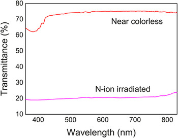
The UV–Vis–NIR spectra showed that the light transmittance for the unirradiated diamond was over 70%, but not 100% because of the absorption through the thickness and the scattering from the faceted diamond (Fig. 2). An absorption peak was observed near 380 nm (∼3.2 eV), not at 5.5 eV of pure diamond, possibly due to the presence of impurity in the tested diamond. Consistent with the optical microscopy observation, the largest reduction in light transmittance was observed for the diamond N-implanted to the highest fluence and annealed, with only about 20% transmittance in the wavelength range from 250 to 850 nm covering the entire range of visible light spectrum. There is an increase in the light transmittance near the wavelength of 825 nm (∼1.5 eV) with an extended tail, suggesting that the bandgap of the diamond is greatly reduced, resembling the absorption characteristics of amorphous semiconductors. However, a complete absorption did not occur because the modified layer is very thin.
UV–Vis–NIR spectra shows that the light transmittance for the unirradiated diamond was over 70% and the largest reduction in light transmittance was observed for the diamond with 3 × 10¹⁷ N ions/cm2 implantation followed by postirradiation heat treatment at 600 °C for 6 h in vacuum, with only about 20% transmittance in the wavelength range from 250 to 850 nm.

About 25% transmittance was seen for the diamond irradiated to a lower fluence and annealed [Fig. 1(b)], and the least reduction in transmittance (30%) was seen for the diamond irradiated to a lower fluence with no annealing [Fig. 1(a)]. However, when the irradiated diamond was annealed for 2 h at 600 °C in air, the black color disappeared restoring to a hazy transparency [Fig. 1(d)], which was accompanied by a weight loss, suggesting a removal of the irradiated layer as CO2 by oxidation. Taking the density of the diamond, the surface area, and the weight loss into account, the average depth of the removed layers was estimated to be ∼3.5μm.
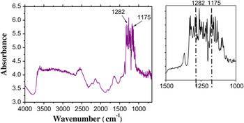
The binding state of the implanted nitrogen was analyzed by FTIR spectroscopy. Figure 3 shows the FTIR spectra for the irradiated and annealed diamond. The absorbance peaks at 1282 and 1175 cm−1 are known to originate from the N–N and symmetrical 4N-vacancy vibrational modes, respectively.Reference Prelas, Popovici and Bigelow5, Reference Collins6 A strong absorption peak near 1700 cm−1 may be due to the C=O carbonyl group. Absorption by C≡C or C≡N bond stretch near 2199 cm−1 was also evident, indicating that a bond breakage and reformation had occurred in the diamond carbon network. A broad O–H absorption band appeared between 3643 and 2450 cm−1 with rather well-defined peak near 2505 cm−1 because of moisture absorption on the sensitized diamond surface after the specimen was exposed to the atmosphere. The results indicate that a considerable atomic rearrangement had occurred in the N-ion-implanted diamond with sp1 and sp2 conjugated carbon bonds, which were incorporated with the implanted nitrogen atoms in various bonding states. Although profuse interstitials, vacancies, and substitutional nitrogen atoms may have been present right after ion implantation, Raman spectra indicates that a significant portion of them disappears after postimplantation annealing by thermally activated atomic rearrangement at elevated temperature (600 °C), to be described later in detail. Although a light absorption associated with nitrogen inclusions and nitrogen-vacancy color centers is known to exist, the photoluminescence from such defect centers is related to the yellow light spectrum in the wavelength range of 560–600 nm, as seen in the nitrogen-containing natural diamond. Since the absorption for N-ion-implanted diamond had occurred in the entire range of visible light spectrum, the major cause for the darkening is most likely due to the formation of conjugated bonds. The π-electrons in the conjugated bonds are loosely bound than those in the covalent bonds and thus easily excited by absorbing the visible light spectrum.
Fourier transform infrared spectroscopic analysis of the black diamond produced by 1 × 10¹⁷ N ions/cm2 implantation followed by heat treatment in 5 × 10⁻⁶ Torr vacuum at 600 °C for 2 h. The absorbance peaks are observed at 1282 and 1175 cm−1, which relate to the characteristics of N–N and symmetrical 4N-vacancy bonds, respectively.

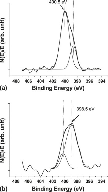
XPS spectra suggest that the as-implanted nitrogen atoms form initially nonstoichiometric C–N compounds (C4N or C5N) in view of the N1s XPS peak appearing mostly near ∼400.3 eV [Fig. 4(a)],Reference Jagielski, Moncoffre, Delichere and Marest7, Reference Ripalda, Diaz, Roman, Galan, Montero, Goldoni, Baraldi, Lizzit, Comelli and Paolucci8 while annealing at 600 °C for 2 h produces a near stoichiometric carbon nitride compound C3N3.75 as evidenced by the N1s peak shifted to ∼398.5 eV [Fig. 4(b)].Reference Jagielski, Moncoffre, Delichere and Marest7 There is a report that the N1s peak at 400.2 eV is due to the presence of substitutional nitrogen in graphite,Reference Ripalda, Diaz, Roman, Galan, Montero, Goldoni, Baraldi, Lizzit, Comelli and Paolucci8 and the N1s peaks at 398.4 and 400.2 eV are attributable to the sp3 and sp2 bonds, respectively, according to Ref. Reference Raole, Mukherjee and John9. It has been reported that the number of sp2 bonds in a diamond increases slightly with a nitrogen doping.Reference Birrell, Gerbi, Auciello, Gibson, Gruen and Carlisle10 It is also known that irradiation produces disordered carbon structures on the surface of a diamond,Reference Lee, Hembree, Rao and Mansur11 which is confirmed with the Raman data in this work. During subsequent thermal annealing, a significant portion of the amorphous layer recrystallized to diamond and the dangling bonds created by irradiation reorganized to sp2 and sp conjugated bonds availing loosely bound π-electrons.
X-ray photoelectron spectroscopy analysis of N1s in diamond: (a) 1 × 10¹⁷ N ions/cm2 implanted diamond shows a broad composite N1s peak near 400.5 eV, indicating that the implanted nitrogen forms mostly nonstoichiometric C–N compounds (C4N or C5N). (b) After annealing at 600 °C for 2 h in vacuum, the composite N1s peak position is shifted to ∼398.5 eV, suggesting that the C–N compound becomes near stoichiometric (C3N3.75).

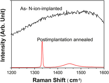
Figure 5 shows Raman spectra of the N-ion-implanted and postimplantation annealed samples in the wave number range of 1200–1600 cm−1, which is taken with an excitation wavelength of 532 nm. Raman spectra indicate that N-ion implantation produced a completely disordered amorphous phase on the surface. This is expected in considering the high dose used in this experiment, a fluence of 1 × 10¹⁷ N ions/cm2, in view of the reported data that showed a complete amorphization at much lower dose although the implanted ion specie and energy are different from this experiment.Reference Hickey, Jones and Elliman12, Reference Prawer13 Nonetheless, in this experiment, it is shown that the color and the electrical conductivity changed further with increasing the fluence to 3 × 10¹⁷ ions/cm2, suggesting that further change in structure had occurred with increasing the fluence, although a complete amorphitization may have occurred at a much lower fluence.
Raman spectra obtained with an excitation wavelength of 532 nm for the N-ion-implanted and postimplantation annealed samples. 1 × 10¹⁷ N ions/cm2 implanted diamond shows completely disordered structure in the range of 1200–1600 cm−1. After annealing at 600 °C for 2 h in vacuum, the first-order crystalline diamond peak appears at 1333 cm−1 and a broad composite peak appears between 1400 and 1500 cm−1.

After annealing at 600 °C, Raman spectra show a well-defined sharp peak at 1333 cm−1, characteristic of sp3 diamond bond,Reference Grimsditch and Ramdas14 and a broad peak centering near 1450 cm−1, a typical composite peak of D (1350 cm−1) and G (1550 cm−1) lines, which was often observed for the diamond-like carbon prepared by chemical-vapor-deposition method.Reference Lee, Hembree, Rao and Mansur11 In spite of the implanted nitrogen, the presence of nitrogen-specific Raman peak is not apparent. However, the range of the composite peak was much wider compared with that observed for a typical disordered carbon, suggesting that the nitrogen inclusion may have modified the vibrational modes. Clearly, further investigation is needed. Surprisingly, the results indicate that once completely disordered structure rearranged to a mixture of good quality diamond and diamond-like carbon, which are connected by sp, sp2, and sp3 bonds. It is thus believed again that the dark color of the irradiated diamond is related largely to the absorption in the visible light spectrum as a result of the excitation of the loosely bound π-electrons in the sp2 and sp conjugated bonds. The enhanced metallic luster observed in the irradiated diamond can be related to the light scattering by the conduction electrons in the sp2 and sp orbitals. The electrical resistivity of pure diamond is known to be in the range of 1012–1016 Ω-cm and that of graphite is in the range of 10−4–10−3 Ω-cm along the basal plane and about 0.2 Ω-cm in the direction perpendicular to the basal plane. A cursory measurement of sheet resistance indicates that the electrical resistivity of the irradiated and annealed diamond is about 5 × 10⁻³ Ω-cm (500 Ω/□ for 1000 Å thick layer), close to a value of highly doped silicon. This result indicates that there is a high concentration of free electrons in the irradiated and annealed diamond, rendering metallic luster.
Colored diamonds are often produced by exposing to ionizing radiation such as gamma rays. However, the color change associated with point defects is temporal and disappears with aging and more rapidly upon thermal exposure. However, the black color produced by ion implantation appears to be permanent in view of the enhanced color by annealing at elevated temperature as seen in this experiment, and the ion-implantation-induced color did not fade away for over 7 years at an ambient temperature in our experiment. It is evident that the introduction of nitrogen into a diamond produces color centers rendering yellow tint, which is overshadowed by the absorption associated with other radiation-induced defects. Nitrogen ion implantation did not produce the color change in cubic zirconia (E g = 7.8 eV), suggesting that of importance is the modification of carbon network structure with unsaturated conjugated bonds, namely sp and sp2 bonds, which is absent in zirconia. There has been a report of color change to black for a diamond subjected to very high temperature above 1000 °C, mainly due to graphitization,Reference Willems, De Corte and Van Tendeloo1, Reference Chen, Sullivan, Friedmann and Gibson15 but such phenomena did not occur at 600 °C.
IV. SUMMARY AND CONCLUSIONS
The optical properties of 70 keV N-ion-implanted diamond are studied in this work. Although a natural diamond with nitrogen inclusions shows a yellow tint, the N-ion-implanted diamond became glossy black with metallic luster, which color was further enhanced by thermal annealing at 600 °C for 2 h in vacuum or inert gas environment, suggesting that the color change is permanent. FTIR, XPS, and Raman studies indicated that the disordered layer of the N-ion-implanted diamond comprised sp, sp2, and sp3 carbon network, with nitrogen atoms in the substitutional sites and some fraction of nitrogen forming a bonding with other nitrogen atoms. The graphitic nature of disordered diamond layer was confirmed by easy oxidation at 600 °C in air in contrast to a pure diamond that is stable even near 1000 °C. However, Raman study revealed that a significant portion of the disordered layer restored to diamond structure by thermal annealing at 600 °C in vacuum and the remainder to much stable diamond-like carbon. UV–Vis–NIR spectra revealed that the insulating diamond became a semiconductor by decreasing the bandgap from 5.5 eV to about 1.5 eV and decreasing the electrical resistivity to the range of 10−3 Ω-cm, a value of highly doped silicon. Optical spectroscopy, however, suggested that some portion of the valence band may have been overlapped with the conduction band. The experimental findings suggest that ion-beam technique may have a potential application in tailoring the bandgap structure of this large bandgap insulator, diamond, and possibly other carbon-based polymeric materials as well.
ACKNOWLEDGMENTS
This work was supported through Proton Engineering Frontier Project from the Ministry of Science and Technology, Republic of Korea.