

1. Portraits of Species
The definition of a newly discovered species in contemporary biology generally occurs through the selection or identification of a suitable – but not necessarily typical – representative (the holotype or type specimen)Footnote 1 and the assignment of a two-part name, whose form is governed by the International Code of Zoological Nomenclature (ICZN) or the International Code of Botanical Nomenclature (ICBN). With all plants and animals that are subject to the rules of these nomenclatures, the act of naming takes the form of the publication of a first description of the type specimen: a written definition of the characteristics of the species,Footnote 2 which is generally accompanied by a graphic and/or photographic representation of the animal and leads to the publication of a binding “portrait” of the species. Solely through a process following the logic of ostensive definition, in other words of definition through the mere act of “pointing out,” holotypes, as philosophers of science have proposed in recent decades, attain the status of binding representatives of their species: “As species are individuals, there is but one rigorous way to define their names: ostensively, in a manner analogous to a christening” (Ghiselin Reference Ghiselin1966).Footnote 3
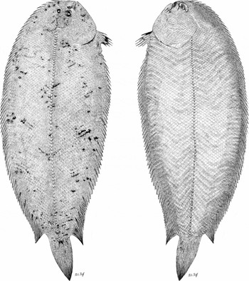
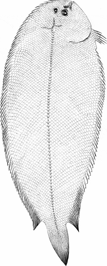
First descriptions and the drawings that accompany them can thus be seen as gestures or indices through which the “christening” of a species is consummated by merely referring to a specimen. If one looks, for instance, into the first description of an Australian sole, which had been caught in 1963 and was named in early 2007, one is struck by the fact that the publication of the Aseraggodes corymbus refers only in a single place to the two drawings of the sole's eye side and blind side (fig. 1): After the abstract and the methodological introduction, the actual first description – entitled “Aseraggodes corymbus, new species” – is immediately followed by a reference to the two drawings and to an x-ray image of the animal. Without anything else linking the images to the text, the metonymic proximity of name and reference to the images defines something that can only be explained in an act of pure ostension: Look, this is the fish that in the future will bear the name of a species!
Nils Hoff: Type drawing of the Australian sole Aseraggodes corymbus, eye side and blind side, 2007. Pencil, pen, and India ink on paper. Museum für Naturkunde, Berlin, © Nils Hoff.

The graphic depiction serves a visual philology designed to facilitate the clearest possible assignment of a name to a certain taxon, in this case to the species, which is represented by a holotype. Such drawings must represent the type specimen as accurately as possible, because in taxonomic practice the illustration assumes the function of a mobile representative of the relatively immobile specimen, in other words, it serves as an “immutable mobile” in the Latourian sense (Latour Reference Latour, Lynch and Woolgar1990). The establishment of a name-bearing type and the unconditional reference of the drawing to this type are necessary to eliminate possible error and doubt that might arise due to an incomplete or inexact first description. Like the specimen itself, the type drawing will in the indeterminate future be subject to (repeated) reevaluation. Its individual quality will later show whether its creator was also able to put down on paper those characteristics of the animal/plant whose relevance was as yet unknown at the time of its creation.
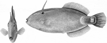
In rare cases, the graphic depiction can even take over the function of the holotype completely; this happens when the first description is drawn up in the absence of a type specimen and makes use of a drawing instead. Many of the hand-colored plates depicting mushrooms in Jean Baptiste François Pierre Bulliard's Herbier de la France functioned as name-giving types (e.g., Agaricus ovinus, in Bulliard 1793, vol. 13, plate 580). Several of the lavish illustrations of hyacinths and narcissuses found in Pierre-Joseph Redouté's Les Liliacées, which was published between 1802 and 1816, achieved the status of so-called iconotypes (cf. Daston Reference Daston2004, 160).Footnote 4 However, this practice applies not only to plants and mushrooms that are difficult to conserve in the herbarium; but also comes into play when the model of a drawing has been lost or when it had never been preserved. The Scottish naturalist Sir John Richardson, for example, named and described the Australian filefish Acanthaluteres brownii exclusively on the basis of a watercolored drawing the Austrian botanic illustrator Ferdinand Lucas Bauer had completed during an expedition in 1801 (fig. 2) (Watts, Pomfrett, and Mabberley Reference Watts, Pomfrett and Mabberley1997, 36–37). Still, one can observe a clear preference for the preserved or dried holotype in the botany and zoology of the last two hundred years (Daston Reference Daston2004, 160). In most instances, drawings do not replace the type specimen; they accompany and interpret it. Despite the competition that it has faced from photography since the late nineteenth century, the black-and-white drawing has maintained its position in many fields of biological taxonomy. It has remained particularly important in the study of smaller organisms (e.g. amphibians, insects and crustaceans), gaining the status of a binding standard of visualization.
Ferdinand Lucas Bauer: Brown's Leatherjacket (Acanthaluteres brownii), 1801. Water colour on paper. © Natural History Museum, London.

Two explanations can be given for the limitation to uncolored (line) drawings: First, monochrome graphics without halftones can be reproduced without any major loss of quality using inexpensive printing technology.Footnote 5 Second, the selection of data associated with hand drawing facilitates the articulation and accentuation of the characteristics relevant for classification. It achieves – and this, for the moment, is my generalized hypothesis – a dovetailing of documentary and subjective access to observed phenomena that is unique to the medium (cf. Krümmel Reference Krümmel2004). The significance of drawing in the context of first descriptions goes far beyond that of illustration in the conventional sense. In most cases it does not synthesize a preexisting verbal catalogue of the type's morphological characteristics, but rather facilitates the examination and recording of these traits.
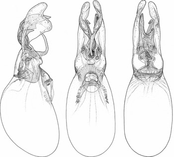
Today (as in earlier eras), specialized scientific illustrators are at work in the field of systematic biology. Biologists themselves also engage in the practice of drawing, although their training is mostly rudimentary at best, having been acquired either in biological illustration courses during their university studies or from one of the many handbooks on drawing created for scientists (for example, Zweifel Reference Zweifel1961; Bethke Reference Bethke1969; Papp Reference Papp1976; Dalby and Dalby Reference Dalby and Dalby1980; Downey and Kelly Reference Downey and Kelly1982; West Reference West1983; Holmgren and Angell Reference Holmgren and Angell1986; Wood Reference Wood1994; Cook Reference Cook1998). Despite most scientists’ deficient or even nonexistent artistic training, hands-on laboratory research often coincides with the production of drawings. For example, the entomologist and taxonomist Johannes Frisch (Museum für Naturkunde, Berlin) spends most of the time devoted to species description drawing at the microscope, so that his drawings are created before any actual description takes place (fig. 3). Tellingly, the beetle specialist considers the subsequent act of writing at his desk to be nothing more than a transcription.Footnote 6
Johannes Frisch: Camera lucida drawings of sexual organs of a male Scopaeus serriae (holotype), front, lateral and rear view, 2006. Pencil and ink on cardboard. Museum für Naturkunde, Berlin, © Johannes Frisch.

The process becomes more complicated when a graphic artist specializing in scientific illustration is commissioned to render images of a species. Professional draftsmen like Nils Hoff, the graphic designer who created the type drawing of Aseraggodes corymbus, are usually on the staff of major museums of natural history; as “invisible technicians,” they support the research process and indeed make it possible (Shapin Reference Shapin1989). Their names rarely appear outside the customary acknowledgments, even though they contribute decisively to the acquisition, recording, and interpretation of data. Here the dialogue between scientist and drawing expands into a complex division of labor in which the perception of a zoologist – a highly stylized perception in the Fleckian sense – must be aligned with an artist's gaze, which has generally not been strongly conditioned by a particular scientific thought style (Fleck Reference Fleck, Bradley and Trenn1979, 125–45). This asymmetry of perception often provoked conflicts that have characterized the relationship between researcher and artist throughout the history of modern scientific drawing.Footnote 7 But the introduction of a second, very differently conditioned pair of eyes also opens an epistemic opportunity: The scientific illustrator can be thought of as a sort of instrument that not only adds to the scientist's perception (since the illustrator generally looks at the organism for a longer period of time and more intensely than the scientist), but also defamiliarizes it – both misleadingly and productively.
Taking this into account, it comes as no surprise that scientists since the late nineteenth century tend to suspect that drawing lacks reliability and precision (Daston and Galison Reference Daston and Galison1992; cf. also Schaffer Reference Schaffer, Jones and Galison1998); still, it was paradoxically the very import of subjective interpretation in the act of drawing that secured its continued vitality after the domain of its legitimate application had been curtailed.Footnote 8 In some – certainly restricted – areas, such as biological or archaeological drawing, the “distanced engagement” (Krümmel Reference Krümmel2004, 14) of the draftsman's pencil remained irreplaceable.
2. Immutable mobiles?
As I have mentioned, published type drawings function as “immutable mobiles” (Latour Reference Latour, Lynch and Woolgar1990), firstly, because they stabilize a specimen by generating and visualizing its description, and secondly, because they mobilize the relatively immobile object, making it available for further treatment and revision. Fellow (and future) taxonomists can return time and again to existing visualizations; they can reframe, reorient, and quote them. According to Latour's case study on the “pédofil of Boa Vista,” graphic visualizations such as drawings do not occupy a special position in the dense fabric that links these images to instruments and practices such as observation and techniques of measurement (Latour Reference Latour and Latour1999): They form an integral part of a series that propels an initially underdetermined phenomenon via a cascade of references toward greater determination and articulateness. Although the object of research is represented in very different ways as it passes through various media, and although every step in the chain of references brings a rupture or discontinuity into play, the object gradually acquires an identity. The reference itself, Latour argues, is established not by the deictic function of each individual representation but by what remains constant through the entire series of transformations. A dialectic of gain and loss characterizes the chain of references, but this dialectic is of no great importance to Latour's theory, since the chain as a whole ensures the reference to the objects of knowledge: “Phenomena, however, are not found at the meeting point between things and the forms of the human mind; phenomena are what circulates all along the reversible chain of transformations, at each step losing some properties to gain others that render them compatible with already-established centers of calculation” (ibid., 71–72; emphasis in the original).
Nevertheless “immutable mobiles” share certain properties that distinguish them from other objects and instruments, amongst them the central feature of generating an optically consistent two-dimensional space on paper. Latour based his discussion of “optical consistency” on the work of William Mills Ivins and Samuel Y. Edgerton. The two art historians had reconstructed the epistemological significance of linear perspective for the sciences and technology of the modern era (Latour Reference Latour, Lynch and Woolgar1990, 26–29). The combination of two media, the geometric “system space,” and the letterpress, thus Latour's summary of Ivins's and Edgerton's findings, effected a comprehensive homogenization of graphic representation, creating the possibility of translating images and plans immediately into real space and vice versa. Since Brunelleschi, that is to say, there is the option of transforming objects on paper without altering their internal properties and proportions. Latour's far-reaching conclusion is that “innovations in graphism are crucial but only insofar as they allow new two-way relations to be established with objects (from nature or from fiction) and only insofar as they allow inscriptions either to become more mobile or to stay immutable through all their displacements” (ibid., 29).
Graphic visualizations or more specifically drawings are, in this perspective, mere vanishing points of the general tendency to render objects manageable and publishable. The different qualities of the various forms of graphic visualizations (such as maps, charts, diagrams, graphs, or photography) seem to be without epistemological import. The attempt to apply the theory of “circulating reference” to the description of drawing as a research technique therefore faces at least two problems: First, Latour's theory does not enable us to distinguish the epistemological benefits of drawing from that of other imaging devices. Second, the practice and conventions of drawing are in contradiction with one essential feature of the “immutable mobiles”: Sketching in the “context of discovery” does not primarily aim at the production of an immutable and authoritative image. Although drawings made in the course of taxonomic research are part of a logistics that documents and mobilizes the specimen, their impact on the production of knowledge is not limited to the benefits of stabilization and transmission. In the course of the preparation of an authoritative portrait of a species, drawings are altered, crossed out, and recommenced, that is to say: it involves the ongoing work of the pencil in which knowledge is processed and transformed. The true knowledge gain, then, can hardly be limited to the act of securing and transferring.
But what precisely are the benefits drawing offers as a research practice, i.e., as a practice enabling researchers to acquire, order, and work through knowledge? What are the techniques of lowering the thresholds between the world, the paper, and the draftsman? In the following, I will direct attention to procedures that enable both a reconfiguration of research objects and one of perception – and that initiate an interaction between both processes. The art-historical scholarship of the past decade has rendered us keenly aware of the specific openness of drawing (Bryson Reference Bryson and de Zegher2003; Bach and Pichler Reference Bach and Pichler2009). The possibility of connecting drawing to other technologies and instruments in an effective way is certainly based on this medium's specific openness or underdetermination. Paper and pencil inform the draftsmen only scantily about their proper use. The skillful handling of those materials must individually be learned through situational experimentation and/or the appropriation of drawing conventions. In the acquisition of a certain manner of drawing, the scientist or artist has to find ways to connect paper and pencil to other instruments and media and he has to develop drawing techniques that anticipate them. In the following, I will describe these techniques of anticipation that render drawing permeable for other media and tools as well as the draftsman's sensory apparatus. I will begin with practices that enable interlinking drawing with other techniques of representation; examine the effects of drawing on the apparatus of perception; and finally scrutinize the processing and analysis of research objects by drawing.
3. Interweaving Media – Reducing Distances
Inspired by the thinking of Roland Barthes, Rüdiger Campe has argued that what is conventionally referred to as writing must be understood as an “unstable ensemble of language, instrumentality, and gesture,” which is subject to considerable historical and individual modification (Campe Reference Campe, Gumbrecht and Pfeiffer1991, 760). I would like to make an analogous claim with regard to drawing, and here, too, the difficulties that beset an investigation of the specificity of various “drawing scenes” is rooted less in the multiplicity of factors involved than in their internal heterogeneity. During the creation of a drawing, the scientist or artist must establish a relationship with a great variety of circumstances: various materials come into play (paper, graphite, ink) as well as various tools (pencil, pen, microscope, and computer), and then there are the various conventions of drawing (perspectival construction, modeling, shading), the artifices of craftsmanship (proportioning, dimensioning, techniques of transferring), and last but not least, the motoric action of drawing itself.Footnote 9
If one understands graphical methods as peculiar interactions between internal and external factors, the analysis of the free-hand drawing becomes more complicated, since the specific style of drawing can no longer be derived directly from the history of graphical conventions. Especially with regard to drawing as a research procedure, its special epistemic function can only be understood through the investigation of the entire setting, since scientific illustration – in contrast to conventional artistic drawing scenes – is anchored in a particularly heterogeneous material ensemble. All of its various devices, methods, and gestures define and limit the artist's activity, so that the success of scientific illustration is quite likely to be dependent on the degree to which it is possible to link the inherent and external conditions of the medium; in other words, to bring together the rules and conventions of drawing with the logic, alien to it, of apparatuses and technically advanced procedures (from the camera lucida to the digital camera, from the microscope to the x-ray machine).
I would like to discuss this hypothesis in greater detail using the example of drawing at the microscope, a technique that has existed since the development of this instrument in the first half of the seventeenth century and is still used today in taxonomic research on small animals. Generally speaking, drawing at the microscope entails dramatic constraints on what is otherwise possible in the medium of drawing – though on the other hand the technique approximates the object of research to the logic of drawing. Biologists have drawn, and still draw, with medium-soft pencils on smooth white paper; the choice of fairly simple material corresponds to the simple graphical means, which are largely limited to outlines and dots; draftsmen avoid most conventional devices such as shading and modeling with dynamic hatching. A decisive reason behind this deliberate limitation of graphical conventions is the fact that the draftsman's data acquisition at the microscope is usually supported by a camera lucida (e.g. in the case of the entomologist Johannes Frisch).Footnote 10 In its simplest construction type this optical instrument consists of a quadrangular prism with a deltoid section that is mounted on the support of the drawing or, in the case of microscopy, permanently integrated in the microscope. The constructions of the different historical and contemporary camerae lucidae differ considerably; but typically a half-silvered prism is installed between the ocular and the objective, in such a way as to enable the observer to see the preparation through the optical axis of the microscope. At the same time a lateral mirror (or a reflective prism) projects the sheet of paper (and the emerging drawing) through the half-silvered prism into the optical path of the microscope and thereby into the eye of the drawing scientist. The camera lucida, that is to say, creates the virtual appearance of the object to be drawn fading into the emerging image.
The drawing practice modified by this device generally differs from free-hand drawing by two particular qualities: First, the camera lucida effectively arrests the draftsman's eye as well as his entire body, since even the smallest motion will undo the coincidence of the object to be drawn and the image that has already been created;Footnote 11 the optical instrument, that is to say, constrains the dynamic play of the act of drawing. Second, this apparatus causes a peculiar kind of partial blindness: the drawing is no longer created in the presuppositionless white of the sheet of paper; instead it is from the very outset produced in the image, which is to say, it continually competes with the specimen to be drawn and the wealth of information it holds. Drawing with the help of the camera lucida bears a structural resemblance to tracing: It enables the draftsman to constantly control the “correctness” of the lines he puts on the paper, which effectively make the contours of the object he traces disappear. However, only linear structures and contours lend themselves to immediate graphical translation in this manner (shading and modeling can hardly be traced), and this emphasis on the outline leads to the characteristic flatness of microscopical drawings.
If the use of the microscope thus reduces drawing to its most basic formal means, the particular preparation it requires also transforms the object under scrutiny into a sort of proto-drawing: The object of research is – if it is not already thin enough – slightly pressed between glass plates, a process that fixes a certain view, transforms it into a surface, and renders it manageable and, as it were, paper-like. Various methods of preparing and optimizing the microscopic image serve to enhance the contrast of the texture under examination and to generate a stable figure-ground relationship. In most cases, the phenomenon of interest would be difficult to distinguish from its background without such intervention. The most important means of contrast enhancement are optical microscopy illumination techniques, particularly those of phase contrast microscopy. Another way of enhancing contrasts, namely the various methods of staining, are only rarely applied in taxonomic research, because the type material should not undergo invasive research procedures. The first step in preparing the specimen, then, consists in transforming the scientific object into a kind of proto-image, which anticipates the transfer of what is observed onto two-dimensional paper. In a second step, techniques of optical illumination (and to a much lesser degree bleaching or staining) let the textures in the specimen emerge with particular distinctness, which is to say, they transform it into a graphic structure.
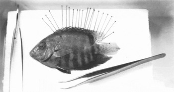
In the macroscopic realm, too, the process of drawing properly speaking begins long before the scientific illustrator picks up paper and pencil. It already begins when a specimen, either conserved in alcohol or dried, is taken from its place of storage and placed in a Petri dish. If the animal does not yet spread out before the draftsman's gaze in sufficiently paper-like fashion, it is fixated with clips, forceps, and pins (fig. 4), if need be against the effects of rigor mortis and the hardening of the body brought on by alcohol. The logic of drawing is thus already in operation before any graphical undertaking has begun.Footnote 12 A form of distance reduction takes place that is more generally characteristic of both the creation of media frameworks and the embedding of drawing in the research process.
Preparation of fishes for drawing, from: The Guild Handbook of Scientific Illustration, 1989, edited by Elaine R. S. Hodges. New York: Van Nostrand Reinhold. This material is reproduced with permission of John Wiley & Sons, Inc.

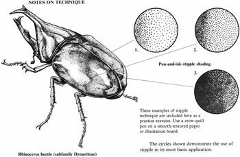
Among the most important craftsman's artifices external to the medium are the above-mentioned flattening of specimens, but also other tricks of the trade mediating between the fairly amorphous and under-differentiated surface of a specimen and the great definitional power of the graphic line. For instance, draftsmen will often simply trace the contours and internal structure of an animal in a photograph, then duplicate the resulting drawing and/or use Photoshop to remove the background. In addition to such medium-external techniques, draftsmen will employ a whole series of artifices inherent to their medium to reduce the distances separating it from other media. The flattening, contrast enhancement, and graphical articulation of the specimen find their equivalent in the limitation of the graphical means to line drawing. In contemporary type drawings, objects are modeled and shaded almost exclusively using techniques of parallel hatching and – predominantly – dotting (fig. 5). In comparison to artistic drawing, the use of these techniques is more strongly conventionalized in scientific illustration. First, the standardization of shading ensures the differentiation of accidental from essential characteristics in the drawn surface, e.g., of phenomena of light and shadow from traits such as texture and patterning (cf. Hodges Reference Hodges1989, 109).Footnote 13 Second, it facilitates the translation of the hand-made drawing into a printed image, as dotting and parallel hatching are typical, even medium-specific, techniques in producing pictures for printing.
Barlowe, Dorothea. 1982. Illustrating Nature: How to Paint and Draw Plants and Animals. New York: Viking Press.

The internal plasticity of drawing, the possibility of reducing it to its rudimentary qualities, and its intrinsic compatibility with other techniques of contrast enhancement that operate directly on the object enable a reduction of distance that – to recall Latour – “draws things together.” Working out Latour's theory of the research process as a chain of references, then, one might speak of a mutual anticipation of representational techniques that effectively lowers the thresholds between different media.
4. Reconfiguring the Gaze
Drawing at the microscope by and large serves a double stratagem: On the one hand, the graphical recording of the preparation propels the generation of the research object and a logistics immediately tied to it. On the other hand, drawing as an activity – as it were, as a special case of observation – is a form of perception training, of professionalizing the gaze.Footnote 14 This second function brings purely self-reflective properties of drawing into play. Science illustration shares these properties with drawing as an artistic practice, yet there is a significant difference between the two: In the visual arts, the self-reflective aspect of drawing characteristically recedes into the background when a draftsman's training is complete. Although artists who have left the academy behind certainly sometimes still draw in order to hone their eye and train their hand, the act of drawing will not or only rarely find its ultimate purpose in this function. In biology, by contrast, drawing frequently served and still serves primarily to support, reinforce, or calibrate another medium, instrument, or activity such as observation (cf. Dennis Reference Dennis1989; de Chadarevian Reference Chadarevian and Mazzolini1993; Bredekamp Reference Bredekamp2000; Schickore Reference Schickore and Geimer2002; Schulze Reference Schulze2005; de Rijcke Reference Rijcke2008; Nasim Reference Nasim and Hoffmann2008; Bredekamp Reference Bredekamp2009).
As Michael Aaron Dennis has shown, the well known pioneer of microscopic research Robert Hooke already considered drawing a form of “disciplined seeing” (Dennis Reference Dennis1989, 345). The pertinent handbooks from the nineteenth century similarly spelled out the essential functions of drawing as a physico-intellectual exercise. For instance, Lionel Beale, the president of the Royal Microscopical Society, writes in 1880: “The student cannot too soon try to delineate what he demonstrates. He will teach himself to observe the more accurately and the more quickly if he records the results of his work in pencil sketches” (Beale Reference Beale1880, 31). In a lecture delivered in 1894, the Swiss geologist Albert Heim describes drawing as a “beneficent moral compulsion” enjoining reflective observation:
All maybeetles seem to us to be identical. The only way for us to gain insight into the differences between individual animals is by drawing. . . . Only by drawing do we force ourselves to see with sufficient awareness, to observe; not merely to look but to attend to what we see with the mind. Drawing from memory is thus a critical examination of our observation, and direct drawing after nature is a school of conscious seeing, a school of observation. (Heim Reference Heim1894, 6)
By the late nineteenth century, the education of the eye by means of the drawing hand has long become a commonplace; it makes a rhetorical appearance in virtually all professional fields where people draw, from engineering to art history (Schulze Reference Schulze2002; Pircher Reference Pircher2005, 103). The authors uniformly emphasize the advantages of drawing: It aids the apperception of detail and the analysis of structure and trains the memory. Even today, writers still characterize drawing as a virtual analysis of what the draftsman sees.Footnote 15 The Guild Handbook of Scientific Illustration published by a professional association of American scientific illustrators, no doubt the most extensive contemporary manual of scientific drawing, states that “drawing a specimen is the best way for either scientist or artist to understand its structure and see its details” (Hodges Reference Hodges1989, 3). The epistemic benefit under discussion here is by no means exhausted by the representational function of drawing. It may well have little to do with the drawing as a product and much more with drawing as a process. And yet what exactly is the relationship between “understanding the structure” or “perceiving the details” and the logic of drawing as a process?
Graphical elaboration impels the object of research under examination toward ever greater definition, a transformation that is probably owing to the processual logic of drawing and the deceleration of observation it entails, which inevitably directs the draftsman's attention to the detail. Unlike writing, drawing is a nonlinear process (cf. Ingold Reference Ingold2007, 120), but it is possible to discern a general tendency within the procedure. Draftsmen inevitably move from the general to the particular, a movement prescribed by the conditions of drawing that lead to a systematization of observation.
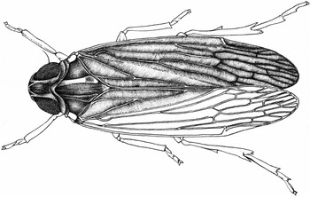
For example, the science illustrator at the Berlin Museum of Natural History, Nils Hoff, preparing a type drawing of a cicada, first measured the animal, determining the proportions of the individual parts of the body, and constructed the insect's shape and outline (fig. 6). He then sketched the structure and organization of the body, and finally added the relief of its surface (i.e., the qualities of its cuticle, exoskeleton, wings, and hairs) and the pigmentation. The graphical recording prescribed a certain sequence of examinations and assured that the illustrator would study every essential morphological feature of the animal. The obligation to render the particular promotes an economy of attention that is advantageous in particular when the phenomena to be examined elude the conventions of everyday seeing due to their amorphous structure and pose a challenge to the observer's visual comprehension. Both graphical conventions such as dotting and instruments such as the camera lucida further abet this deceleration, curbing the spontaneity and speed of the hand; so they too must be regarded as agents constraining the illustrator's creative liberty (in ways that turn out to be quite productive). Deceleration and the attention to detail force the draftsman to make distinctions, even when the phenomenon under observation is highly amorphous. In this regard, drawing can be understood as a productive provocation to the illustrating scientist. The importance of this activity within the research process can hardly be overstated, for drawing, by constantly forcing the illustrator to make decisions, effectively engenders the scientific object.
Nils Hoff: Unfinished drawing of a cicada. Pencil, pen and India ink on paper. Museum für Naturkunde, Berlin, © Nils Hoff.

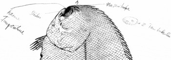
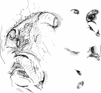
Let me offer another example from the day-to-day work of the science illustrator Nils Hoff in Berlin: While working on a type drawing of the holotype of the above-mentioned Australian sole (Aseraggodes corymbus), he noticed an unclear spot on the fish's blind side immediately to the right of the mouth. What was initially merely a problem of representation unexpectedly led to a morphological question, which could only be answered through further inspection and study of the fish (fig. 7). Originally articulated next to the drawing as a question framed as a rebus (a sketch of the unclear structure and the written note “+2 tentacles”), the feature was subsequently identified – the ichthyologist Peter Bartsch lent his assistance – under the microscope and with the aid of a hair used as a probe as the fish's nasal tube. In a first, adventurously dynamic sketch (fig. 8, top left) the illustrator outlined the organ's tube-like structure. Then, after closer observation, he refined the image into a more detailed version with dotted shading (fig. 8, top right). Finally, this last version was integrated directly into the overall plan of the fish (fig. 1). As in the classical artistic workshop practice, the illustrator's work here displays an apparent division of function between the overall and the detail study: The science illustrator produced a tentative, though already highly detailed, plan of the entire animal that records its structure, while study sketches of selected parts enabled the exploration of what had not yet been understood.
Nils Hoff: Aseraggodes corymbus, detail of blind side, 2006. Printout of a scanned drawing with notes. Museum für Naturkunde, Berlin, © Nils Hoff.

Nils Hoff: Aseraggodes corymbus, sketches of nasal tube and mouth, 2006. Ink on paper. Museum für Naturkunde, Berlin, © Nils Hoff.

The graphical recording of details such as the nasal tube, however, accomplishes an articulation of what is seen that goes far beyond what the naked eye perceives, beyond even the information offered by a photograph of the fish: The structure of this organ was revealed only under the binocular and after the introduction of a probe. This fact suggests that what the finished drawing (fig. 1) presents to the viewer as a homogenous texture of lines and dots contains considerable depths of visibility.Footnote 16 The central epistemic benefit of drawing is probably based on this methodical alternation between the disintegration of the comprehensive image and the reintegration of detail. The skilled draftsman must repeatedly fracture the harmony of his first perception of the object in order to create an image; the drawn image accordingly corresponds to no single homogeneous perception of an object. In fact, it synthesizes perceptions that concern the integrated totality of the organism, its structure, with others that regard particular features or organs.
Yet drawing not only promotes the articulation of the research object, it also has significant retroactive effects on the draftsman's sensory apparatus. The virtual analysis entailed by the act of drawing can be regarded as a process of ongoing exchange between the drawn object and the working model the draftsman forms in his mind, a model that undergoes constant revision as the drawing proceeds. The art historian James Elkins has very accurately described this mechanism of interference between cognitive and graphical operations as follows:
The game is intricate enough with its slant rhymes between the feel of the model in my mind and the feel of the paper. It is made more difficult because drawn lines have the power to remake my own imagination. Every line I draw reforms the figure on the paper, and at the same time it redraws the image in my mind. And what is more, the drawn line redraws the model, because it changes my capacity to perceive. (Elkins Reference Elkins and Savage2005, 112; emphasis in the original)
Drawing introduces a mechanism of articulation into the research process that is neither utterly pre-conceptual nor entirely subsumable under the rubric of conceptualization, even though it promotes the conceptual penetration of what the draftsman sees. It accomplishes a reconfiguration of perception, a process in which primary and secondary effects of drawing can be distinguished. On the one hand primary properties of drawing become effective, on the other secondary effects can be observed that might be traced back to changes in the situation of observation brought about by the act of drawing. Such secondary effects of drawing include the deceleration of observation, its subjection to discipline and systematization; among the primary effects of the medium, the dissociation of detail and whole should be mentioned and, more fundamentally, the necessity to introduce distinctions, as the definitional power of the line continually forces the illustrator to decide: What are the precise contours of an organ? Which structural plan organizes the organism? What is fact, and what artifact?
In the act of drawing, the draftsman's sensory apparatus thus relates to the object of research. Besides operations of separating and bringing differences to the fore, the medium of drawing enables a sequentialization and processing of the phenomena under observation. In the following, I will turn to another epistemological effect of drawing, namely the manipulation of research objects through continuous graphical re-interpretation. I will examine the parallel habit of some draftsmen to use different drawing techniques, and I will finally demonstrate how this constant switching between modes of drawing can constitute a research strategy in its own right.
5. Processing by Drawing
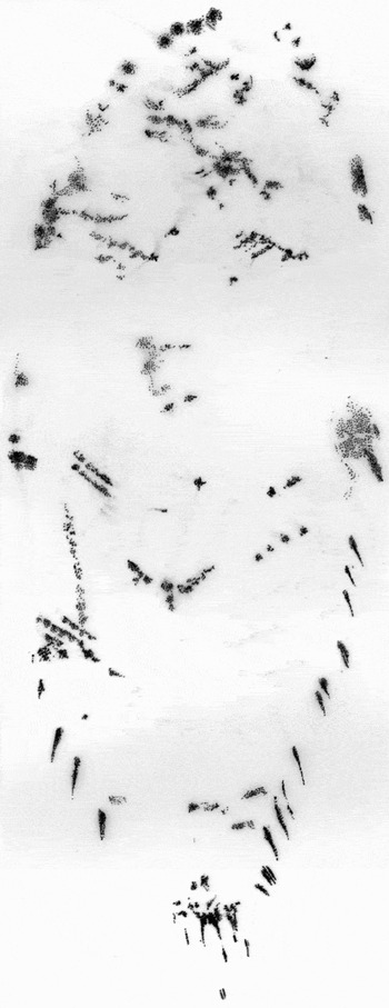
In the case of the Australian sole Aseraggodes corymbus, the gradual processing of observations occurred in a series of precise “topographical” overall views of different levels that were ultimately compiled using Photoshop into a binding image of the species. Here the fish's various traits, such as the arrangement of its scales (fig. 9), the structure of its muscles, and its pigmentation (fig. 10), attained visual independence, with each set of characteristics displaying its own order. The seemingly passive and unpremeditated process of recording visual data through drawing already contains a great deal of reconstruction. As I mentioned earlier, the artist's study of the various levels of the sole's topography in successive stages was interrupted on a number of occasions when he paused to sketch detail studies of uncertain or problematic features. For example, he had to clarify the architecture of the tubular nostril or the lateral lineFootnote 17 – particularly the relationship between this nerve channel running along the middle of both sides of the fish and the surrounding scales – before these organs could be integrated into the context of the fish's overall anatomy.Footnote 18 In other words, the draftsman moved – as it were, on the fly – between different modes of graphical elaboration: between different perspectives, between detail and overall views, between exploratory sketches and meticulous studies after nature.
Nils Hoff: Scale plan of Aseraggodes corymbus, eye side, 2006. Pencil, pen and India ink on paper. Museum für Naturkunde, Berlin, © Nils Hoff.

Nils Hoff: Aseraggodes corymbus, eye side, pigmentation, 2006. Pencil on paper. Museum für Naturkunde, Berlin, © Nils Hoff.

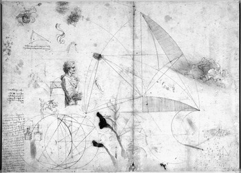
The epistemic possibilities of constant switching between different modes of drawing have first been realized in the workshops of Italian artists of the early Renaissance. Sheets such as the drawing by Leonardo now at Windsor Castle (32 by 44 cm; fig. 11) served primarily in the generation of new inventions, motives and forms, but the masters also used them to collect materials and to transmit them to apprentices and assistants. These paper archives thus formed a reservoir of future undertakings. Leonardo's sheet, too, evinces characteristic traits indicating that drawing had around 1500 already successfully internalized its function as an “immutable mobile,” directly aiming at the mobilization of the sketched objects and designs. The drawing shows a significant weakening of the image as a “tout-ensemble,” i.e., as a compositional unity transcending its parts.Footnote 19 In contradistinction to easel painting, the figures and objects drawn here do not necessarily relate to the format and the frame of their material support. Leonardo's sheet contains various studies and notations in more or less random sequence, with some of them even overlapping. The sheet's margin is no more than a loose frame holding them together, without integrating the individual observations and projects into compositional unity. The weakening of the frame is accompanied by a dynamic relation to the pictorial ground: The white surface of the paper now functions as a mere two-dimensional support (thus in the geometric constructions), now becomes legible as the space behind a plant or figure. The weak determination of frame and ground must be understood as a formal interiorization of the workshop drawing's central function, as it facilitates the copying, quoting, or cutting-out of individual felicitous trouvailles that can then be elaborated or used elsewhere.
Leonardo da Vinci: Various studies, before 1489–1490. Pen and brown ink on paper. Windsor Castle, the Royal Collection, 2011, © Her Majesty Queen Elizabeth II.

This weak determination enables, on the one hand, the close interweaving of writing and image, for what is not considered background for the purposes of an evocation of atmosphere can be written on, labeled, or scrawled on without confusing the beholder. On the other hand, it promotes a conception of the sheet of paper as a scene of open projects: Leonardo employs a great variety of graphic modes, working more or less simultaneously in several of them and using them in constant alternation. He thus turns the sheet into a sort of biotope of parallel observations or findings defined by a dialectic between openness and shortage of space, a dialectic that directly points to the economic conditions of artistic production: Since the late thirteenth century, the production of paper in Italy had made a material available that was cheap enough to serve the recording even of preliminary sketches and notes but still too expensive to be put aside after only partial use and thus wasted.
In addition to weakening the frame, the multiplication of graphic modes of handling phenomena enables a processing of research objects that no longer requires moving between different media. In contrast to painting, the medium of free-hand drawing – as established during the Renaissance – knows not just one grammar and one vocabulary; it allows for different and modally differentiated forms of access. Drawing as a design practice as it emerges in the Italian Renaissance workshop enters an alliance with linear perspective; however, the system space does not elicit here the homogenizing effect one can observe in contemporary easel and wall painting, nor does it exclude other or older modes of representation. Against Latour's argument, based on the work of Ivins and Edgerton, about the “optical consistency” of modern graphic visualization, the advantage of the workshop drawing would seem to consist in keeping a situation open in which the inscription can relate in different ways to the objects of knowledge or observation. The drawing as a study after nature can aid the visual meditation on a specific phenomenon, as in Leonardo's cloud study, where it materializes the reflection on the morphological or spatial qualities of things that gain stability and permanence in the recording. One can distinguish from this mode that of drawing in the register of possibility; one example would be the sketch of a press whose construction Leonardo ponders in the bottom left corner of the Windsor sheet. Sketches such as this one do not imitate an object that preexists them, although they certainly draw on what has already been tested and is already known; rather, they seek to anticipate a new solution. Finally, Leonardo's sheet contains geometric constructions that examine the transformation of segments of circles, presupposing a shift in the use of the instruments. They do not serve the morphological description of existing objects, nor the anticipation of future objects, but rather the mathematical penetration of the world. On the one hand, each of the three modes opens in a different way toward the world and its objects. On the other hand, each evinces a different integration of the rules of geometry: the purely mathematical construction of circle segments; the depiction of a press according to the rules of linear perspective; a cloud study entirely unconcerned by the rules of perspective.
The skilled draftsman, however, alternates between these modes of drawing on the fly, and so a permeability emerges between perceived and invented figures, or between regular and irregular figures that leads to the core of what is conventionally called the creativity of drawing. The rapid alternation between the modes of drawing creates zones of indistinguishability and semantic instability. In Leonardo's drawing, two trees stand before the man in vaguely antique dress depicted in profile; the one in front bears leaves, whereas the one further back, entirely bare, virtually grows into the man's body, where it suddenly takes on the appearance of blood vessels. The falling cascade of the toga's folds recalls the geometric construction in the man's back, while a small mountain massif grows out of the spandrels of the circle segment subdivided by triangles as a variation of the basic shape of the geometric figure. Leonardo's graphical transmutations effect the development of a polysemy that, seizing on a specific point or element, reinterprets the system of representation.Footnote 20 Leonardo treats not only the geometric figures in accordance with defined rules of transformation; the entire process of drawing elaborates the logic of a geometry of transformation avant la lettre.Footnote 21
True, none of the natural science draftsmen and drawing biologists discussed here study anything like the wealth of disparate projects Leonardo engaged in at the same time. And yet we can detect the effectiveness of the “Leonardo principle” even on the desks of contemporary science illustrators. Remember, for instance, the above-mentioned sequential procedure used by Nils Hoff. During his morphological exploration of the Australian sole, he alternated between meticulously recording the fish's overall topography and the study of questionable or unclear details in rough sketches and several perspectives. Hoff used now dynamic hatching, now disciplined dots; and perhaps even more fundamentally: He alternated between pencil-strokes moving forward at times tentatively, at times rapidly, and lines providing exaggerated articulation.Footnote 22 This explorative translation of what he saw into different modes of drawing extended almost to the point of dissociating the research object: The exaggerated articulation of the nasal tube and the lateral line are owing to this disintegration of detail and whole – they could not have been perceived in the holotype as they appear in the type drawing.
The “Leonardo principle” is most evident, however, in the production of the so-called scale plan (fig. 9). This detailed graphical recording was the basis for a count of the scales and an examination of their structure – a mapping that was of some scientific relevance, since the number and arrangement of the scales entered the animal's first description as species-specific. Not only does the type specimen display marks of its violent death, such as the indentation in the rays on its edge caused by the fishing net (see fig. 12); the ventral and dorsal fins were also damaged and made brittle during preservation. The sole's surface displays numerous cracks, indentations and other defects. The first scale plan (fig. 13), created after a close inspection of both the specimen (under the microscope) and the photograph (which served as an instrument of analysis thanks to its possibilities of artificial contrast enhancement), clearly evinces the difficulties of an activity Michael Lynch has referred to as “upgrading geometricity” (Lynch Reference Lynch1985, 54–55; cf. Lynch Reference Lynch, Lynch and Woolgar1990): a (weak) form of geometricization that allows one to discover regularities in visual data and fill in missing information. In a first step, Nils Hoff placed transparent paper on a printout of the digital photograph and traced diagonal lines along the rows of scales, which were less than clearly articulated. By integrating the rows of scales into a grid, he sought – as he himself describes it – to gain “some idea of order,”Footnote 23 which, in this first sketch, is not so rigid as to be condemned to failure on account of the irregularities in the scale surface. Between this first attempt and the final, definitive version lay a long and laborious process of balancing the reconstruction of a linear “blueprint” and the individual deviations from it.
Nils Hoff: Aseraggodes corymbus, eye side. Digital photograph, 2006. Museum für Naturkunde, Berlin, © Nils Hoff.

Nils Hoff: Aseraggodes corymbus, eye side, first sketch for the scale plan, 2006. Pencil on tracing paper. Museum für Naturkunde, Berlin, © Nils Hoff.

This quest for a geometry of what is seen, a rather bureaucratic process in comparison to Leonardo's sketches, owes no less to the variety of graphical modes and the possibility of spontaneous reinterpretation it entails. The draftsman's line, which – unlike the line as defined in geometry – is not a set of points but always already a pencil-stroke in motion, propels a category leap between morphological description, constructive sketch, and the geometrical penetration of the world that can be put to productive use. This polysemy ultimately rests on the fact that any pencil-stroke dynamically determines its relation to the ground, defining the latter as mere horizontal support or work-surface, as in geometric construction, or as a background possessing (illusionistic) depth, as in European easel painting. If one considers drawing in the perspective of this polysemy rooted in the medium itself, it can no longer be described as a simple method of mobilization in the sense of the “immutable mobiles.” The epistemic function of the act of drawing, in other words, transcends the realm of procedures of transmission, however broadly defined; it instead implies an active conversation between what is known or seen and the possibilities and limitations of pencil and paper. Drawing on Latour's work but also seeking a critical revision of his claims, this study has sought to show that, first, mobilization does not come into play only when a fact or finding moves from one place or medium to another one; it is already active in essential ways within the medium. It should have become clear, second, that while the invariance of the “immutable mobiles” is a central virtue of the logistics of “paperwork,” the recovery of graphic knowledge requires a vastly more dynamic activity – a sequential processing that brings out differences by translating the phenomenon under examination into various techniques of graphic representation.
Acknowledgment
I thank Gerrit Jackson for translating this article from the German.