INTRODUCTION
Haemoproteus spp. (Haemosporida, Haemoproteidae) are widespread in birds all over the world, and some species cause disease, sometimes even lethal in avian hosts (Valkiūnas, Reference Valkiūnas2005; Mehlhorn, Reference Mehlhorn2015). Many studies have reported reduced survival of wild birds infected with Haemoproteus parasites and negative effects of this infection on immunity indices, body condition, and reproductive success of the hosts (Marzal et al. Reference Marzal, de Lopes, Navarro and MØller2005; Valkiūnas et al. Reference Valkiūnas, Žičkus, Shapoval and Iezhova2006; La Puente et al. Reference La Puente, Merino, Toma, Moreno, Morales, Lobato, Garcia-Fraile and Belda2010). Additionally, there is growing evidences for a trade-off between reproductive effort and resistance to parasites, particularly when food resources are limited (Merino et al. Reference Merino, Moreno, Jose-Sanz and Arriero2000; Garvin et al. Reference Garvin, Homer and Greiner2003; Atkinson, Reference Atkinson, Atkinson, Thomas and Hunter2008; Mehlhorn, Reference Mehlhorn2015). However, little is known about the effects of Haemoproteus infections on blood-sucking insects.
Experimental studies showed that high parasitaemia of Haemoproteus spp. is lethal to Culicoides midges. Liutkevičius (Reference Liutkevičius2000) and Valkiūnas and Iezhova (Reference Valkiūnas and Iezhova2004) reported high mortality of Culicoides impunctatus infected with Haemoproteus balmorali, Haemoproteus belopolskyi, Haemoproteus dolniki, Haemoproteus fringillae, Haemoproteus lanii and Haemoproteus tartakovskyi. However, mechanisms of mortality remain unclear. It was speculated (Valkiūnas and Iezhova, Reference Valkiūnas and Iezhova2004) that mortality of biting midges might be due to the damage caused by ookinetes and/or developing oocysts, which injure the epithelial cells of the midgut and might cause associated inflammatory reactions. However, histological observations are absent in biting midges. Valkiūnas et al. (Reference Valkiūnas, Kazlauskienė, Bernotienė, Bukauskaitė, Palinauskas and Iezhova2014) reported numerous Haemoproteus ookinetes migrating throughout entire body of exposed mosquitoes Ochlerotatus cantans. The parasites damaged tissues in abdomen, thorax and even head of this insect. That also might be the case in biting midges, which are the main vectors of Haemoproteus parasites belonging to subgenus Parahaemoproteus (Bukauskaitė et al. Reference Bukauskaitė, Žiegytė, Palinauskas, Iezhova, Dimitrov, Ilgūnas, Bernotienė, Markovets and Valkiūnas2015; Žiegytė et al. Reference Žiegytė, Bernotienė, Palinauskas and Valkiūnas2016).
Because mechanisms of mortality in Culicoides biting midges due to Haemoproteus infections are unknown, the aim of this study was to follow the survivorship and pathology caused by post-feeding on high-intensity infection of H. lanii (cytochrome b lineage hRB1) in experimentally infected biting midges C. impunctatus. This biting midge is widespread in Europe and willingly takes blood meal on birds (Glukhova, Reference Glukhova1989; Glukhova and Valkiūnas, Reference Glukhova and Valkiūnas1993; Blackwell, Reference Blackwell1997; Žiegytė et al. Reference Žiegytė, Palinauskas, Bernotienė, Iezhova and Valkiūnas2014; Bukauskaitė et al. Reference Bukauskaitė, Žiegytė, Palinauskas, Iezhova, Dimitrov, Ilgūnas, Bernotienė, Markovets and Valkiūnas2015). We allowed wild-caught females of this species to feed on a naturally infected bird with high parasitaemia, collected dead insects and examined them histologically.
MATERIALS AND METHODS
Study site, selection of experimental birds and collection of blood samples
The work was carried out at the Biological Station of the Zoological Institute of the Russian Academy of Sciences on the Curonian Spit in the Baltic Sea (55°05′N, 20°44′E) in June 2014. Birds were caught with mist nets. About 30 µl of blood was collected with heparinized microcapillaries by puncturing the brachial vein and stored in SET buffer (Hellgren et al. Reference Hellgren, Waldenström and Bensch2004) for molecular analysis. The samples were held at ambient temperature in the field and later at –20 °C in the laboratory. One drop of blood was used to make two blood films, which were fixed in absolute methanol and stained with Giemsa (Valkiūnas, Reference Valkiūnas2005). One red-backed shrike (Lanius collurio) naturally infected with H. lanii (hRB1) was selected as a donor of mature gametocytes (Fig. 1A, B) to infect C. impunctatus. Haemoproteus spp. uninfected red-backed shrikes were not found, and all tested birds (21 individuals) were infected with different levels of parasitaemia. One uninfected juvenile common crossbill (Loxia curvirostra) was used to feed a control group of biting midges. At our study site, the breeding period of crossbills takes place in the end of winter and beginning of spring when transmission of haemosporidians is absent. Juvenile crossbills remain uninfected with haemosporidians on the Curonian Spit in May–June (Valkiūnas, Reference Valkiūnas2005). Both experimental birds were selected using microscopic and polymerase chain reaction (PCR)-based examination of their blood samples (see below). The birds were kept indoors in a vector-free room under controlled conditions [55–60% relative humidity (RH), 20 ± 1 °C, the natural light–dark photoperiod (L/D) 17:7 h] and fed with standard diets for seed eating or insectivorous bird species. They survived to the end of this study and were then released.
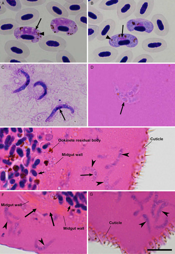
Mature microgametocytes (A), macrogametocytes (B) and ookinetes (C–G) of Haemoproteus lanii (lineage hRB1) in the circulation of red-backed shrike Lanius collurio (A, B), midgut contents (C) and histological sections (D–G) of abdomen (E–G) and thorax (D) of experimentally infected biting midges Culicoides impunctatus. Long simple arrows – nuclei of parasites, triangle arrowheads – pigment granules, simple arrowheads – ookinetes in haemocoel, short simple arrow – nun-digested red blood cell in the midgut, long triangle arrows – midgut wall damage caused by massive protrusion of ookinetes in haemocoel. Note presence of numerous ookinetes in haemolymph of the haemocoel of abdomen area; the parasites are often seen in large clumps, which lead to interruption of the haemolymph circulation (E, G). Scale bar = 10 µm.

Experimental design and making preparations of parasites
Experimental infections were performed near the Lake Chaika, located close to the village Rybachy, were density of C. impunctatus was high (Bukauskaitė et al. Reference Bukauskaitė, Žiegytė, Palinauskas, Iezhova, Dimitrov, Ilgūnas, Bernotienė, Markovets and Valkiūnas2015). Wild-caught biting midges were exposed by allowing them to take blood meals on selected birds as described by Valkiūnas (Reference Valkiūnas2005). Briefly, the feathers from the birds’ head were gently plucked off from a surface of about 1 cm2. Birds were kept in hands covered with rubber gloves at a site with high density of biting midges, which were allowed to feed naturally on feather-free areas. The birds were exposed to bites of biting midges between 10 and 12 pm. When several females began taking blood meals on a bird head, the head with feeding insects was carefully placed into unzipped insect cage (12×12 × 12 cm3) made of fine-mesh bolting silk. The engorged females fly off after feeding. The cage with engorged biting midges was then closed using a zipper. Cages with engorged flies were placed in plastic packs and transported to the laboratory. Forty-six females of C. impunctatus took infected blood meals, and 17 females took blood meals from uninfected bird. The latter group served as a negative control. Pads of cotton-wool moistened with 10% saccharose solution were placed on the top of each insect cage. Both infected and control groups of biting midges were held in standard conditions (16–18 °C, 70 ± 5% RH and L/D photoperiod of 17:7 h).
To determine survivorship of experimental and control groups, dead biting midges were collected from insect cages 12 and 24 h post-exposure (PE), and then daily until 7 day PE. They were identified morphologically according to Gutsevich (Reference Gutsevich1973), counted, and some of them were dissected in a drop of 0·9% normal saline to examine midgut contents for ookinetes. The midgut was extracted from abdomen and gently crushed to prepare a thin smear using dissecting needles, which were disinfected in fire after each dissection to prevent contaminations. The smears were air dried, fixed in absolute methanol and stained with Giemsa the same way as blood films (Valkiūnas, Reference Valkiūnas2005). We examined midgut contents of six dead midges for ookinetes 12 h PE. After dissection, all residual parts of insects were fixed in 96% ethanol and tested by PCR (see below) in order to confirm the presence of parasite lineages in exposed biting midges.
Entire bodies of 13 experimentally infected midges were fixed in 10% neutral formalin in order to use them for histological examination. Formalin-fixed heads, thoraxes and abdomens of biting midges were embedded in paraffin separately. In all, 104 histological sections of 4 µm were obtained, stained with haematoxylin–eosin and examined under a light microscope (see below). Eight experimental and 17 control insects were tested for presence of parasites by PCR-based methods (see below).
Infected and control biting midges were processed individually, and they were examined using the same methods.
Microscopic examination of preparations and parasite morphology
All preparations were examined with Olympus BX-43 light microscope equipped with Olympus SZX2-FOF digital camera and imaging software QCapture Pro 6·0, Image-Pro plius (Tokyo, Japan). Blood films were examined at low magnification (×400) for approximately 15 min, and then at least 100 fields were studied at high magnification (×1000). Intensity of parasitaemia in birds was estimated just after exposure of biting midges. It was determined as a percentage by actual counting of the number of mature gametocytes (Fig. 1A, B) per 1000 red blood cells. All vector preparations were first examined at low magnification (×100, ×600) and then at high magnification (×1000). Representative preparations of gametocytes (accession no. 48894 NS) and ookinetes in midgut content (48895, 48896 NS) and histological sections (48897, 48898 NS) were deposited in Nature Research Centre, Vilnius, Lithuania. The statistical analysis was carried out using Statistica 7 package. Percentages of survived control and experimental insects were compared by Fisher's exact test. A P value of 0·05 or less was considered significant.
PCR and sequencing
Total DNA was extracted from all samples using the standard ammonium acetate extraction method (Richardson et al. Reference Richardson, Jury, Blaakmeer, Komdeur and Burke2001). For genetic analysis of parasites, a nested PCR protocol was used as described by Bensch et al. (Reference Bensch, Stjernman, Hasselquist, Ostman, Hansson, Westerdahl and Pinheiro2000) and Hellgren et al. (Reference Hellgren, Waldenström and Bensch2004). We amplified a segment of parasite cyt b gene using two pairs of primers. In the first PCR, we used HaemNFI and HaemNR3 for detection of Haemoproteus, Plasmodium and Leucocytozoon species. For the second PCR, we used primers HAEMF and HAEMR2, which are specific to Haemoproteus and Plasmodium parasites. All amplifications were evaluated by running 1·5 µl of the final PCR product on a 2% agarose gel. One negative control (nuclease-free water) and one positive control (Haemoproteus sp. infected blood sample, which was positive by microscopic examination of blood films) were used. No cases of false positive or negative amplifications were found. All PCR positive samples were sequenced from both directions. The genetic analyser ‘Basic Local Alignment Search Tool’ (National Centre of Biotechnology Information website: http// www.ncbi.nlm.nih.gov/BLAST) was used to determine lineages of detected DNA sequences. Sequences were edited and aligned using the program BioEdit (Hall, Reference Hall1999) and deposited in GenBank (accessions KU529941, KU529942).
Because we used wild-caught C. impunctatus in experiments, we determined prevalence of possible natural Haemoproteus infection in biting midges. Unfed biting midges were collected using an entomological net at the same study site where we exposed donor birds to bites of C. impunctatus (see above). In all, 108 wild-caught unfed females were tested by PCR amplification. DNA was extracted from 27 pools of biting midges, each containing four midges.
DNA extracted from individual flies was used to confirm the species identification of C. impunctatus used in our experiments. For this purpose, the insect-specific primers LCO149 and HCO2198 were applied to amplify a fragment of cytochrome oxidase subunit I of mitochondrial DNA (Folmer et al. Reference Folmer, Black, Hoeh, Lutz and Vrijenhoek1994). The amplicons were sequenced in both directions.
Ethical statement
The experiments described herein comply with the current laws of Lithuania and Russia. Experimental procedures were approved by the International Research Co-operation Agreement between the Biological Station Rybachy of the Zoological Institute of the Russian Academy of Sciences and the Institute of Ecology of Nature Research Centre (25–05–2010). All efforts were made to minimize handling time and potential suffering of birds. None of the experimental birds suffered apparent injury during experiments, and all birds were released after experiments.
RESULTS
According to the PCR-based analysis, no natural infection was found in wild-caught biting midges (n = 108) collected at the study site, indicating that the probability to use naturally infected insects was low in our experiment. Morphological identification and PCR-based testing confirmed that all experimental (n = 46) and control (n = 17) biting midges belonged to C. impunctatus. All obtained DNA sequences of insects (n = 13) were identical to the sequence of C. impunctatus with GenBank accession KJ627800, and they were of 99% similarity with other corresponding sequences of the same insect species available in GenBank.
Parasites were not detected in control biting midges (n = 17) using PCR-based testing, but the presence of the lineage hRB1 of H. lanii was confirmed in experimentally infected insects (n = 8).
The survivorship of control and infected groups differed significantly (Fisher's exact test, P < 0·001). Twelve hours PE, sudden death was reported among 45 of 46 (98%) infected insects, but all midges in control group (n = 17, 100%) were alive (Fisher's exact test, P < 0·001). All experimental biting midges were dead 24 h PE, but all control insects were alive at the same time (Fisher's exact test, P < 0·001). First dead control insects were reported 2 days PE, and four insects survived until 7 day PE (observation time).
Massive infection of mature H. lanii ookinetes was seen in all preparations of midgut contents and histological sections of dead midges 12 h PE. Ookinetes were numerous in midgut contents (Fig. 1C). Numerous ookinete residual bodies possessing clumps of brown pigment granules were seen in the midgut contents, in which non-digested red blood cells were readily visible (Fig. 1E, F). Mature ookinetes were numerous in histological section of abdomen (Fig. 1E–G) and thorax (Fig. 1D), but were not seen in histological sections of head. Examination of histological sections revealed the massive ookinetes infection in haemocoel both in abdomen (Fig. 1E–G) and thorax (Fig. 1D). The parasites damaged these parts of body. Massive eruption of ookinetes from midgut content to haemocoel leaded to the damage of midgut wall (Fig. 1F). Numerous large clumps of ookinetes (up to ten parasites, Fig. 1F, G) were often seen in haemocoel both in abdomen and thorax; that likely lead to interruption of the haemolymph circulation. Oocysts were not seen. This study shows that numerous ookinetes migrate from the midgut to the haemocoel and then overfill both abdomen and thorax within 12 h PE causing insect mortality before parasites reach the heads of the midges. Because mortality occurs rapidly after infected blood meal, oocysts have no time to develop. Ookinetes, but not oocysts are responsible for mortality in biting midges after blood meal on intensely infected birds.
DISCUSSION
Species of Culicoides are natural vectors of avian haemoproteids (Garnham, Reference Garnham1966; Valkiūnas, Reference Valkiūnas2005; Levin et al. Reference Levin, Valkiūnas, Santiago-Alarcon, Cruz, Iezhova, O'Brien, Hailer, Dearborn, Schreiber, Fleischer, Ricklefs and Parker2011; Bukauskaitė et al. Reference Bukauskaitė, Žiegytė, Palinauskas, Iezhova, Dimitrov, Ilgūnas, Bernotienė, Markovets and Valkiūnas2015). The key results of this study are that: (1) blood meals with high numbers of H. lanii gametocytes (parasitaemia 5·2%) led to a 100% mortality of Culicoides biting midges; (2) these insects die rapidly, the majority within 12 h PE; and (3) the main cause of death is migrating ookinetes, which penetrate midgut wall, damage the wall, overfill haemocoel and cause interruption of the haemolymph circulation and might damage mechanically organs in abdomen and thorax.
Marked mortality of biting midges and mosquitoes due to Haemoproteus (Parahaemoproteus) infections has been reported in blood-sucking Culicoides biting midges and bird-biting Culicidae (Liutkevičius, Reference Liutkevičius2000; Valkiūnas and Iezhova, Reference Valkiūnas and Iezhova2004; Valkiūnas et al. Reference Valkiūnas, Kazlauskienė, Bernotienė, Bukauskaitė, Palinauskas and Iezhova2014). Mature gametocytes of these parasites (Fig. 1A, B) are completely prepared for exflagellation. Gametogenesis, fertilization and ookinetes development readily occur even in vitro, without adding any additional medium, except anticoagulants (Garnham, Reference Garnham1966; Valkiūnas, Reference Valkiūnas2005). This is not a case in Plasmodium parasites, which require the presence of additional stimuli (xanthurenic acid and host blood-derived factors) for exflagellation and development of ookinetes (Arai et al. Reference Arai, Billker, Morris, Panico, Delcroix, Dixon, Ley and Sinden2001; Sinden, Reference Sinden2009). It is thus not unexpected to observe numerous mature Haemoproteus spp. ookinetes within several hours after blood meal both in susceptible (Culicoides spp.) and non-susceptible (mosquitoes) insects and also in vitro (Valkiūnas, Reference Valkiūnas2005). Mature ookinetes of different Haemoproteus species develop within 2–12 h after exposure to air both in vitro and in vivo at 16–20 °C (Valkiūnas, Reference Valkiūnas2005; Valkiūnas et al. Reference Valkiūnas, Palinauskas, Križanauskienė, Bernotienė, Kazlauskienė and Iezhova2013; Žiegytė et al. Reference Žiegytė, Palinauskas, Bernotienė, Iezhova and Valkiūnas2014; Bukauskaitė et al. Reference Bukauskaitė, Žiegytė, Palinauskas, Iezhova, Dimitrov, Ilgūnas, Bernotienė, Markovets and Valkiūnas2015). During normal life cycle, Haemoproteus spp. ookinetes migrate through the epithelial layer of the midgut of the vector and round up under the basal lamina giving rise to oocysts (Valkiūnas, Reference Valkiūnas2005). Our study shows that Haemoproteus spp. ookinetes can rapidly penetrate though the midgut epithelial layer, reach the haemocoel, migrate in haemolymph (Fig. 1E) forming large clumps of the parasites, which likely interrupt haemolymph circulation and probably damage mechanically organs in abdomen and thorax in biting midges (Fig. 1D–G).
Available experimental studies show that high parasitaemia with H. tartakovskyi and H. balmorali are lethal for Ocherotatus cantans and probably for other bird-biting mosquitoes. Ookinetes were reported in abdomen, thorax and head of infected mosquitoes (Valkiūnas et al. Reference Valkiūnas, Kazlauskienė, Bernotienė, Bukauskaitė, Palinauskas and Iezhova2014). We show that the same is true for Culicoides biting midges infected with H. lanii. Additionally, mortality is even more severe in tiny biting midges, which rapidly die before ookinetes reach the head. Migrating ookinetes and even oocyst-like bodies of haemoproteids have been reported in heads of infected mosquitoes (Valkiūnas et al. Reference Valkiūnas, Kazlauskienė, Bernotienė, Bukauskaitė, Palinauskas and Iezhova2014), but they were not seen in heads of biting midges during this study. It is worth mentioning that ookinetes of Plasmodium gallinaceum, the avian malaria parasite, move actively in the body of non-vector insects, resulting in partial or ectopic sporogonic development in unnatural hosts (Schneider and Shahabuddin, Reference Schneider and Shahabuddin2000). Actually, the records of haemosporidian ookinetes and oocysts outside the midgut wall of blood-sucking insects is not unexpected because Hepatocystis kochi (Haemoproteidae), a haemosporidian parasite of African monkeys, completes sporogony and produces viable sporozoites in the head of the biting midge Culicoides adersi, the natural parasite vector, in which transit ookinetes are also present in the thorax (Garnham, Reference Garnham1966). However, we show that H. lanii ookinetes do not reach heads of biting midges likely due to rapid death of infected insects.
This study is in accordance with the former experimental observation about mortality caused by H. lanii in C. impunctatus biting midges sampled in the same population on the Curonian Spit (Valkiūnas and Iezhova, Reference Valkiūnas and Iezhova2004). However, mechanisms of mortality remained unknown and survivorship was higher in the latter observation, during which 48% biting midges survived between 1 and 2 day PE, and 18% of the midges survived until 7–8 day PE. Higher survivorship likely is due to lighter parasitaemia, which was 2·2% in study published by Valkiūnas and Iezhova (Reference Valkiūnas and Iezhova2004), but was 5·2% in our study. The high virulence of H. lanii in biting midges is not an exception. The available experimental data (Liutkevičius, Reference Liutkevičius2000; Valkiūnas and Iezhova, Reference Valkiūnas and Iezhova2004) indicate that blood meals on birds with high parasitaemia (>1%) of H. balmorali, H. belopolskyi, H. dolniki and H. fringillae also resulted mortality (48%) of C. impunctatus within 48 h PE, but mortality was not reported in midges fed on uninfected birds. Additionally, it was shown that survivorship depends on intensity of parasitaemia in birds (Liutkevičius, Reference Liutkevičius2000; Valkiūnas and Iezhova, Reference Valkiūnas and Iezhova2004). Mature ookinetes of H. lanii are worm-like bodies reaching 17 µm in length on average (Valkiūnas et al. Reference Valkiūnas, Palinauskas, Križanauskienė, Bernotienė, Kazlauskienė and Iezhova2013). Direct mechanical damage by large number of these relatively big organisms is the main reason of mortality. The death occurs rapidly (within 12 h PE), i.e. before development of oocysts, which develop several days later, if insects survive (Žiegytė et al. Reference Žiegytė, Palinauskas, Bernotienė, Iezhova and Valkiūnas2014; Bukauskaitė et al. Reference Bukauskaitė, Žiegytė, Palinauskas, Iezhova, Dimitrov, Ilgūnas, Bernotienė, Markovets and Valkiūnas2015). Thus, the main reason of sudden biting midge mortality is migrating ookinetes, but not oocysts of the parasite.
Haemoproteus spp. sporogony readily completes in Culicoides biting midges, which are effective vectors (Atkinson, Reference Atkinson1991; Valkiūnas, Reference Valkiūnas2005), and the majority of infected females survive until development of sporozoites in case of blood meal with low parasitaemia (Žiegytė et al. Reference Žiegytė, Palinauskas, Bernotienė, Iezhova and Valkiūnas2014). For experimental vector research, we recommend using infected birds with Haemoproteus parasitaemia of <1%. Experimental studies show that many exposed biting midges survived such Haemoproteus spp. infections and can act as vectors (Žiegytė et al. Reference Žiegytė, Palinauskas, Bernotienė, Iezhova and Valkiūnas2014; Bukauskaitė et al. Reference Bukauskaitė, Žiegytė, Palinauskas, Iezhova, Dimitrov, Ilgūnas, Bernotienė, Markovets and Valkiūnas2015; Žiegytė et al. Reference Žiegytė, Bernotienė, Palinauskas and Valkiūnas2016).
Light parasitaemia of Haemoproteus spp. (<1%) predominate in wildlife (Valkiūnas, Reference Valkiūnas2005; Asghar et al. Reference Asghar, Hasselquist and Bensch2011). That is probably an evolutionary adaptation of parasites to survive in vectors during sporogonic development because light infections provide more chances for the infected insects to live long enough to feed again. However, high parasitaemia (between 1 and 10%) is also common in wildlife, particularly during bird breeding period when active transmission occurs and juvenile birds get infected (Valkiūnas, Reference Valkiūnas2005). Because the prevalence of Haemoproteus spp. infections is often high (Valkiūnas et al. Reference Valkiūnas, Iezhova and Shapoval2003; Pérez-Tris et al. Reference Pérez-Tris, Helgren, Križanauskienė, Waldenström, Secondi, Bonneaud, Fjeldså, Hasselquist and Bensch2007) and the parasitaemia of over 1% often causes mortality in biting midges, these parasites might be a possible factor influencing density of bird-biting midges. The same is true for Leucocytozoon infections, which cause mortality in simuliid flies (Desser and Yang, Reference Desser and Yang1973; Allison et al. Reference Allison, Desser and Whitten1978). The importance of Haemoproteus spp. and Leucocytozoon spp.–vector interactions and the rate of influence of these infections on density of blood-sucking insects remain unclear in wildlife populations; it is worth doing additional investigation because these parasites are widespread and prevalent in birds and are virulent in bird-biting insects. It is worth mentioning that species of Plasmodium requires more additional stimuli for exflagellation and ookinetes production than related species of Haemoproteus and Leucocytozoon. Plasmodium parasites produce ookinetes mainly in representatives of certain genera of Culicidae mosquitoes (Sinden, Reference Sinden2009; Palinauskas et al. Reference Palinauskas, Žiegytė, Ilgūnas, Iezhova, Bernotienė, Bolshakov and Valkiūnas2015) and are less virulent for insects than Haemoproteus (Ferguson and Read, Reference Ferguson and Read2002; Valkiūnas et al. Reference Valkiūnas, Kazlauskienė, Bernotienė, Bukauskaitė, Palinauskas and Iezhova2014). Additional studies are needed for the better understanding molecular mechanisms of high virulence of avian Haemoproteus parasites in dipteran insects.
This study and former experimental research (Liutkevičius, Reference Liutkevičius2000; Valkiūnas and Iezhova, Reference Valkiūnas and Iezhova2004; Valkiūnas, Reference Valkiūnas2005) showed that high Haemoproteus spp. parasitaemia is the cause of mortality in biting midges. It is thus probable that lower blood meal parasitaemia should be preferable for effective Haemoproteus parasite transmission. Additional experiments are needed for better understanding this issue.
ACKNOWLEDGEMENTS
We are grateful to the director of the Biological Station ‘Rybachy’ Casimir V. Bolshakov for providing facilities for the experimental research and Rita Žiegytė, Mikas Ilgūnas and Vaidas Palinauskas, for participation in field work.
FINANCIAL SUPPORT
This study was partly funded by the Research Council of Lithuania (no. MIP-045/2015 to G. V.), and also supported by the Open Access to research infrastructure of the Nature Research Centre under Lithuanian open access network initiative (grant awarded to Nature Research Centre).