Introduction
Oocytes of the pig enter meiosis between days 40 and 48 post conception (Black & Erickson, Reference Black and Erickson1968; Mauléon & Mariana, Reference Mauléon, Mariana, Cole and Cupps1977) and become arrested in the diplotene stage of the first meiotic prophase (germinal vesicle stage, GV) around the time of birth (Mauléon & Mariana, Reference Mauléon, Mariana, Cole and Cupps1977). In adult animals several fully grown oocytes reinitiate meiosis during each oestrous cycle following the preovulatory gonadotrophin surge (Ainsworth et al., Reference Ainsworth, Tsang, Downey, Marcus and Armstrong1980). Under suitable in vitro conditions, pig oocytes resume meiosis (Edwards, Reference Edwards1965), which is characterized morphologically by an initial condensation of chromatin in the intact GV followed by germinal vesicle breakdown (GVBD) and continued chromatin condensation, accompanied by microtubule reorganization (Motlik & Fulka, Reference Motlik and Fulka1976). These events lead to the metaphase spindle organization and subsequent completion of the first reduction division, after which oocytes enter the second meiotic division and become arrested at the meta phase II (MII) stage until fertilization. Biochemically these cellular changes are mediated by specific signal transduction pathways. One of the key molecules regulating the resumption of meiosis is mitogen-activated protein kinase (MAPK), which is also termed extracellular regulated kinase (ERK). MAPK has been reported to be involved in oocyte maturation in Xenopus (Gotoh et al., Reference Gotoh, Nishida, Matsuda, Shina, Kosaka, Shiokawa, Akiyama, Ohta and Sakai1991), mice (Sobajima et al., Reference Sobajima, Aoki and Kohmoto1993), pigs (Inoue et al., Reference Inoue, Naito, Aoki, Toyoda and Sato1995), goats (Dedieu et al., Reference Dedieu, Gall, Crozet, Sevellec and Ruffini1996), cows (Fissore et al., Reference Fissore, He and Van de Woude1996), rats (Zernicka-Goetz et al., Reference Zernicka-Goetz, Verlhac, Géraud and Kubiak1997), horses (Goudet et al., Reference Goudet, Belin, Bézard and Gérad1998) and humans (Sun et al., Reference Sun, Breitbart and Schatten1999a). Oocytes exhibit two isoforms of MAPK, MAPK1 (44 kDa) and MAPK2 (42 kDa). MAPK belongs to the group of serine/threonine kinases; its active form is dually phosphorylated on threonine and tyrosine residues by MAPK kinase (MAPKK or MEK) (Campbell et al., Reference Campbell, Seger, Graves, Graves, Jensen and Krebs1995). MAPK seems to be essential for meiotic resumption of oocytes, the organization of microtubules and arrest at the MII stage (Sun et al., Reference Sun, Blumenfeld, Rubinstein, Goldman, Gonen and Breitbart1999b).
The phosphorylation pattern of MAPK during in vitro maturation (IVM) depends on the species and the maturation conditions. MAPK is unphosphorylated in GV-stage oocytes, becomes phosphorylated around GVBD and remains at high phosphorylation levels during arrest at MII until fertilization. MAPK activity was detected in mice about 4 h before GVBD during FSH-induced meiotic resumption in vitro (Su et al., Reference Su, Rubinstein, Luria, Lax and Breitbart2001). In contrast, when mouse oocytes underwent spontaneous resumption of meiosis, activated MAPK was detected after GVBD (Verlhac et al., Reference Verlhac, Pennart, Maro, Cobb and Clarke1993). Phosphorylation of MAPK has been reported in pig oocytes shortly before (Inoue et al., Reference Inoue, Naito, Nakayama and Sato1998; Wehrend & Meinecke, Reference Wehrend and Meinecke2001), during (Motlik et al., Reference Motlik, Pavlok, Kubelka, Kalous and Kalab1998; Ye et al., Reference Ye, Flint, Luck and Campbell2003) and even after GVBD (Inoue et al., Reference Inoue, Naito, Aoki, Toyoda and Sato1995; Liang et al., Reference Liang, Li-Jun, Zhong, Chen, Schatten and Sun2005). GVBD was inhibited or reduced when MAPK phosphorylation was inhibited by MEK inhibitors (PD98059 or U0126), during IVM of COCs (e.g. Kagii et al., Reference Kagii, Naito, Sugiura, Iwamori, Ohashi, Goto, Yamanouchi and Tojo2000; Meinecke & Krischek, Reference Meinecke and Krischek2003). This suggests that MAPK has to be phosphorylated in oocytes to overcome arrest at the GV-stage. However, GVBD proceeded undisturbed when the germ cell-specific MAPK kinase kinase Mos (oncogene, Moloney sarcoma virus) was inhibited by using Mos knockout mice (Colledge et al., Reference Colledge, Carlton, Udy and Evans1994; Hashimoto et al., Reference Hashimoto, Watanabe, Furuta, Tamemoto, Sagata, Yokoyama, Okazaki, Nagayoshi, Takeda and Ikawa1994; Su et al., Reference Su, Wigglesworth, Pendola, O'Brien and Eppig2002) and by injecting c-mos antisense RNA into pig oocytes (Ohashi et al., Reference Ohashi, Naito, Sugiura, Iwamori, Goto, Naruoka and Tojo2003). These observations strongly support the view that MAPK activation in oocytes is not involved in the initiation of the resumption of meiosis. Furthermore, spontaneous meiotic resumption of denuded oocytes has been reported in medium supplemented with U0126 in the mouse (Tong et al., Reference Tong, Fan, Chen, Song, Schatten and Sun2003) and pig (Fan et al., Reference Fan, Tong, Lian, Li, Gao, Cheng, Chen, Schatten and Sun2003).
Phosphorylated MAPK was detected in mouse and pig cumulus cells of COCs immediately following recovery from follicles (Su et al., Reference Su, Wigglesworth, Pendola, O'Brien and Eppig2002; Fan et al., Reference Fan, Tong, Lian, Li, Gao, Cheng, Chen, Schatten and Sun2003). A significant increase in MAPK activity in porcine cumulus cells was observed after 2 h (Fan & Sun, Reference Fan and Sun2004) and 8 h incubation (Shimada et al., Reference Shimada, Maeda and Terada2001) and activity peaked after 20 h of IVM (Shimada et al., Reference Shimada, Maeda and Terada2001).
The purpose of the present study was to investigate in further detail the phosphorylation of MAPK both in the oocyte itself and in the cumulus cells during the initial phases of FSH/LH-induced IVM.
Materials and Methods
Unless otherwize stated, chemicals and reagents were obtained from Sigma-Aldrich. Porcine FSH and LH were obtained from the National Hormone and Peptide Program of the Harbor–UCLA Medical Center (Torrance, CA, USA). The following antibodies were purchased form Santa Cruz Biotechnologies: polyclonal rabbit anti-ERK1 antibody (K-23), recognising unphosphorylated and phosphorylated MAPK1/ERK1 and to a less extent MAPK2/ERK2, and monoclonal mouse anti-p-MAPK antibody (E-4), recognising phosphorylated MAPK1/ERK1 and MAPK2/ERK2.
Collection and IVM of cumulus–oocyte complexes
Ovaries of peripuberal gilts (age: 6–7 months, bodyweight: 90–120 kg) were collected at a local abattoir and placed in a thermos flask with 0.9% (w/v) saline solution (at between 30 and 33 °C) and transported to the laboratory within 1.5 h of slaughter of the animals. Individual follicles (between 3 and 5 mm in diameter) were sliced with a scalpel blade and flushed with 0.9% saline containing 1% (v/v) heat-inactivated fetal calf serum (FCS) by means of a 20 ml syringe connected to a 26-gauge needle. COCs with at least five compact cumulus cell layers were selected and washed three times in Dulbecco's phosphate-buffered saline (PBS) containing 10% (v/v) FCS. For IVM, 25 COCs were placed in each well of a 4-well multidish (Nunc) with 0.5 ml culture medium (TCM199, 20 μg/ml insulin, 80 μg/ml l-glutamine, 50 μg/ml gentamycin, 20% (v/v) FCS, 2.5 μg/ml FSH, 5.0 μg/ml LH). The MEK inhibitor U0126 (Favata et al., Reference Favata, Horiuchi, Manos, Daulerio, Stradley, Feeser, Van Dyk, Pitts, Earl, Hobbs, Copeland, Magola, Scherle and Trzaskos1998) was dissolved in dimethylsulphoxide (DMSO) and a 5.0 mM stock solution was stored in dark flasks at 4 °C and used within a month at a final concentration of 10 μM. Control medium was prepared identically except that it contained no U0126 and 0.2% DMSO (v/v).
IVM was carried out at 39 °C in 5% CO2 in air and humidified atmosphere for the appropriate time.
Determination of nuclear maturation stages
After IVM, COCs were treated when necessary with 0.25% (w/v) hyaluronidase (bovine testes) and denuded by repeated pipetting with a fine bore Pasteur pipette. Denuded oocytes (DOs) were prepared as whole mounts, fixed in ethanol/acetic acid (3:1) for at least 24 h and stained with 2% (w/v) aceto-orcein solution. Nuclear maturation stages were determined under a phase-contrast microscope (BX 60, Olympus) according to the method of Motlik & Fulka (Reference Motlik and Fulka1976). In the course of IVM 3.7 ± 3.0% of COCs and 8.1 ± 6.8% of DOs degenerated (shrunken cytoplasm, clumped chromatin mass). These oocytes were discarded and not included in the results.
SDS-PAGE and immunoblot analysis
Cumulus cells and oocytes of COCs were stored separately in 4 μl PBS at –80 °C until use. The degree of phosphorylation of MAPK proteins of oocytes and cumulus cells was determined by modified sodium dodecyl sulphate polyacrylamide gel electrophoresis (SDS-PAGE) (Lämmli, Reference Lämmli1970). The probes were heated for 5 min in SDS sample buffer at 95 °C, centrifuged for 1 min at 12 000 g and cooled on ice. The probes were separated by gel electrophoresis with a 4.5% stacking gel and a 10.5% separation gel for 10 min at 100 V and for 55 min at 200 V. The proteins were transferred from the gel onto a nitrocellulose membrane (Hybond-ECL, Amersham Biosciences) for 2 h at 1 mA/cm2 and the membrane was blocked overnight at 4 °C in a blocking buffer comprised of 50 mM Tris, 150 mM NaCl, 0.1% (v/v) Tween 20 and 5% Teleostean gelatine. Depending on the experiment, the membrane was incubated at room temperature overnight with antibodies against ERK1 (+2) or with p-MAPK diluted 1:333 and 1:500 in TBST (50 mM Tris, 150 mM NaCl, 1% Tween 20), respectively. After washing three times for 10 min in TBST HRPO-conjugated goat anti-rabbit immunoglobulin (IgG) (Perbio) or HRPO-conjugated rabbit anti-mouse IgG (Perbio) diluted 1:667 in TBST was placed on the membrane for 1 h. Then the membrane was intensively washed, six times for 10 min in TBST, and processed by an enhanced chemiluminescence detection system (SuperSignal® West Dura Extended Duration Substrate, Perbio). A Kodak® X-Omat AR Film was used for visualization. The membrane was placed for reprobing in a stripping buffer comprised of 100 mM 2-mercaptoethanol, 2% (w/v) SDS and 62.5 mM Tris–HCl, pH 6.7, for 2 h at room temperature in order to remove the antibody from the membrane.
Experimental design
Experiment 1
COCs were incubated in the presence of FSH/LH for 0, 21, 25, 29 and 45 h. At the end of incubation, the degree of expansion of the COCs was recorded under a stereomicroscope by a photographic unit and the nuclear maturation status of the oocytes was determined. The phosphorylation pattern of MAPK in both oocytes and cumulus cells was determined by immunoblot. In further studies, the phosphorylation pattern of MAPK in cumulus cells was investigated following incubation of COCs in the presence or absence of FSH/LH for 0, 0.5 and 2 h. To prevent FCS in the culture medium from influencing the phosphorylation pattern of MAPK, this experiment was repeated using medium with 0.3% (w/v) polyvinylpyrrolidone (PVP) instead of FCS.
Experiment 2
COCs were first incubated in the presence of FSH/LH for 0, 3, 5, 8 and 10 h, then washed three times and transferred into medium without FSH/LH and subsequently incubated for further 23, 21, 18 and 16 h, respectively. The nuclear maturation status of the oocytes was examined at the end of the 26 h incubation period.
Experiment 3
Here we examined the role of cumulus cells in mediating the effect(s) of MEK inhibition on oocyte nuclear maturation pattern. COCs were first incubated in culture supplemented with 10 μM U0126 for 24 h, then washed three times in inhibitor-free medium and cultured in the absence of U0126 for further 24 h. At the end of the culture period nuclear maturation status was determined. Furthermore, COCs were denuded immediately after recovery from the follicles and cultured in inhibitor-free medium. Intact COCs were incubated in medium supplemented with U0126. At the end of the 26 h incubation period, the nuclear maturation status was determined.
In a third series of experiments, COCs were first cultured for 5 h in medium without U0126 then transferred to medium containing U0126 and incubated for another 21 h as intact COCs. In subsequent experiments, COCs were first cultured for 5 h in the absence of U0126, then denuded and transferred into medium with or without U0126 and cultured as DOs for a further 21 h.
To investigate the phosphorylation pattern of the MAPK under the influence of U0126 during IVM, COCs were incubated in the presence of U0126 for 0, 2, 21, 26, 30 and 45 h. Following incubation both oocytes and cumulus cells were analysed separately by protein blotting.
Statistical analysis
All experiments were repeated at least three times. Nuclear maturation results were analysed statistically (http://faculty.vassar.edu/lowry/VassarStats.html) using chi-squared analysis. A probability of p < 0.01 was considered to be statistically significant.
Results
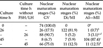
There was no expansion of the cumulus cell complexes during incubation of COCs in medium without FSH/LH, whereas there was a moderate degree of expansion of these COCs after 21 h incubation in the presence of FSH/LH; these became fully expanded by the end of incubation (45 h). Immediately after recovery from follicles, all (74/74) of the oocytes were in the GV stage (Table 1). Following incubation with FSH/LH, 81.9% (122/149) of the oocytes had undergone GVBD after 26 h of IVM and 87.4% (104/119) of the oocytes had reached AI–MII stages at the end of the incubation period (45 h). In contrast, after IVM without FSH/LH for 26 and 45 h, 90.7% (88/96) and 75.0% (66/88), respectively, of the oocytes remained in the GV stage (Table 1). This indicates that only a small proportion of oocytes resumed meiosis spontaneously under the described conditions.
Nuclear maturation stages of COCs cultured in medium with or without FSH/LH for 26 or 45 h

GV, germinal vesicle stage; Di/MI, diakinesis/metaphase I; AI–MII, anaphase I–metaphase II.
a–dValues of rows with different letters differ significantly (p < 0.01).
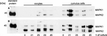
Both isoforms of MAPK were unphosphorylated in oocytes recovered immediately after liberation from follicles (0 h, Fig. 1). About half of the MAPK molecules became phosphorylated following a 25 and 29 h incubation, which corresponded to the time of GVBD. The degree of phosphorylation continued to increase until the end of incubation (45 h), when nearly all the MAPK was phosphorylated. In contrast, a minimal amount of MAPK was already phosphorylated in cumulus cells at the time of recovery from follicles (0 h, Fig. 1); after 21 h of incubation, the cumulus cells contained more phosphorylated than unphosphorylated MAPK isoforms. After 25 and 29 h of incubation, the proportion of phosphorylated and unphosphorylated MAPK was well balanced, while at the end of culture (45 h), the phosphorylated forms were greatly reduced and almost all the MAPK was dephosphorylated.
MAPK phosphorylation in oocytes and cumulus cells during IVM of COCs. Twenty-five oocytes and cumulus cells of five COCs were loaded per lane. The same membrane was first incubated with the anti-p-MAPK antibody (A) and then stripped with the anti-ERK1 (+2) antibody (B). (A) Phosphorylation of MAPK2 occurred in oocytes at 25, 29 and 45 h of IVM, whereas there was only weak phosphorylation of MAPK1 by the antibody. Minimal amounts of MAPK1/2 were already phosphorylated in cumulus cells at 0 h; phosphorylation peaked at 21 h and decreased until 45 h. (B) MAPK1/2 were partially phosphorylated in oocytes at 25 and 29 h and became fully phosphorylated at 45 h of IVM. Minimal amounts of MAPK1/2 were already phosphorylated in cumulus cells at 0 h; phosphorylation peaked at 21h and decreased until 45 h.

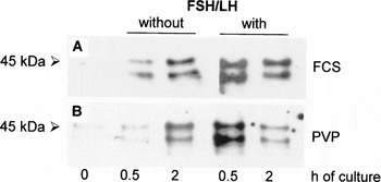
There were minimal amounts of phosphorylated MAPK in cumulus cells of COCs before culture in medium supplemented with FSH/LH (at 0 h) (Fig. 2A). There was a marked increase in phosphorylation of MAPK after 0.5 and 2 h. Cumulus cells of COCs incubated in medium without FSH/LH showed the same pattern of MAPK phosphorylation, although there was less phosphorylation of MAPK at 0.5 h. After 2 h of incubation there was no difference between the two groups in the degree of phosphorylation of MAPK proteins. Replacement of FCS by PVP led to the same degree of MAPK phosphorylation (Fig. 2B), suggesting that FCS growth factors are not involved in MAPK phosphorylation under the present conditions.
MAPK phosphorylation in cumulus cells during the first 2 h of IVM. Cumulus cells of 10 COCs were loaded per lane and the anti-p-MAPK antibody was used. In medium supplemented with FSH/LH and FCS (A) or with FSH/LH and PVP (B) amplification of MAPK phosphorylation was detectible as early as after 0.5 h incubation. Phosphorylation of MAPK decreased until the end of cultivation (2 h).

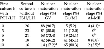
When COCs were cultured in the absence of FSH/LH, 90.7% (88/97) of the oocytes did not resume meiosis (Table 2). Most of the COCs (88.0%, 81/92) incubated first in medium supplemented with FSH/LH for 3 h before transferred into medium without FSH/LH and cultured for further 23 h remained in the GV stage. Priming the COCs in medium supplemented with FSH/LH for 5, 8 and 10 h before the transfer into medium without FSH/LH and a further incubation for 21, 18 or 16 h led to a significant increase in the resumption of meiosis in 26.6% (21/79), 53.9% (49/91) and 82.8% (67/81) of the oocytes, respectively. These results indicate that the presence of FSH/LH is required for at least 5 h in the culture medium to induce a significant proportion of oocytes to resume meiosis during the IVM period in the absence of gonadotrophins. Therefore, 5 h was chosen as the priming time for the following experiments.
Nuclear maturation stages of COCs cultured first in medium with and subsequently in medium without FSH/LH for the indicated times

GV, germinal vesicle stage; Di/MI, diakinesis/metaphase I; AI–MII, anaphase I–metaphase II.
a–dValues of rows with different letters differ significantly (p < 0.01).
The majority of the COCs cultured first in medium supplemented with 10 μM U0126 for 24 h and subsequently transferred in to inhibitor-free medium and cultured for further 24 h resumed meiosis (GV: 20.6%, 32/155; Di/MI: 69.7%, 108/155; AI–MII: 9.7%, 15/155). This demonstrates that the inhibitory action of U0126 on nuclear maturation progression of COCs is completely reversible.
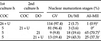
When COCs were denuded immediately after recovery from the follicles and cultured in medium without U0126 for 26 h, 50.0% (52/104) of the oocytes resumed meiosis. As 24% (25/104) and 26.0% (27/104) of the oocytes reached Di–MI and AI–MII stages, respectively, this indicates that half of the denuded oocytes resumed meiosis spontaneously under the above conditions. Conversely, COCs incubated for 26 h in medium supplemented with U0126 remained in the GV stage (97.4%, 114/117), showing that MEK inhibition by U0126 in intact COCs prevented resumption of meiosis (Table 3). When intact COCs were first cultured for 5 h in inhibitor-free medium and then transferred and incubated in medium supplemented with U0126 for a further 21 h, the oocytes also remained in the GV stage (96.4%, 81/84), which shows that U0126 prevented resumption of meiosis even when the COCs underwent an initial 5 h of inhibitor-free culture. After denudation of COCs following a first culture period in U0126-free medium for 5 h and after another 21 h in the presence or absence of U0126, the oocytes resumed meiosis in spite of the presence of the MEK inhibitor (Table 2). This demonstrates that the presence of cumulus cells around oocytes is required to prevent the resumption of meiosis by U0126. Regardless of the presence or absence of the inhibitor, removal of surrounding cumulus cells induced an accelerated progression through meiotic maturation and significantly more DOs reached AI–MII stages during the 26 h incubation period than did intact COCs (cf. Tables 1 and 2).
Effect of U0126 (+U) on nuclear maturation stages of COCs cultured first as intact COCs and subsequently as denuded oocytes (DO) for the indicated times

GV, germinal vesicle stage; Di/MI, diakinesis/metaphase I; AI–MII, anaphase I –metaphase II.
Values of rows with different letters (a, b, c, d) differ significantly (p < 0.01).
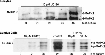
Immunoblot analysis of oocytes and cumulus cells collected after culture of COCs in media supplemented with U0126 for 0, 2, 26, 30 and 45 h demonstrated that MAPK phosphorylation in oocytes was prevented (Fig. 3). Both isoforms were unphosphorylated when tested and no double bands or mobility shift were detected. By contrast, treatment of COCs with U0126 reduced MAPK phosphorylation in cumulus cells only during the first 2 h of culture (Fig. 3), but there were double bands and mobility shifts at 26 h of incubation, even in the presence of 50 instead of 10 μM of U0126 in the medium. These results indicate that MAPK phosphorylation in oocytes responded well to inhibition by U0126, whereas MAPK phosphorylation in cumulus cell exhibited only a transient and limited sensitivity against MEK repression.
MAPK phosphorylation in oocytes and cumulus cells during IVM of COCs in medium supplemented with U0126. Forty oocytes and cumulus cells of 10 COCs were loaded per lane. The anti-p-MAPK antibody was used. U0126 prevented phosphorylation of MAPK in oocytes. In cumulus cells U0126 only reduced MAPK phosphorylation, even at a concentration of 50 μM.

Discussion
The present study investigated the role of the MAPK in pig oocytes and in cumulus cells during gonadotrophin-induced resumption of meiosis in vitro. Evaluation of the current IVM model revealed that expansion of cumulus oophorus and resumption of meiosis following FSH/LH stimulation occurred at high maturation rates (GVBD > 80%) at regular kinetics (GVBD 25–29 h). Resumption of meiosis was prevented in the majority of the oocytes in the absence of gonadotrophins after 26 and 45 h (90.7 and 75.0%, respectively) of culture. It must be pointed out that these in vitro conditions do not reflect the physiological situation in adult cycling animals, but this model makes it possible to discriminate between spontaneous and gonadotrophin-induced resumption of meiosis. Furthermore, it is in agreement with a previously reported model (Motlik et al., Reference Motlik, Nagai and Kikuchi1991) using pig cumulus-enclosed oocytes attached to a portion of membrana granulosa.
The pattern of MAPK phosphorylation in oocytes during FSH/LH-induced maturation observed in the present study (Fig. 1) confirms earlier reports (Kagii et al., Reference Kagii, Naito, Sugiura, Iwamori, Ohashi, Goto, Yamanouchi and Tojo2000; Kanayama et al., Reference Kanayama, Miyano and Lee2002). The current study further demonstrated that minor amounts of MAPK in cumulus cells are already phosphorylated when COCs are recovered from follicles. In contrast, MAPK in oocytes did not start to become phosphorylated until after 25 h of incubation. The former finding corresponds to observations in the mouse, where MAPK phosphorylation was also detected after 30 min of IVM (Su et al., Reference Su, Denegre, Wigglesworth, Pendola, O'Brien and Eppig2003). MAPK phosphorylation in pig cumulus cells has been reported to occur after 1, 2, 8 and 12 h of IVM (Shimada et al., Reference Shimada, Maeda and Terada2001; Fan et al., Reference Fan, Tong, Lian, Li, Gao, Cheng, Chen, Schatten and Sun2003; Fan & Sun, Reference Fan and Sun2004; Liang et al., Reference Liang, Li-Jun, Zhong, Chen, Schatten and Sun2005). These differences may depend in part on the different hormones, growth factors and time schedules used to induce resumption of meiosis in vitro. In the present experiments, phosphorylation of MAPK in cumulus cells peaked at between 25 and 29 h and disappeared after 45 h of IVM, revealing a phosphorylation pattern, which differed conspicuously from that of the oocyte itself. Phosphorylation of MAPK in cumulus cells rose within 30 min after IVM was begun (Fig. 2A, B). It must be pointed out that MAPK kinase phosphorylation in cumulus cells also increased in the absence of FSH/LH, although to a lesser degree than in the presence of gonadotrophins during the first 2 h of IVM. It is assumed that this MAPK activation occurred spontaneously, as replacement of FCS, which is a rich source of growth factors, by PVP did not change the phosphorylation pattern (Fig. 2B). There was a marked increase in the phosphorylation of MAPK after 0.5 h culture in FSH/LH-supplemented medium; this indicates that the gonadotrophin signal led to additional phosphorylation of MAPK. This finding is supported by studies on primary cultures of porcine granulosa cells in which FSH and LH selectively activated cytosolic ERK1 above basal levels. Furthermore, activation of ERK1 by LH was considerably more rapid (maximum at 5 min) and transient than activation of ERK1 by FSH (maximum between 15 and 60 min) (Cameron et al., Reference Cameron, Foster, Bukovsky and Wimalasena1996). Taken together, the data support a rapid (within minutes) MAPK phosphorylation and activation in granulosa and cumulus cells following gonadotrophin stimulation. However, the precize regulation and functional roles of MAPK activation in cumulus cells remain unknown (Fan & Sun, Reference Fan and Sun2004).
To obtain a more definitive analysis of the action of gonadotrophins on the cumulus cells, COCs were exposed to FSH/LH-containing medium for different times and then transferred to gonadotrophin-free medium and cultured for a total of 26 h. This experiment showed that COCs required between 5 and 10 h of exposure to FSH/LH to resume meiosis in the absence of gonadotrophins. This means that all intercellular and intracellular steps required for meiotic progression took place during the first 5 to 10 h of IVM. During that time, the protein synthesis essential for GVBD occurred (Fulka et al., Reference Fulka, Motlik, Fulka and Jilek1986), but there were no visible nuclear signs of maturation, since pig oocytes require about 13 h to reach the GV-II stage under these conditions (Motlik & Fulka, Reference Motlik and Fulka1976; Wehrend & Meinecke, Reference Wehrend and Meinecke2001).
To elucidate the role of the MAPK in more detail, we suppressed the upstream kinase (MEK) of the MAPK with U0126. The present results demonstrate that 10 μM U0126 prevented phosphorylation of MAPK in the oocytes matured as intact COCs (Fig. 3). These findings also indicate that the absence of MAPK phosphorylation in the oocytes was accompanied by an arrest at the GV stage (97.4%, Table 3). These observations agree with previous results for U0126 (Kagii et al., Reference Kagii, Naito, Sugiura, Iwamori, Ohashi, Goto, Yamanouchi and Tojo2000, Fan et al., Reference Fan, Tong, Lian, Li, Gao, Cheng, Chen, Schatten and Sun2003) and confirm earlier studies employing the MEK inhibitor PD 98059 (Meinecke & Krischek, Reference Meinecke and Krischek2003). However, above studies reported that 23.6, 42.0 and 33.6% of the oocytes underwent GVBD although the COCs had been cultured in the presence of the inhibitors for 44, 48 and 30 h, respectively. These differences might be due to the incubation of different selected COCs, by the use of an inhibitor-free collection medium before the COCs were transferred into inhibitor supplemented medium and by the different inhibitors used, respectively.
It is of particular interest to note that phosphorylation of MAPK was prevented in oocytes by the MEK inhibitor (Fig. 3), whereas in the cumulus cells U0126 reduced the MAPK phosphorylation only during the first 2 h of IVM and that more intense bands of phosphorylated MAPK appeared even in the presence of 50 μM U0126 after 26 h of culture (Fig 3). This confirms and extends the findings of Fan et al., (Reference Fan, Tong, Lian, Li, Gao, Cheng, Chen, Schatten and Sun2003), who reported a pronounced reduction of MAPK phosphorylation in cumulus cells following inhibition by U0126. When COCs were cultured in U0126-supplemented medium, the MAPK of oocytes remained unphosphorylated and the cells stayed in the GV stage despite the weak phosphorylation of MAPK in cumulus cells. This observation indicates that the MEK inhibitor is unable to prevent phosphorylation of MAPK in cumulus cells but acts concomitantly on phosphorylation events of MAPK in oocytes. Furthermore, prevention of MAPK phosphorylation in oocytes depends on the presence of the intact cumulus oophorus during culture, as DOs progressed through nuclear maturation when incubated in U0126-supplemented medium (Kagii et al., Reference Kagii, Naito, Sugiura, Iwamori, Ohashi, Goto, Yamanouchi and Tojo2000; Su et al., Reference Su, Wigglesworth, Pendola, O'Brien and Eppig2002; Fan et al., Reference Fan, Tong, Lian, Li, Gao, Cheng, Chen, Schatten and Sun2003; current study). Taken together, these data suggest that MEK does not induce phosphorylation of MAPK in cumulus cells, but is responsible for MAPK phosphorylation in oocytes. This implies that the MAPK of the cumulus cells may be phosphorylated by one or more alternative pathways and exerts an as yet undefined influence, which may not be directly involved in the induction of resumption of meiosis. This assumption is supported by the finding that early phosphorylation of MAPK occurred in cumulus cells in the absence of FSH/LH and even after replacement of FCS by PVP in the culture medium (Fig. 2B). Under such conditions, most of the oocytes remained in the GV stage (Table 1) despite the spontaneous MAPK phosphorylation in cumulus cells.
After denudation of COCs, significantly more oocytes reached the AI–MII stages during 26 h culture than did non-denuded COCs. This phenomenon was even more pronounced when the COCs were subjected to 5 h gonadotrophin priming before denudation (Table 3). The accelerated resumption of meiosis following denudation of pig oocytes is known to be due to a shortening of the latent period until GVBD occurs (Mattioli et al., Reference Mattioli, Galeati, Bacci and Barboni1991). This suggests strongly that cumulus cells influence, by one or more yet unknown mechanisms, the activation in the oocyte of maturation promoting factor (MPF), which is responsible for induction of nuclear breakdown.
In summary, the data from an IVM model that allows a distinction between spontaneous and gonadotrophin-induced resumption of meiosis indicate there are at least two different MAPK phosphorylation events in cumulus cells and oocytes following gonadotrophin stimulation. In cumulus cells, FSH/LH induced an early and rapid U0126-insensitive phosphorylation of MAPK, while U0126-susceptible MAPK phosphorylation occurred around the time of GVBD in the oocyte itself.
Acknowledgement
The authors gratefully acknowledge the skilled technical assistance of Edita Podhajsky, Monika Guemmer and Christian Lipke and Ronald H.F. Hunter for critical reading of the manuscript.