Cystic fibrosis is an autosomal recessive multisystem disease detected in 1/3400 live births and is prevalent in Caucasians. The disease occurs because of structural and functional defects in the cystic fibrosis transmembrane conductance regulator protein, which encodes chloride channels in the apical membrane of epithelial cells. Reference Kosorok, Wei and Farrell1 Thus, ion transport in the epithelial cell plasma membrane is impaired. This situation causes a reduction in the volume of fluids secreted in all organs (lungs, pancreas, intestines, sweat glands, etc.) and thickening of the mucosa. Consequently, inflammation occurs in these organs. The most conspicuous clinical manifestations occur in the lungs. Over time, recurrent infections that are deteriorated by chronic inflammation in the lungs of patients with cystic fibrosis cause a severe decrease in the ability of the lung parenchyma and functions. Lung failure usually occurs in patients with cystic fibrosis. Parallel to the progressive deterioration of the lungs, the right ventricular function of patients with cystic fibrosis is also impaired. Reference Fraser, Tullis, Sasson, Hyland, Thornley and Hanly2–Reference Ionescu, Ionescu, Payne, Obieta-Fresnedo, Fraser and Shale5
Cor pulmonale is the deterioration of the anatomy (hypertrophy or dilatation) and function of the right ventricle due to diseases that affect the function and/or structure of the lung. In the physiology of Cor pulmonale developed due to cystic fibrosis, the incriminating factors are both chronic hypoxemias resulting from progressive lung injury and pulmonary arterial hypertension caused by cystic fibrosis. In addition, low partial pressure of oxygen pressure level is thought to contribute to the deterioration of myocardial contractions. Reference Bridges and Bradbury6,Reference Hamutcu and Woo7 The clinical diagnosis of Cor pulmonale and right ventricular failure in patients with cystic fibrosis is arduous, because symptoms of pulmonary disease are present in the foreground. In daily practice, right ventricular function and anatomy are not frequently evaluated in transthoracic echocardiography examinations because of the difficult geometry of the right ventricle. Also, it remains challenging to demonstrate the development of pulmonary hypertension in patients with cystic fibrosis using conventional echocardiographic measurements.
Cardiac MRI is a rapid and reliable method for evaluating the right ventricular morphology, volume, and function. According to the literature, cardiac MRI has proven superior to other imaging methods (transthoracic echocardiography, CT, and heart catheter) in evaluating the right ventricle. It is considered the best method because it allows for a comprehensive evaluation of cardiac morphology and physiology. Reference Geva8 The most crucial advantage of cardiac MRI is that it provides time-lapsed three-dimensional visualisation with a high-resolution image.
It has been scientifically proven in many adolescents and adult patient studies that both anatomical (dilatation and/or hypertrophic change) and functional (systolic and/or diastolic) disorders of the right ventricle can develop in patients with moderate and severe cystic fibrosis using cardiac MRI. Reference Matthay, Berger and Loke3,Reference Ionescu, Ionescu, Payne, Obieta-Fresnedo, Fraser and Shale5,Reference Alfakih, Plein, Bloomer, Jones, Ridgway and Sivananthan9,Reference Grothues, Moon and Bellenger10 Unfortunately, to the best of our knowledge, there are no studies showing the presence of right ventricular failure, cor pulmonale, and pulmonary arterial hypertension in children with mild cystic fibrosis using cardiac MRI. This study aimed to assess the ventricular anatomy, function of the right ventricle and the haemodynamic findings of pulmonary artery of children with mild cystic fibrosis through cardiac MRI.
Materials and methods
This study included patients who had been followed up by paediatric pulmonology and paediatric cardiology departments at Bezmialem Vakif University Hospital. The patient group of this prospective study consisted of 32 children with mild cystic fibrosis. The patient group was clinically stable, with no respiratory exacerbation in the previous 6 months. Spirometry was performed in all patients with cystic fibrosis. All patients had genotype and sweat test confirmation, and only children with mildly reduced forced expiratory volume in 1 s (FEV1) were selected for this study. Ethics approval was granted by the Ethics Committee of Bezmialem Vakif University, and a consensus form was obtained from each patient participating in the study.
Our patients and their age-matched controls were aged from 6 to 17 years and 7 to 15 years, respectively. All patients who participated in the study had mild cystic fibrosis. Height, weight, body mass index, body surface area, and heart rate were measured in all patients. Cardiac MRI was used to assess ventricular volumes, anatomy, and function and quantify the flow vessel over the pulmonary artery in the control and study groups. Right ventricular geometry was determined using the right ventricular anterior wall thickness (mm), ratio of body surface area (right ventricular anterior wall thickness/body surface area (mm/m2), right ventricular end-diastolic volume (ml), right ventricular end-diastolic volume/body surface area (mm/m2), right ventricular end-systolic volume (ml), and right ventricular end-systolic volume/body surface area (ml/m2)). Systolic function was determined using the ejection fraction [(%), systolic volume (ml), cardiac output (L/ml), and ratio of body surface area (cardiac output/body surface area (L/ml/m2), systolic volume/SBA (ml/m2), tricuspid annular plane systolic excursion]. Haemodynamic findings of pulmonary artery were determined using PA peak flow (ml/s), PA time to peak flow (ms), PA peak velocity (cm/sn), PA time to peak velocity (ms), PA average area (cm2), and PA systolic pressure (mmHg). All data of children with mild cystic fibrosis were compared with those of 30 age-matched healthy control group participants.
MRI
In this study, cardiac MRI was used to estimate ventricular volume and function and quantify the flow vessel over the pulmonary artery. All examinations were conducted at the Bezmialem Vakif University Hospital in the Radiology Department using a 1.5-Tesla scanner (Magnetom Avanto; Siemens Medical Systems, Erlangen, Germany) with a six-channel body coil. Cardiac MRI scans were performed without contrast, None of the patients were sedated during the procedure. The procedure was explained to the patients, and informed constent was obtained from the patients’ parents. The patients who were too young to adapt to the condition of cardiac MRI were excluded from the study. The study protocol consisted of ECG-triggered steady-state free precession balanced cine magnetic resonance images of the heart with end-expiration breath holds obtained in the usual planes: long vertical axis, four-chamber, short-axis (SA), and right ventricular outflow tract. Two imaging planes are commonly used to determine volumetric function: the short-axis oblique orientation (SA) or the axial (transverse) orientation (AX). We used SA orientation to determine right ventricular functions. The SA stack covered both ventricles from the base to the apex. We used the following sequence parameters: repetition time (TR), 42.24; echo time (TE), 1.11; and 109 × 192 matrix. Pulmonary artery pressure was calculated from peak velocity using a pulmonary trunk velocity map. Velocity mapping was performed using a velocity-encoding range of 150 cm/s. Velocity encoding was adjusted by aliasing. In addition, the tricuspid annular plane systolic excursion was measured to study right ventricular function.
Image analysis
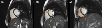
The following parameters were quantified in the right ventricle: end-diastolic volume and end-systolic volume. Estimations were determined from balanced steady-state free precession cine magnetic resonance sequences in SA planes. The volumes of all right ventricular slices at the maximum relaxation and contraction stages were estimated to obtain end-diastolic volume and end-systolic volume, respectively. The criteria used to manually draw the contours of the right ventricle are detailed in Fig 1. Three patients showed inadequate compliance during the MRI and were excluded from the study. Proper quality was achieved in all patients included in the study. Endocardial contouring was performed in a blinded manner without knowledge of the clinical status of the patient, indication for the cardiovascular MRI examination, or previous measurements. The contours were carefully drawn with particular emphasis on avoiding misinterpretation of the basal slices. Therefore, right ventricle segmentation is technically feasible. The basal slices near the valve plane were carefully revised to avoid including the right atrium. As Alfakih et al. Reference Alfakih, Plein, Bloomer, Jones, Ridgway and Sivananthan9 indicated regarding the inflow part of the right ventricle, the blood volume is excluded from the right ventricle volume if the surrounding wall is thin and not trabeculated because it is in the right atrium.
Two-chamber short-axis (SA) images of right ventricle segmentation at end-diastolic phases and at basal (a), mid-cavity (b), and apical slices (c). The lines represent the contours of the right ventricle including trabeculations.
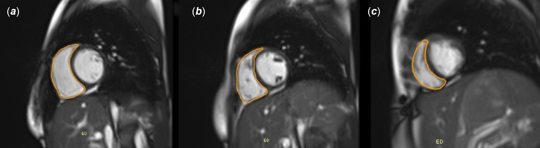
The right ventricle trabeculae were included in the volume-cavity calculations in the SA series. Special care was taken to ensure inclusion of the outflow tract. Ventricular function was analysed using a dedicated workstation (Syngo Via; Siemens Medical Solutions, Erlangen, Germany) equipped with cardiac MRI-certified software. The user manually selected all end-diastolic and end-systolic phases in the SA planes (Fig 1). The phases of both end diastole and end systole were independently defined for each ventricle. The program computed the end-diastolic and end-systolic volumes indexed to the body surface area according to endocardial tracing. Phase-velocity magnetic resonance was performed. A conventional phase-sensitive gradient-echo sequence was used in a double-oblique plane perpendicular to the dominant flow direction in the main pulmonary artery.
Pulmonary function tests
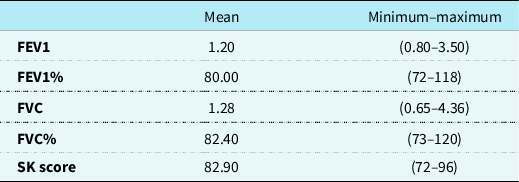
Spirometry was performed in all patients with cystic fibrosis. They were clinically classified using the FEV1 value modified by the Shwachman–Kulczycki score. Thus, 32 children with mild cystic fibrosis values were included in the study. Height and weight measurements were obtained from the patients, and spirometry (Winspiro Pro, Medical International Research, Italy) was performed for patients aged >8-year-old. The forced vital capacity and FEV1 were measured. The best test measurement was recorded for the third time using a spirometer. Forced vital capacity and FEV1 values were expressed as a percentage of the patient’s age, height, and weight according to the prediction equations, which were standardised in our institution. Mild cystic fibrosis evaluation was calculated based on 70% and above the expected FEV1 according to the European Respiratory Society classification. Similarly, modified streptokinase was calculated, and patients were classified according to American Thoracic Society/ERS guidelines. All patients who had a score of 71 or above were considered to be in the patient group (Table 1).
Pulmonary function tests and clinical score findings of patient with cystic fibrosis.
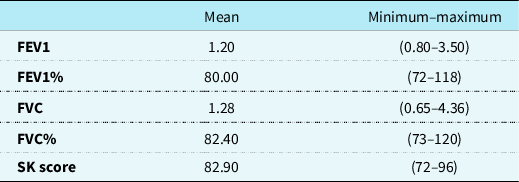
FEV1: Forced expiratory volume in 1 s, FVC: Forced vital capacity, SK: Modified Shwachman–Kulczycki.
Statistical analysis
Statistical analyses were performed using the SPSS software (version 20.0; IBM Corp., Armonk, NY, USA). Measurement data were presented as mean ± standard deviation. Pearson’s correlation and paired sample t-tests were used to compare the two groups for all parameters. Statistical significance was set at p < 0.05.
Results
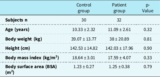
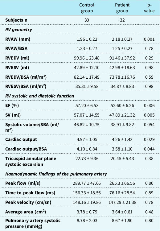
Our patients and their age-matched controls were aged from 6 to 17 years and 7 to 15 years, respectively. All patients had mild cystic fibrosis. The baseline characteristics are shown in Table 1. No statistical differences were observed in age, height, weight, body mass index, body surface area, or heart rate between the two groups (Table 2). Right ventricular anterior wall thickness/body surface area (mm/m2), right ventricular end-diastolic volume (ml), end-systolic volume (ml), right ventricular end-diastolic volume/body surface area (ml/m2), right ventricular end-systolic volume/body surface area (ml/m2), and tricuspid annular plane systolic excursion were not significantly different between the groups (Table 3). Right ventricular anterior wall thickness (mm), ejection fraction (%), cardiac output (L/ml), cardiac output (L/ml/m2), systolic volume (ml/m2), and longitudinal axis in diastole (mm) were significantly different between the two groups (p < 0.01). The right ventricular anterior wall thickness (mm) was significantly higher in children with cystic fibrosis (p = 0.01). Ejection fraction (%), cardiac output (L/ml), cardiac output (L/ml/m2), and systolic volume (ml/m2) were significantly lower in patients with cystic fibrosis (p < 0.01). Our study showed that there was no significant statical difference in PA peak flow (ml/s), PA time to peak flow (ms), PA peak velocity (cm/sn), PA time to peak velocity (ms), PA average Area (cm2), and PA systolic pressure (mmHg) between the two groups (Table 3).
Demographic characteristics of patient and control groups.
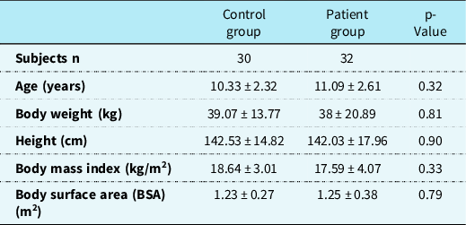
Values are expressed as mean ± SD; (*): significant.
Comparison of cardiovascular MRI findings of patients and control groups.
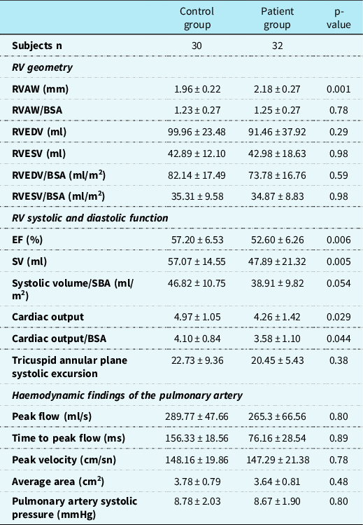
BSA = body surface area; EF = ejection fraction; RV = right ventricle; RVAW = right ventricular anterior wall thickness; RVEDV = right ventricular end-diastolic volume; RVESV = right ventricular end systolic volume; SV = systolic volume
Discussion
Transthoracic two-dimensional echocardiography is a fast, repeatable, non-invasive diagnostic tool for evaluating the size and function of the right ventricle in a normal individual. Unfortunately, although it has few limitations, transthoracic echocardiography produces an inadequate image quality in patients with advanced lung disease. Reference Ionescu, Ionescu, Payne, Obieta-Fresnedo, Fraser and Shale5,Reference Alfakih, Plein, Bloomer, Jones, Ridgway and Sivananthan9,Reference Grothues, Moon and Bellenger10 Cardiac MRI is a non-invasive radiologic procedure for evaluating cardiac functions and volumes that do not use geometric assumptions.
Cardiac MRI provides clear quantitative and qualitative information about the diameters, volumes, transvalvular blood flow and the functions of both ventricles. Also, it shows the contractility, viability, and fibrosis development of myocardium. Therefore, cardiac MRI appears to be the most accurate and easily reproducible imaging technique for analysing cardiac anatomy. Conversely, cardiac MRI has become the gold standard utility in assessing right ventricular visualisation and measurement. Reference Grothues, Moon and Bellenger10–Reference Robertson, Kohler and Hoskins13 Right ventricular end-diastolic volume and right ventricular end-systolic volume of the right ventricle can be measured from two different windows using an SA view and AX windows via cardiac MRI. Alfakih et al. Reference Alfakih, Plein, Bloomer, Jones, Ridgway and Sivananthan9 compared the axial plane with an SA and concluded that the volumes derived from the axial slices were lower than those derived from the SA (notably, the right ventricular end-diastolic volume was 4.8% lower). Previous studies stated that the values estimated in the axial plane were more reproducible than those estimated in the SA; therefore, they proposed using the axial plane to estimate and assess right ventricular function. Reference Marchesseau, Ho and Totman14–Reference Jimenez-Juan, Joshi and Wintersperger16 Many studies have reported that right ventricular function can be measured accurately using these two planes, both in the normal and dilated hearts. Reference Grothues, Moon and Bellenger10 Mooij et al. studied patients with right ventricular disease and found good reproducibility in the segmentation results of the SA. Reference Mooij, de Wit, Graham, Powell and Geva17 Clarke et al. did a study on the observer variability of right ventricular volume measurements between different images which were obtained by using the short-axis plane in 50 children with CHD. The intra- and interobserver reliability of right ventricular end-diastolic volume, end-systolic volume, and stroke volume measurements was excellent for both contouring methods. In most measurements, observer reliability was not influenced by the imaging plane except for right ventricular end-systolic volume, which slightly favoured the axial plane (p = 0.047). Verma et al. stated that the addition of an axial stack does not provide any usefulness over the short-axis orientation in the assessment of single right ventricular function and volumes in functionally single right ventricle hearts. Reference Sanam, Khoo and Noga18 In our study, we obtained the volume measurements using SA which are reported as a reliable method in the literature.
In our study, right ventricular anterior wall thickness, right ventricular anterior wall thickness/body surface area (mm/m2), right ventricular end-diastolic volume (ml), right ventricular end-diastolic volume/body surface area (mm/m2), right ventricular end-systolic volume (ml), and right ventricular end-systolic volume/body surface area (ml/m2)) parameters were used to measure right ventricular geometry via cardiac MRI. Systolic function was determined using the ejection fraction [(%), systolic volume (ml), cardiac output (L/ml), and ratio of body surface area (cardiac output /body surface area (L/ml/m2), systolic volume/SBA (ml/m2)], and tricuspid annular plane systolic excursion. In our study, we compared the right ventricular systolic functions and geometry results obtained with the results of chronic obstructive lung disease and cystic fibrosis patients reported in the literature.
There are some previous studies evaluating changes in the right ventricular anatomy and function in patients with chronic obstructive lung disease and pulmonary fibrosis by using cardiac MRI. Buklioska-Ilievska et al. Reference Buklioska-Ilievska, Minov and Kochovska-Kamchevska19 stated that right ventricular remodelling already occurs in the early stages of the disease and even before PH development, so a deeper evaluation of right ventricular function is warranted from the very beginning of chronic obstructive lung disease. More specifically, the mid RVEDd was increased in chronic obstructive lung disease patients compared to a control group (28 ± 4.8 mm versus 24.4 ± 4.3 mm).
Vonk-Noordegraaf A et al. Reference Vonk-Noordegraaf, Marcus, Holverda, Roseboom and Postmus20 concluded that concentric right ventricular hyperthropy is usually the first sign that right ventricular pressure overload in patients with chronic obstructive lung disease. This clinical manifestation does not change the functions of the heart. Kroft et al. Reference Kroft, Simons, van Laar and de Roos21 found impaired right ventricular diastolic function by using MRI in patients with mild to moderate pulmonary fibrosis, whereas left ventricular diastolic and biventricular systolic functions were preserved. However, few studies evaluate changes in the right ventricular anatomy in patients with cystic fibrosis lung disease using CMR imaging. Schenk et al. Reference Mooij, de Wit, Graham, Powell and Geva17 reported a significant increase in right ventricular end-diastolic volume and a decrease in MRI-derived right ventricular ejection fraction in patients with far-advanced lung disease.
As a result, the studies which were conducted in patients with chronic obstructive pulmonary disease in the literature reported volume changes and concentric hypertrophy findings due to the development of right ventricular remodelling even in the early stages. In our study, we did not detect any change in right ventricular volume in the patient group compared to the control group, but we found an increase in right ventricular wall thickness in line with the literature. This made us think that changes in the geometry of the right ventricle in patients with mild cystic fibrosis begin in early childhood.
There are few previous studies evaluating changes in the right ventricular anatomy and function in patients with moderate and severe cystic fibrosis lung disease by using cardiac MRI. Lagan et al. Reference Lagan, Naish and Bradley22 investigated myocardial manifestations in adults with moderate and severe cystic fibrosis, both in a stable state and during an acute respiratory exacerbation, and investigated the relationship between cardiac and pulmonary disease with contrast-enhanced cardiac MRI. They showed that stable cystic fibrosis is associated with adverse myocardial remodelling, including left ventricular systolic dilatation and hypertrophy, driven by myocardial fibrosis. There was also an association with a significant reduction in right ventricle ejection fraction, mediated by an increase in right ventricular end-systolic volume. Right ventricle ejection fraction improved in the stable stage but remained borderline reduced compared to controls. However, there was a significant increase in right ventricular end-systolic volume in the stable stage.
Since, no study in the literature evaluates right ventricular anatomical changes in patients with mild cystic fibrosis with cardiac MRI. Our study didn not find a change in ventricular volume, but the right ventricular wall size was changed and, consistent with the literature, This conclusion made us think that right ventricular geometry in patients with mild fibrosis starts from childhood and first with right ventricular hypertrophy. The literature reports that right ventricular systolic functions are impaired primarily in moderate and severe cystic fibrosis patients. Contrary to the literature, our study proves that right ventricular systolic functions were impaired even in mild cystic fibrosis patients. In addition, to the best of our knowledge, this is the first study that proves the deterioration in right ventricular systolic functions started from childhood.
The contractile pattern of the right ventricle differs from that of the left ventricle because the former relies mainly on longitudinal shortening rather than circumferential contraction. Tricuspid annular plane systolic excursion measures the distance of systolic extension along the longitudinal plane of the right ventricular ring segment from a standard apical four-chamber view representing the longitudinal function of the right ventricle. When various markers of right ventricular systolic function were examined in a general population study, tricuspid annular plane systolic excursion was the only right ventricular function parameter that predicted survival. Reference Pavlicek, Wahl and Rutz23 The advantages of tricuspid annular plane systolic excursion include its simplicity, lack of dependence on the optimal image quality, and repeatability. In addition, it can be performed with basic two-dimensional echocardiography or cardiac MRI and does not require any advanced equipment or software. Current guidelines recommend the routine use of this simple method to assess right ventricular function and accept <16 mm as an impaired value. Reference Rudski, Lai and Afilalo24 Unlike in the literature, the tricuspid annular plane systolic excursion values of the children in our patient group were not statistically significant when compared with the values of volunteers in the control group.
Another cardiac MRI approach is velocity-encoded imaging for the assessment of pulmonary hypertension. The analysis of these images allows for the identification of changes or irregularities in pulmonary blood flow in pulmonary hypertension. Previous studies using this technique detected highly inhomogeneous velocity profiles, a large volume of retrograde flow, and decreased distensibility of the main pulmonary artery in patients with pulmonary hypertension. Reference Bogren, Klipstein and Mohiaddin25,Reference Kondo, Caputo and Masui26 From the quantitative analysis of the pulmonary flow profile, non-invasive indices (e.g., acceleration time, defined as the time from the onset of flow to the peak velocity, and acceleration volume) were derived for post-void residual assessment. Reference Mousseaux, Tasu and Jolivet27 Peak blood flow velocity in the main pulmonary artery was lower in patients with pulmonary hypertension and showed an inverse correlation with mean pulmonary artery pressure and post-void residual. Examination of patients with chronic thromboembolic pulmonary hypertension showed that the values after pulmonary endarterectomy were significantly higher than those before the surgical intervention but did not reach the normal range. Patients with pulmonary hypertension secondary to cystic fibrosis have demonstrated a significant decrease in peak velocity in both the right and left pulmonary arteries. Reference Kreitner, Ley and Kauczor28,Reference Ley, Puderbach and Fink29 Ley et al. Reference Ley, Puderbach and Fink29 evaluated systemic and pulmonary artery circulation dynamics in patients with cystic fibrosis using MRI. Phase-contrast flow measurements were performed in the ascending aorta, pulmonary trunk, and left and right pulmonary arteries, resulting in the following parameters: peak velocity (cm/s), time to peak velocity (ms), and average area (cm Reference Fraser, Tullis, Sasson, Hyland, Thornley and Hanly2 ). In addition, they calculated the blood flow ratio between the broncho-systemic shunt and the left and right lungs. Their study found that no parameter was significant for the ascending aorta or pulmonary trunk in either population. In addition, they detected a significant difference (p < 0.001) in the patient group compared to the volunteers. Wolf et al. Reference Mousseaux, Tasu and Jolivet27 stated that screening of cystic fibrosis patients for the development of PH using MRvenc (velocity encoding) of the MPA is not possible. In later stages of disease, the quantification of acceleration time, MFG (mean systolic blood velocity) and distensibility in the MPA may be use ful for the detection, follow-up, and control of therapy of PH. MRvenc of the MPA completes the MRI-based follow-up of lung parenchyma damage in patients suffering from cystic fibrosis. Our study showed that there was no significant statical difference in PA peak flow (ml/s), PA time to peak flow (ms), PA peak velocity (cm/sn), PA time to peak velocity (ms), PA average area (cm Reference Fraser, Tullis, Sasson, Hyland, Thornley and Hanly2 )., and PA systolic pressure (mmHg) in the patient group.
Study limitations
Our study has some limitations. The first limitation is the small size of the cohort study. Secondly, we could only obtain the volume measurements only in axial axis due to the dystechnia. The third limitation of this study is that no transthoracic echocardiography was done on the patients to compare the results with cardiac MRI findings, and further studies are needed to adress this gap.
Conclusion
In our study, cardiac MRI was utilised to investigate whether the right ventricle was affected functionally and anatomically in children with mild cystic fibrosis. Statistically significant differences were detected in the right ventricular anterior wall thickness (mm), ejection fraction (%), cardiac output (L/ml), cardiac output (L/ml/m2), and systolic volume (ml/m2) values, which were used to evaluate right ventricular anatomy and systolic functions, in the patient group than those the control group. In other right ventricular-derived tests, right ventricular anterior wall thickness/body surface area (mm/m2), right ventricular end-diastolic volume (ml), right ventricular end-diastolic volume/body surface area (ml)/m2), right ventricular end-systolic volume (ml), right ventricular end-systolic volume/body surface area (ml/m2), and tricuspid annular plane systolic excursion were not statistically different between the patient and control groups. In addition, no significant difference was observed between the parameters measured from in phase-contrast flow examinations performed for the pulmonary artery. Our study showed that both anatomical (only hypertrophic change) and haemodynamic (systolic) disorders of the right ventricle can develop in children with mild cystic fibrosis. This study proves that cardiac MRI can be used to assess the alterations in the geometry, systolic functions, and haemodynamic stability of right ventricle in children with mild cystic fibrosis.
Financial support
This research received no specific grant from any funding agency or commercial or not-for-profit sectors.
Conflicts of interest
None.
Ethical standards
This article contains human participants, so Institutional Review Board approval was required for this research article and was obtained from the hospital’s local ethical committel ethical committee.
Credit authorship contribution statement
Hafize Otcu Temur: Conceptualization, Methodology, Formal analysis, Investigation, Data curation, Writing – original draft, Visualization. Alpay Alkan: Conceptualization, Methodology, Investigation, Writing – review and editing. Can Yılmaz Yozgat: Conceptualization, Methodology, Investigation, Writing – review and editing, Supervision, Project administration. Erkan Cakır: Investigation, Writing – review and editing. Hakan Yazan: Investigation, Data curation, Writing – original draft, Visualization. Dilek Hacer Cesme: Investigation, Writing – review and editing. Fatma Celik Yabul: Conceptualization, Methodology, Investigation, Writing – review and editing. Yilmaz Yozgat: Conceptualization, Investigation, Methodology, Writing – review and editing, Supervision, Project administration.