INTRODUCTION
Emotional incontinence (EI) is defined as virtually uncontrollable episodes of laughter, crying, or both (Kim & Choi-Kwon, Reference Kim and Choi-Kwon2000). Laughing or crying occurs without a clear relationship to a stimulus, or the outbursts can be triggered by minor, nonspecific stimuli. In addition, patients may not experience any concomitant or subsequent change in feeling. EI is a distressing and embarrassing complaint for the patient; it is often socially disabling (Andersen et al., Reference Andersen, Vestergaard and Riis1993) and may interfere with rehabilitation. Other terms that have been used for this clinical syndrome include emotionalism, pathological laughing and crying, pseudobulbar affect, and pathological emotionality (Schiffer & Pope, Reference Schiffer and Pope2005). EI occurs in a variety of neurological conditions including multiple sclerosis (MS), amyotrophic lateral sclerosis (ALS), Alzheimer’s disease, traumatic brain injury, and stroke (Dark et al., Reference Dark, McGrath and Ron1996). Common evaluation methods of EI are clinical interviews, chart reviews, the Pathological Laughing and Crying Scale (Robinson et al., Reference Robinson, Parikh, Lipsey, Starkstein and Price1993), and other questionnaires (Schiffer & Pope, Reference Schiffer and Pope2005).
EI affects 11–52% of all stroke survivors (Schiffer & Pope, Reference Schiffer and Pope2005; Tang et al., Reference Tang, Chan, Chiu, Ungvari, Wong, Kwok, Wong, Richards and Ahuja2004). Poststroke emotional incontinence (PSEI) usually starts within weeks of a stroke (Kim et al., 1997), may last from 1 week to a few years (Ceccaldi et al., Reference Ceccaldi, Poncet, Milandre and Rouyer1994; Kim, 1997), and has a fluctuating clinical course (Dark et al., Reference Dark, McGrath and Ron1996). It may respond to selective serotonin reuptake inhibitors (Choi-Kwon et al., 2006).
The pathogenesis of PSEI is poorly understood. A neurochemical hypothesis posits that frontal lesions disrupt the cortical serotonerg system, and the resulting neurotransmitter dysfunction produces PSEI (Andersen et al., Reference Andersen, Vestergaard and Lauritzen1994; Brown et al., Reference Brown, Sloan and Pentland1998; Kim & Choi-Kwon, 2000, 2002; Murai et al., Reference Murai, Barthel, Berrouschot, Sorger, von Cramon and Muller2003).
The neuropathological substrate of EI has not been determined. Wilson (Reference Wilson1924) proposed that it was caused by bilateral corticobulbar motor tract lesions. Recently, Parvizi et al. (Reference Parvizi, Anderson, Martin, Damasio and Damasio2001) hypothesized that disconnection in the cortico-pontin-cerebellar pathways may also be an important link in the pathogenesis. Black (Reference Black1982) suggested three levels of injury (cortical, bulbar, and hypothalamic) as the possible causes of pathological laughter, with the common factor being disinhibition in the upper brain stem level. House et al. (1989) proposed that frontal lesions may produce disinhibition of emotional controls similar to the way in which frontal lobe damage produces disinhibition of social behavior.
Several lines of evidence suggest that the frontostriatal circuit is involved in the development of EI. The frontal–subcortical circuits are known to mediate emotion and affective behavior (Cummings, Reference Cummings1993). Pathologically altered behavior due to disturbed mood and affect regulation has long been associated with frontal lobe lesions (Malloy et al., Reference Malloy, Bihrle and Duffy1993). EI has been found to co-occur with frontal release signs such as grasp reflex (Langworthy & Hesser, Reference Langworthy and Hesser1940). Neuroimaging studies have shown that EI may be related to specific frontal areas, such as the anterior cingulate and dorsolateral prefrontal cortices (Lopez et al., Reference Lopez, Zivkovic, Smith, Becker, Meltzer and DeKosky2001), and the medial inferior frontal areas (Ghaffar et al., Reference Ghaffar, Chamelian and Feinstein2008). PSEI is associated with anterior cortical (Kim & Choi-Kwon, Reference Kim and Choi-Kwon2000; Morris et al., Reference Morris, Robinson and Raphae1993) and frontal lobe lesions (Ross & Stewart, Reference Ross and Stewart1987) and lenticulocapsular lesions (Kim & Choi-Kwon, Reference Kim and Choi-Kwon2000), particularly those that involve the dorsal globus pallidus (Kim, 2002). The brain stem and parietal lesions can also generate EI (Asfora et al., Reference Asfora, DeSalles, Abe and Kjellberg1989; Ghaffar et al., Reference Ghaffar, Chamelian and Feinstein2008; Kim & Choi-Kwon, 2000).
There have been preliminary reports on impaired executive functions in EI among patients with MS (Feinstein et al., Reference Feinstein, Connor, Gray and Feinstein1999) and ALS (McCullagh et al., Reference McCullagh, Moore, Gawel and Feinstein1999). However, neither of these studies evaluated the lesion sites.
Although frontal and basal lesions are known to produce executive dysfunction (Cummings, Reference Cummings1995) and may also be responsible for PSEI, no report has simultaneously evaluated executive function and frontal and basal ganglia lesions in patients with EI. The aim of this case–control study was to assess the relationship between executive functions, PSEI, and frontal and basal ganglia infarcts in stroke survivors. We hypothesized that patients with PSEI, compared to those without PSEI, would have (1) more frontal and basal ganglia infarcts and (2) poorer executive functions. In addition, correlations between the severity of PSEI, executive functions, and frontal and basal ganglia infarcts were examined.
METHOD
Subjects
Five hundred and sixteen participants were recruited from patients consecutively admitted with first-ever or recurrent stroke to the Acute Stroke Unit of the Prince of Wales Hospital from December 2004 to June 2007. The Prince of Wales Hospital is a university-affiliated general hospital serving a population of 800,000 in Hong Kong. The inclusion criteria for the study were (1) Chinese ethnicity, (2) age 18 years or above, (3) well-documented (clinical presentation and computed tomography scan of the brain) first or recurrent acute stroke occurring within 7 days before admission, (4) Cantonese dialect as the primary language, and (5) ability to give consent. The exclusion criteria included (1) transient ischemic attack, cerebral hemorrhage, subdural hematoma, or subarachnoid hemorrhage; and (2) history of a central nervous system disease such as tumor, trauma, hydrocephalus, MS, Parkinson’s disease, etc. All 516 patients were screened for PSEI by a psychiatrist (WKT). Thirty-nine (7.6%) patients had PSEI. The ratio of PSEI to non-PSEI patients was 1:12.2. A control group (n = 39), matched in terms of age, sex, education, global cognitive functions, and neurological impairment, was recruited among those in the same cohort of stroke patients who did not present with PSEI. The recruitment of both the PSEI and control groups was conducted simultaneously. Specifically, having identified a patient with PSEI, all subsequent non-PSEI patients were screened to determine if they matched the PSEI patient just identified. This process continued until all 39 PSEI patients and their matched controls were recruited. The study protocol was approved by the Clinical Research Ethics Committee of the Chinese University of Hong Kong. All participants signed a written consent form.
Collection of Demographic and Clinical Data
A research nurse, who was blind to the psychiatrist’s diagnoses, collected the demographic data (age, sex, and education level) and assessed the severity of stroke with the National Institute of Health Stroke Scale (NIHSS; Brott et al., Reference Brott, Adams, Olinger, Marler, Barsan, Biller, Spilker, Holleran, Eberle, Hertzberg, Rorick, Moomaw and Walker1989).
Assessment of PSEI
Psychiatric interviews were conducted 3 months after the stroke at a research clinic. A psychiatrist (WKT) administered a brief questionnaire to both patients and their relatives to establish the diagnosis of PSEI according to Kim’s criteria (Choi-Kwon et al., Reference Choi-Kwon, Han, Kwon, Kang, Choi and Kim2006). PSEI was considered to be present if the patients exhibited excessive or inappropriate laughing, crying, or both compared with their premorbid state. If both the patient and relative(s) who lived with the patient agreed that excessive or inappropriate laughing or crying occurred on two or more occasions since the latest episode of stroke, the diagnosis of PSEI was established. The overall severity of PSEI was also assessed by the psychiatrist by means of a 5-point scale (very mild, mild, moderate, severe, and very severe). The rating of the severity scale was based on the frequency, inappropriateness, and the lack of control of the crying or laughing episodes. A research assistant (RA), trained by the principal author, administered the Mini-Mental State Examination (MMSE; Chiu et al., Reference Chiu, Lee and Chung1994) to measure the participants’ global cognitive function.
Psychometric Assessment
The RA, trained by a neuropsychologist, administered the following tests of prefrontal cortex functions. (1) Chinese Frontal Assessment Battery (CFAB; Mok et al., Reference Mok, Wong, Lam, Fan, Tang, Kwok, Hui and Wong2004), which assesses the presence and severity of a dysexecutive syndrome affecting both cognition and motor behavior. It is a simple bedside battery comprising six subtests exploring conceptualization, mental flexibility, motor programming, sensitivity to interference, inhibitory control, and environmental autonomy. The score of each item ranges from 0 to 3. A higher score denotes better performance. The total score was obtained by adding the score of all six items. The battery has good internal consistency (Cronbach’s alpha was .78), concurrent validity (Spearman’s rho with the Wisconsin Card Sorting Test was .77), and discriminant validity, allowing the user to distinguish between patients with frontal or subcortico-frontal cognitive impairment and healthy control subjects (Dubois et al., Reference Dubois, Slachevsky, Litivan and Pillon2000). (2) Stroop Test (Stroop, Reference Stroop1992), in which participants were presented with the names of colors written in different colors and asked to name the color written and not the color named by the word. The time to complete the task and the number of errors made were recorded. This test examined the participants’ mental flexibility and the capacity to inhibit overlearned responses in favor of performing novel behavior. (3) Modified Card Sorting Test (MCST; Heaton et al., Reference Heaton, Chelune, Talley, Kay and Curtiss1993). Participants were requested to discern through trial and error three sorting parameters. Feedback of right or wrong attempts was provided for each card sorted. After six correct sorts, the relevant sorting parameter changed. The number of categories completed and the number of preservative responses and total errors were measured. (4) Modified Verbal Fluency Test (MVFT; Chiu et al., Reference Chiu, Chan, Lam, Ng, Li, Wong and Chan1997). The participants were asked to recite as many examples as they could think of in the semantic categories of animals, fruits, and vegetables, in 1 min each. The total number of examples generated in 3 min constituted the final score. (5) Go–NoGo test, which assessed response selection/inhibition. This test is an adaptation of the Schacher and Logan’s (Reference Schacher and Logan1990) task developed by Rubia et al. (Reference Rubia, Russell, Overmeyer, Brammer, Bullmore, Sharma, Simmons, Williams, Giampietro, Andrew and Taylor2001). A motor response (pressing a button as fast as possible) is either initiated (Go) or inhibited (NoGo) depending on whether a green square (Go) or red square (NoGo) stimulus appears on a computer screen. Visual stimuli appeared in a random order for 200 ms, with an intertrial interval of 1600 ms. To increase the difficulty of the task, 70% of the stimuli were the green square (Go stimulus) and 30% the red square (NoGo stimulus). The task was administered in two blocks of 90 trials following an initial practice block to ensure adequate understanding of the task. Errors of omission (misses), errors of commission (false alarms), and the reaction time needed to correct trials during the experimental condition were recorded.
Radiological Examination
Magnetic resonance imaging (MRI), including diffusion-weighted imaging and sequence of proton density, was performed on each participant with a 1.5-T system (Sonata; Siemens Medical, Erlangen, Germany) within 7 days of admission.
A neurologist (YKC), who was blind to the psychiatric diagnoses, assessed the MRIs. The MRI assessment included the following. (1) Brain infarcts: The presence of acute infarcts affecting the frontal, temporal, parietal, and occipital lobes; basal ganglia; thalamus; and infratentorial region (Mok et al., Reference Mok, Wong, Lam, Fan, Tang, Kwok, Hui and Wong2004). (2) White matter lesions: The extent of white matter lesions was graded using Fazekas’ 4-point scale (Fazekas et al., Reference Fazekas, Chawluk, Alavi, Hurtig and Zimmerman1987). Periventricular lesions and deep white matter lesions were scored on proton density images. Inter-rater and intra-rater reliability tests were performed on 20 participants, and the agreements were good (kappa = .70–1.00).
All human data included in this study were obtained in compliance with regulations of the Chinese University of Hong Kong.
Statistical Analysis
All statistical tests were performed by SPSS for Windows (Release 14.0; SPSS Inc., Chicago, IL). Demographic and clinical variables (age, gender, educational level, and NIHSS and MMSE scores) and the scores of the psychometric tests of the PSEI subjects were compared with those of matched controls using the χ2 test, Fisher’s Exact test, Student’s t test, and the Mann–Whitney U test, as appropriate. The Spearman’s rho between the severity of PSEI and executive function and the presence of frontal and/or basal ganglia infarcts was calculated. Power calculations were also performed. The level of significance was set at .05.
RESULTS
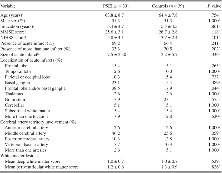
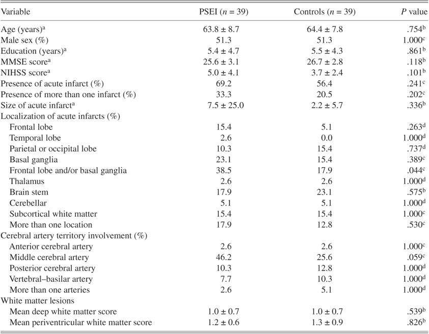
The demographic and MRI characteristics and stroke-related data stratified by PSEI status are shown in Table 1. There were no significant differences between the two groups with respect to age, sex, education, MMSE, NIHSS total score, the presence and size of acute infarcts, and the extent of white matter lesions. The PSEI group had significantly more acute infarcts in the frontal lobe and/or basal ganglia. In addition, there was a trend whereby the involvement of the middle cerebral artery territory was more frequent in patients with PSEI.
Demographic, clinical, and MRI variables of subjects with PSEI and their controls
a Mean ± standard deviation.
b Mann–Whitney U test.
c χ2 test.
d Fisher Exact test.
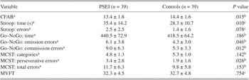
Executive functions stratified by PSEI status are displayed in Table 2. The PSEI group had a significantly lower CFAB score, required more time to complete the Stroop Test, and made more omission and commission errors in the Go–NoGo test. There was a trend for PSEI patients to make more errors in the Stroop Test and perseverative errors in the MCST. There was no significant difference between groups in terms of verbal fluency as measured by the MVFT.
Comparison between the PSEI and control groups with respect to executive functions

a Mean ± standard deviation.
b Mann–Whitney U test.
c t Test.
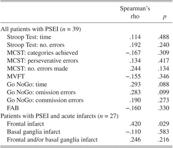
The severity of PSEI was very mild, mild, moderate, severe, and very severe in 5.2%, 51.3%, 28.2%, 3.7%, and 3.7% of subjects, respectively. The correlations between the severity of PSEI and all psychometric data were less than .3, and the Go–NoGo time and omission errors were of borderline significance. There was a modest but significant correlation between frontal infarct and PSEI severity (Table 3).
Correlation between the severity of PSEI and the location of acute infarcts and executive function

DISCUSSION
To the best of our knowledge, this is the first case–control study that has examined executive functions and frontal infarcts in stroke patients with EI.
In this study, PSEI was associated with frontal and/or basal ganglia lesions. This finding provides further support to the hypothesis that frontal–subcortical circuits may be involved in the development of PSEI. The common feature of these circuits is that they originate in frontal cortex, project to the striatum (caudate, putamen, ventral striatum), connect to the globus pallidus and substantia nigra, and from there connect to the thalamus (Cummings, Reference Cummings1993). These circuits have been implicated in a variety of neuropsychiatric disorders, including depression, obsessive compulsive disorder, schizophrenia, and substance abuse (Tekin & Cummings, Reference Tekin and Cummings2002). The pathways of these circuits might explain why lesions in different cortical and subcortical structures, such as frontal (Ghaffar et al., Reference Ghaffar, Chamelian and Feinstein2008; House et al., Reference House, Dennis, Molyneux, Warlow and Hawton1989; Lopez et al., Reference Lopez, Zivkovic, Smith, Becker, Meltzer and DeKosky2001), lenticulocapsular (Kim & Choi-Kwon, Reference Kim and Choi-Kwon2000), and dorsal globus pallidus (Kim, Reference Kim2002), could all lead to EI.
In the present study, brain stem infarcts were not found to be associated with PSEI. Although brain stem lesions may be involved in EI in nonstroke populations (Ghaffar et al., Reference Ghaffar, Chamelian and Feinstein2008), the evidence of their role in PSEI is more controversial as both positive (Asfora et al., Reference Asfora, DeSalles, Abe and Kjellberg1989; Kim et al., 2000) and negative (Tang et al., Reference Tang, Chan, Chiu, Ungvari, Wong, Kwok, Wong, Richards and Ahuja2004) results have been reported.
Our findings suggest that executive functions are impaired in patients with PSEI. Feinstein et al. (Reference Feinstein, Connor, Gray and Feinstein1999) evaluated executive functions in 13 MS patients who also presented with EI. The EI group performed worse in the Stroop Test than did the matched controls. In addition, the EI group performed more poorly in a verbal fluency test but not on a card sorting test. Similarly, in a controlled study, McCullagh et al. (Reference McCullagh, Moore, Gawel and Feinstein1999) examined executive functions in 10 ALS patients with EI and found that the EI group made more total errors in the Wisconsin Card Sorting Test than the matched control group. There was also a similar trend for perseverative errors. However, the performance of the EI group in a gambling task and a smelling test was similar. Piamarta et al. (Reference Piamarta, Iurlaro, Isella, Atzeni, Grimaldi, Russo, Forapani and Appollonio2004) found no association between PSEI status and Frontal Assessment Battery (FAB) score. No data have been published on the performance of patients with EI in the Go–NoGo test. The reasons for the discrepancy in the results could be differences in the nature of diseases, the diagnostic criteria of EI, the selection of executive function tests, small sample sizes, and the resulting type II error. It is interesting to note that as well as the more time-consuming psychometric tests, a brief bedside screening test such as the CFAB is able to detect the impairment of executive functions in patients with PSEI.
Piamarta et al. (Reference Piamarta, Iurlaro, Isella, Atzeni, Grimaldi, Russo, Forapani and Appollonio2004) reported no significant correlation between PSEI and FAB scores in 33 elderly stroke patients with supratentorial lesions. We could not find any significant correlation between the severity of PSEI and executive functions. The sample size of our study has an 80% power for a correlation ≥0.43 or more, hence modest correlations cannot be ruled out. Executive dysfunction associated with PSEI can be explained by the excess of lesions in the frontal–subcortical circuits. The dorsolateral prefrontal circuit, one of the frontal–subcortical circuits, is mainly responsible for executive functions. Lesions affecting the frontal–subcortical circuits were more frequent in stroke patients presenting with executive dysfunction (Vataja et al., Reference Vataja, Pohjasvaara, Mantyla, Ylikoski, Leppavuori, Leskela, Kalska, Hietanen, Aronen, Salonen, Kaste and Erkinjuntti2003).
We found significant correlation between the severity of PSEI and frontal infarcts. To the best of our knowledge, this correlation has not been reported previously. It suggests that frontal infarct is indeed involved in the development of PSEI or at least aggravates PSEI.
The main limitation of this study is its relatively small sample size, which resulted in a low power in detecting the potential differences between groups in terms of stroke severity (39%), frontal lobe infarcts (14%), and basal ganglia infarcts (32%). It also reduced the statistical power in determining the difference between groups in the executive functions in certain tests, such as the MVFT, and the correlation between the severity of PSEI and executive functions. Furthermore, the assessment of PSEI was made only once, at the 3-month follow-up. As patients who had died before the 3-month follow-up were not included in the study, the sample was biased to an unknown degree. Another potential weakness of the study is that MMSE was used to measure the overall cognitive function. MMSE is known to correlate with more comprehensive cognitive measures such as the Wechsler Adult Intelligence Scale (Folstein et al., Reference Folstein, Folstein and McHugh1975). It has been used to detect cognitive impairment in Chinese stroke survivors (Mok et al., Reference Mok, Wong, Lam, Fan, Tang, Kwok, Hui and Wong2004; Tang et al., Reference Tang, Chan, Chiu, Ungvari, Wong, Kwok, Mok, Wong, Richards and Ahuja2006). The PSEI and normal group had similar MMSE scores, which were well above the cutoff for dementia in Chinese populations (Chiu et al., Reference Chiu, Lee and Chung1994). Finally, the volume of old infarcts was not measured because, unlike a new infarct, their sizes shrunk with time (Caplan, Reference Caplan2000).
In summary, this study adds to the recently published data on the association between PSEI, executive functions, and frontal lobe involvement. A possible explanation for the three main findings, namely the association between PSEI and (1) frontal and/or basal ganglia lesions and (2) impairment in executive functions, and the (3) correlation between the severity of PSEI and frontal infarct, is that frontal–subcortical infarcts cause both PSEI and executive dysfunction. The above explanation, while biologically plausible, should be regarded as speculative at this point. Further exploration of executive functions in PSEI by functional imaging studies is warranted. Follow-up studies are needed to examine the temporal relationship between executive functions and PSEI.
ACKNOWLEDGMENTS
This work was supported by a Direct Grant for Research 2005/2006 from the Chinese University of Hong Kong.