Disease and clinical symptoms induced by bovine herpesvirus type 1 (BHV-1)
BHV-1 is an α-herpesvirinae subfamily member that causes significant economical losses to the cattle industry (Turin et al., Reference Turin, Russo and Poli1999). Three BHV-1 subtypes, BHV-1.1 (1), BHV-1.2a (2a) and BHV-1.2b (2b), have been identified based on antigenic and genomic analysis (Metzler et al., Reference Metzler, Matile, Gasman, Engels and Wyler1985). Subtype 1 virus isolates are the causative agents of infectious bovine rhinotracheitis (IBR) and are frequently found in the respiratory tract as well as aborted fetuses. Subtype 1 strains are prevalent in Europe, North America and South America. Subtype 2a is frequently associated with a broad range of clinical manifestations in the respiratory and genital tracts such as IBR, infectious pustular vulvovaginitis (IPV), balanopostitis (IPB) and abortions (Oirschot, Reference Oirschot1995). Subtype 2a is prevalent in Brazil and was present in Europe prior to the 1970s (Oirschot, Reference Oirschot1995). Subtype 2b strains are associated with respiratory disease and IPV/IPB, but not abortion (Oirschot, Reference Oirschot1995; D'Arce et al., Reference D'Arce, Almedia, Silva, Spilki, Roehe and Ams2002). Subtype 2b strains are less pathogenic than subtype 1 and are frequently isolated in Australia or Europe, but not Brazil (Edwards et al., Reference Edwards, White and Nixon1990).
In feedlot cattle, the respiratory form of BHV-1 is the most common (subtype 1 strains). In breeding cattle, abortions or genital infections tend to be more common. Genital infections can occur in bulls (IPB) and cows (IPV) within 1–3 days of mating or close contact with an infected animal. Transmission can also occur in the absence of visible lesions and through artificial insemination with semen from sub-clinically infected bulls.
The incubation period for the respiratory and genital forms of BHV-1 is 2–6 days (Yates, Reference Yates1982). With respect to respiratory disease, clinical symptoms include high fever, anorexia, coughing, excessive salivation, nasal discharge, conjunctivitis with lacrimal discharge, inflamed nares, and dyspneae if the larynx becomes occluded with purulent material. Nasal lesions consist of numerous clusters of grayish necrotic foci on the mucous membrane of septal mucosa. In the absence of bacterial pneumonia, recovery typically occurs 4–5 days after the onset of clinical symptoms. Abortions can occur at the same time as respiratory disease, but may also be seen up to 100 days after infection, which is likely due to reactivation from latency.
With respect to genital infections, the first clinical signs are frequent urination and a mild vaginal infection (Yates, Reference Yates1982). It is also common to observe swollen vulva or small papules followed by erosions and ulcers on the mucosal surface. In bulls, similar lesions occur on the penis and prepuce. If secondary bacterial infections occur, there may be inflammation of the uterus and transient infertility with purulent vaginal discharge for several weeks. In the absence of bacterial infections, animals usually recover within 2 weeks after infection. Regardless of the involvement of secondary bacterial infection, BHV-1 establishes lifelong latency following acute infection.
The seroprevelance of BHV-1 ranges from 14 to 60% in Africa and from 36 to 48% in Central and South America (Straub, Reference Straub and Morin1990). In The Netherlands, 49% of cows that give birth to their first calf are seropositive, whereas 91% of older cows are seropositive (Wuyckhuise et al., Reference Wuyckhuise, Van Bosch, Franken, Hage, Verhoeff and Zimmer1994). Serological testing and removal of infected animals has been successfully used to eliminate BHV-1 from Denmark, Switzerland and Austria (Ackermann and Engels, Reference Ackermann and Engels2005). In these countries, cattle populations are relatively small and movement of cattle can be controlled. With respect to the United States and other countries, eradication would be difficult, perhaps impossible, and very expensive.
The seroprevalence of BHV-1 in bison raised on a ranch is 43.8% (Sausker and Dyer, Reference Sausker and Dyer2002), indicating that infection can readily occur in bison. One would assume that BHV-1 is readily transmitted from buffalo to cattle and vice versa. Since more buffalo meat is being consumed each year in the United States, BHV-1 infections will have an impact on the emerging bison industry. It is not clear what the percentage of seropositive animals is due to vaccination. Nevertheless, it is clear that BHV-1 is widely disseminated in the field.
Relationship between BHV-1-induced immune suppression and bovine respiratory disease
Bovine respiratory disease complex (BRDC), also referred to as ‘shipping fever’, costs the US cattle industry at least $3 billion year−1 (Tikoo et al., 1995a, b; National Agricultural Statistics Service (NASS), 1996; Bowland and Shewen, Reference Bowland and Shewen2000). In addition to the clinical symptoms described above for BHV-1, BHV-1 can initiate BRDC by transiently suppressing the immune system of infected cattle (Yates, Reference Yates1982). BHV-1-induced immune suppression leads to secondary bacterial infections (Pasteurella haemolytica, Pasteurella multocida and Haemophilus somnus for example) that can cause pneumonia (Yates, Reference Yates1982). Increased susceptibility to secondary infection correlates with depressed cell-mediated immunity after BHV-1 infection (Griebel et al., Reference Griebel, Qualtiere, Davis, Gee, Bielefeldt Ohmann, Lawman and Babiuk1987a, Reference Griebel, Ohmann, Lawman and Babiukb, Reference Griebel, Ohmann, Lawman and Babiuk1990; Carter et al., Reference Carter, Weinberg, Pollard, Reeves, Magnuson and Magnuson1989). CD8+ T-cell recognition of infected cells is impaired by repressing expression of major histocompatibility complex (MHC) class I and the transporter associated with antigen presentation (Hariharan et al., Reference Hariharan, Nataraj and Srikumaran1993; Nataraj et al., Reference Nataraj, Eidmann, Hariharan, Sur, Perry and Srikumaran1997; Hinkley et al., Reference Hinkley, Hill and Srikumaran1998). CD4+ T-cell function is impaired during acute infection of calves because BHV-1 infects CD4+ T-cells and induces apoptosis (Winkler et al., Reference Winkler, Doster and Jones1999). Two viral genes, bICP0 and UL41.5, can inhibit specific immune responses in the absence of other viral genes. A discussion of these genes and how they inhibit immune responses is provided below.
The bICP0 protein activates viral gene expression and inhibits interferon (IFN) signaling pathway
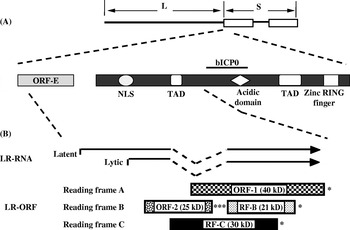
The bICP0 protein is the major transcriptional regulatory protein because it activates expression of all viral promoters (Everett, Reference Everett2000), and the bICP0 transcript is constitutively expressed during productive infection (Fraefel et al., Reference Fraefel, Zeng, Choffat, Engels, Schwyzer and Ackermann1994). The ICP0 homologues encoded by BHV-1 and herpes simplex virus type 1 (HSV-1) contain a well-conserved C3HC4 zinc RING finger near their respective N-termini (Fig. 1A). Mutational analysis has demonstrated the importance of the C3HC4 zinc RING finger domain of bICP0 and ICP0 (Everett, Reference Everett1987, Reference Everett1988; Everett et al., Reference Everett, Barlow, Milner, Luisi, Orr, Hope and Lyon1993; Inman et al., Reference Inman, Zhang, Geiser and Jones2001c). ICP0 (Maul et al., Reference Maul, Guldner and Spivack1993; Maul and Everett, Reference Maul and Everett1994; Everett et al., Reference Everett, Meredith, Orr, Cross, Kathoria and Parkinson1997, Reference Everett, Earnshaw, Findlay and Lomonte1999a, Reference Everett, Lomonte, Sternsdorf, van Driel and Orrb) and bICP0 (Parkinson and Everett, Reference Everett2000; Inman et al., Reference Inman, Zhang, Geiser and Jones2001c) colocalize with and disrupt the proto-oncogene promyelocytic leukemia protein-containing nuclear domains. Disruption of the bICP0 zinc RING finger prevents trans-activation of a simple viral promoter (Zhang and Jones, Reference Zhang and Jones2005) and impairs the ability of bICP0 to stimulate plaque formation following transfection of BHV-1 DNA into cultured bovine cells (Inman et al., Reference Inman, Zhang, Geiser and Jones2001c; Geiser and Jones, Reference Geiser and Jones2003). The bICP0 protein contains two transcriptional activation domains (TAD) and a nuclear localization signal (NLS) that is necessary for efficient transcriptional activation (Zhang and Jones, Reference Zhang and Jones2005) (Fig. 1A). Unlike most transcription factors, bICP0 or ICP0 does not apparently bind to specific DNA sequences, suggesting that bICP0 activates transcription by interacting with cellular transcriptional machinery. Support for this prediction comes from the finding that bICP0 associates with chromatin remodeling enzymes, histone deacetylase 1 (HDAC1) (Zhang, Reference Zhang and Jones2001) and p300, a histone acetyl transferase (Zhang and Jones, Reference Zhang and Jones2001). The ability of bICP0 to associate with HDAC1 and p300 correlates with activating viral transcription.
Schematic of bICP0, LR gene and ORF-E genes within the BHV-1 genome. (A) Schematic of BHV-1 genome, location of bICP0 and ORF-E. Domains within bICP0 include an NLS (nuclear localization sequence), TAD (transcriptional activation domain), acidic domain and the C3HC4 zinc RING finger. (B) The start sites for LR-RNA transcription during latency and productive infection were previously described (Devireddy and Jones, Reference Devireddy and Jones1998; Hossain et al., Reference Hossain, Schang and Jones1995). The C-termini of bICP0 and ORF-E overlap the LR gene and are antisense with respect to bICP0 and ORF-E. Reading frames B and C (RF-B and RF-C) each contain an open reading frame (ORF) that lacks an initiating Met. The (*) denotes the position of stop codons that are in frame with the respective ORF.

In the absence of other viral genes, bICP0 inhibits IFN signaling (Henderson et al., Reference Henderson, Zhang and Jones2005). bICP0 (directly or indirectly) reduces IRF3 (IFN regulatory factor 3) protein levels in human or bovine cells, which consequently leads to reduced IFN-β promoter activity (Saira et al., Reference Saira, Zhou and Jones2007). In addition, bICP0 inhibits the ability of IRF7 to activate IFN-β promoter activity, but bICP0 does not dramatically reduce IRF7 protein levels (Saira et al., Reference Saira, Zhou and Jones2007). The RING finger of ICP0 (Van Sant et al., Reference Van Sant, Hagglund, Lopez and Roizman2001; Boutell et al., Reference Boutell, Sadis and Everett2002; Boutell and Everett, Reference Boutell and Everett2003) and bICP0 (Dia et al., Reference Dia, Zhang, Fan, Gao, Sun, Yang, Xin, Jin, Geng and Wang2005) is an E3 ubiquitin ligase, suggesting that the bICP0 RING finger mediates IRF3 degradation. The finding that a functional proteasome is necessary for bICP0-induced IRF3 degradation (Saira et al., Reference Saira, Zhou and Jones2007) supports the notion that the bICP0 RING finger mediates IRF3 degradation. The RING finger is not the sole component necessary for bICP0-induced IRF3 degradation because specific mutations near the C-terminus of bICP0 are also important (Saira et al., Reference Saira, Zhou and Jones2007). Additional studies are necessary to understand how bICP0 inhibits IRF7 induction of IFN-β promoter activity as well as the mechanism by which bICP0 induces IRF3 degradation.
What role does IRF3 play during IFN production, and why is it important for bICP0 to target IRF3 functions? Following virus infection, two cellular protein kinases, IKK-ε or TBK-1, phosphorylate serine residues at the C-terminus of IRF3, which induces IRF3 homodimerization and nuclear translocation (Fitzgerald et al., Reference Fitzgerald, McWhirter, Faja, Rowe, Latz, Golenbock, Coyle, Liao and Maniatis2003; Sharma et al., Reference Sharma, ten Oever, Grandvaux, Zhou, Lin and Hiscott2003). Nuclear IRF3 associates with other transcriptional activators resulting in direct binding and stimulation of IFN-β promoter activity (Wathelet et al., Reference Wathelet, Lin, Parekh, Ronco, Howley and Maniatis1998). IRF3 also directly binds several consensus DNA-binding sites, including ISRE (IFN response elements), and can consequently stimulate transcription of IFN-stimulated genes in the absence of IFN (Guo et al., Reference Guo, Peters and Sen2000; Mossman et al., Reference Mossman, Macgregor, Rozmus, Goryachev, Edwards and Smiley2001). IRF3 activation is an immediate early regulator of the IFN response, whereas IRF7 is believed to be a component of the early response (Yoneyama et al., Reference Yoneyama, Suhara, Fukuhara, Fukuda, Nishida and Fujita1998; Yuang et al., Reference Yuang, Lowther, Kellum, Au, Lin, Hiscott and Pitha1998; Sato et al., Reference Sato, Taniguchi and Tanaka2001). Surprisingly, IRF7 is more important with respect to inhibiting viral infection when IRF3 versus IRF7 knockout mice are compared (Honda et al., Reference Honda, Yanai, Negishi, Asagiri, Saton, Mizutani, Shimada, Ohba, Takaoka, Yoshida and Taniguchi2005). Since BHV-1 does not grow in mice unless they lack IFN receptors (Abril et al., Reference Abril, Engels, Limman, Hilbe, Albini, Franchini, Suter and Ackerman2004), it is clear that the ability of BHV-1 to inhibit innate immune responses, IFN for example, is crucial for pathogenesis in cattle and host range.
UL49.5
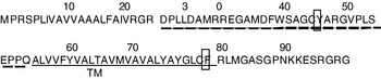
The BHV-1 UL49.5 open reading frame (ORF), also known as glycoprotein N (gN), encodes a 96-amino-acid protein with an apparent molecular mass of 9 kDa (Liang et al., Reference Liang, Tang, Manns, Babiuk and Zamb1993). The UL49.5 ORF contains a signal peptide (N-terminal 22 amino acids), an extracellular domain of 32 amino acids, a transmembrane region of 25 amino acids and a 17 amino acids long cytoplasmic tail (Liang et al., Reference Liang, Tang, Manns, Babiuk and Zamb1993) (Fig. 2). The UL49.5 protein is expressed as a non-glycosylated type I membrane protein (Liang et al., Reference Liang, Tang, Manns, Babiuk and Zamb1993, Reference Liang, Chow, Raggo and Babiuk1996).
Schematic of the BHV-1 gN protein. Predicted amino acid sequences of the BHV-1 gN (UL49.5) protein (120). Predicted signal sequence amino-terminal 22 amino acids (shaded letters), luminal domain (dashed line) and transmembrane domain (underlined) are indicated. Conserved cysteine residue (C42) within the luminal domain and a cysteine residue within the transmembrane domain are boxed.

All gN herpesvirus orthologues studied to date form complexes with glycoprotein M (gM), which is encoded by the UL10 ORF (Jöns et al., Reference Jöns, Dijkstra and Mettenleiter1998; Wu et al., Reference Wu, Zhu and Letchworth1998; Rudolph et al., Reference Rudolph, Seyboldt, Granzow and Osterrieder2002). gM is important for secondary envelopment because in PRV secondary envelopment in the Golgi and subsequent egress require gM, in the absence of gE (Brack et al., Reference Brack, Dijkstra, Granzow, Klupp and Mettenleiter1999). The gN homologues encoded by PRV and BHV-1 inhibit transporter-associated antigen processing (TAP)-mediated transport of cytosolic peptides into ER, which consequently blocks the assembly of peptide-containing ternary MHC-I complexes in vitro in virus-infected cells (Koppers-Lalic et al., Reference Koppers-Lalic, Reits, Ressing, Lipinska, Abele, Koch, Rezende, Admiraal, van Leeuwen, Bienkowska-Szewczyk, Mettenleiter, Rijsewijk, Tampé, Neefjes and Wiertz2005; Lipińska et al., Reference Lipińska, Koppers-Lalic, Rychlowski, Admiraal, Rijsewijk, Bieńkowska-Szewczyk and Wiertz2006). Furthermore, the BHV-1 gN targets the TAP complex for proteosomal degradation (Koppers-Lalic et al., Reference Koppers-Lalic, Reits, Ressing, Lipinska, Abele, Koch, Rezende, Admiraal, van Leeuwen, Bienkowska-Szewczyk, Mettenleiter, Rijsewijk, Tampé, Neefjes and Wiertz2005). The TAP complex consists of a TAP1/TAP2 heterodimer, both of which are members of the ATP-binding cassette transporter superfamily (Androlewicz et al., Reference Androlewicz, Anderson and Cresswell1993; van Endert et al., Reference van Endert, Saveanu, Hewitt and Lehner2002; Koch et al., Reference Koch, Gruntrum, Heintke, Kyritisis and Tampe2004).
Peptide transport by TAP is a critical step in MHC class I antigen presentation (Ahn et al., Reference Ahn, Meyer, Uebel, Sempe, Djaballah, Yang, Peterson, Fruh and Tampe1996; Hughes et al., Reference Hughes, Hammond and Cresswell1997; Ambagala et al., Reference Ambagala, Gopinath and Srikumaran2004; Koppers-Lalic et al., Reference Koppers-Lalic, Reits, Ressing, Lipinska, Abele, Koch, Rezende, Admiraal, van Leeuwen, Bienkowska-Szewczyk, Mettenleiter, Rijsewijk, Tampé, Neefjes and Wiertz2005). In the absence of a functional TAP transporter, most MHC class I molecules are not loaded with peptides (Hughes et al., Reference Hughes, Hammond and Cresswell1997). However, they are retained within the ER and ultimately directed for degradation by the proteasome (Hughes et al., Reference Hughes, Hammond and Cresswell1997). Structurally, both gM and TAPs are similar because they both contain multiple membrane spanning segments (Wu et al., Reference Wu, Zhu and Letchworth1998; Koch et al., Reference Koch, Gruntrum, Heintke, Kyritisis and Tampe2004). A gN mutant that lacks the cytoplasmic tail can still bind to the TAP complex and block peptide transport, but this mutant gN protein does not degrade TAP (Koppers-Lalic et al., Reference Koppers-Lalic, Reits, Ressing, Lipinska, Abele, Koch, Rezende, Admiraal, van Leeuwen, Bienkowska-Szewczyk, Mettenleiter, Rijsewijk, Tampé, Neefjes and Wiertz2005). However, when the transmembrane domain in addition to the cytoplasmic tail is truncated, gN no longer blocks TAP function (Koppers-Lalic et al., Reference Koppers-Lalic, Reits, Ressing, Lipinska, Abele, Koch, Rezende, Admiraal, van Leeuwen, Bienkowska-Szewczyk, Mettenleiter, Rijsewijk, Tampé, Neefjes and Wiertz2005). Therefore, sequences within the gN transmembrane domain are likely to interact with TAP. The phenotype of a viral strain expressing the various gN mutant proteins has not been tested in the context of a virus and/or virally infected cells. In summary, the ability of gN to inhibit TAP is hypothesized to prevent virus-infected cells from being killed by CD8+ T-cells.
Viral encoded structural proteins mediate cell entry and pathogenesis
To fully understand the role that BHV-1 plays in BRDC, one must understand how BHV-1 initiates productive infection. Consequently, this section summarizes what we know about how the virus enters cultured cells and disseminates in vivo.
α-Herpesvirus glycoproteins are required for viral particles to bind and penetrate permissive cells. In addition, a subset of viral glycoproteins is necessary for virus release, cell fusion and cell-to-cell spread. Ten of the 12 BHV-1 envelope proteins (gB, gC, gD, gE, gG, gH, gI, gL, gM and gK) are glycosylated and two (gN and Us9) are non-glycosylated (Van Drunen Littel-van den Hurk and Babiuk, Reference Van Drunen and Babiuk1986; Liang et al., Reference Liang, Tang, Manns, Babiuk and Zamb1993; Baranowski et al., Reference Baranowski, Dubuisson, van Drunen, Babiuk, Michel, Pastoret and Thiry1995, Reference Baranowski, Keil, Lyaku, Rijsewijk, Van Oirschot, Pastoret and Thiry1996; Khattar et al., Reference Khattar, van Drunen, Attah-Poku, Babiuk and Tikoo1996; Schwyzer and Ackermann, Reference Schwyzer and Ackermann1996; Brideau et al., Reference Brideau, Banfield and Enquist1998; Meyer et al., Reference Meyer, Hanon, Georlette, Pastoret and Thiry1998; Mettenleiter, Reference Mettenleiter2000, Reference Mettenleiter2002; König et al., Reference König, Giesow and Keil2002; Nakamichi et al., Reference Nakamichi, Matsumota and Otsuka2002; Chowdhury et al., Reference Chowdhury, Mahmood, Simon, Al-Mubarak and Zhou2006). Five envelope proteins (gB, gD, gH, gL and gK) are essential for growth in cultured cells, whereas the remaining seven (gC, gE, gG, gI, gM, gN and Us9) are not essential for growth in cultured cells (Schwyzer and Ackermann, Reference Schwyzer and Ackermann1996; Brideau et al., Reference Brideau, Banfield and Enquist1998; Butchi et al., Reference Butchi, Jones, Perez, Doster and Chowdhury2007).
All α-herpesvirus gC homologues (including BHV-1) facilitate attachment of virus particles to the cell surface by binding to heparan sulfate proteoglycan (Okazaki et al., Reference Okazaki, Matsuzaki, Sugahara, Okada, Hasebe, Iwamura, Ohnishi, Kanno, Shimizu, Honda and Kono1991). In the absence of gC, gB complements the attachment function of gC by binding to heparan sulfate (Li et al., Reference Li, Van Drunen, Babiuk and Liang1995; Mettenleiter, Reference Mettenleiter2002). Following initial attachment mediated by gB and/or gC, the next step in viral entry requires the interaction of gD with the cellular receptor nectin 1, also known as HveC (Spear, Reference Spear1993, Reference Spear2004; Campadelli-Fiume et al., Reference Campadelli-Fiume, Cocchi and Menotti2000, Reference Campadelli-Fiume, Amasio, Avitabile, Cerretani, Forghieri, Gianni and Menotti2007; Spears et al., Reference Spear, Eisenberg and Cohen2000; Mettenleiter, Reference Mettenleiter2002; Spear and Longnecker, Reference Spear and Longnecker2003; Campadelli-Fiume and Gianni, Reference Campadelli-Fiume, Gianni and Sandri-Goldin2006), or HVEM, a member of the tumor necrosis factor family of proteins (Montgomery et al., Reference Montgomery, Warner, Lum and Spear1996). Nectin 1 belongs to a family of intercellular adhesion molecules whose ectodomain contains three immunoglobulin-like domains (Ogita and Takai, Reference Ogita and Takai2006). Certain members of the tumor necrosis factor family can induce apoptosis (Schmitz et al., Reference Schmitz, Kirchhoff and Krammer2000), suggesting that an interaction between gD and HVEM induces apoptosis in certain cell types (Hanon et al., Reference Hanon, Meyer, Vanderplasschen, Dessy-Doize, Thiry and Pastoret1998). Following gD-receptor binding, gB and/or a gH–gL complex interacts with cellular membrane protein(s) leading to fusion of the viral envelope to the cell membrane (Pertel et al., Reference Pertel, Fridberg, Parish and Spear2001; Mettenleiter, Reference Mettenleiter2002; Spear et al., Reference Spear, Manoj, Yoon, Jogger, Zago and Myscofski2006; Campadelli-Fiume et al., Reference Campadelli-Fiume, Amasio, Avitabile, Cerretani, Forghieri, Gianni and Menotti2007; Reske et al., Reference Reske, Pollara, Krummenacher, Chain and Katz2007). The mechanism of viral and cellular membrane fusion is not well understood other than the fact that gD-receptor binding must occur prior to the fusion process (Mettenleiter, Reference Mettenleiter2002; Spear et al., Reference Spear, Manoj, Yoon, Jogger, Zago and Myscofski2006). Viral entry into an adjacent cell (cell-to-cell spread) also requires gD because gD-deleted viruses are incapable of cell-to-cell spread (Fehler et al., Reference Fehler, Herrman, Saalmuller, Mettenleiter and Keil1992). In stably transformed cells expressing gD, cell-to-cell spread of wild-type (wt) virus is restricted (Dasika and Letchworth, Reference Dasika and Letchworth1999), suggesting that gD binds to putative cellular receptors, which are present on the apical cell membrane at the cell surface and lateral cell membrane located at intercellular junctions (Dasika and Letchworth, Reference Dasika and Letchworth1999; Campadelli-Fiume et al., Reference Campadelli-Fiume, Cocchi and Menotti2000, Reference Campadelli-Fiume, Amasio, Avitabile, Cerretani, Forghieri, Gianni and Menotti2007; Sakisaka et al., Reference Sakisaka, Taniguchi, Nakanishi, Takahashi, Miyahara, Ikeda, Yokoyama, Peng, Yamanishi and Takai2001; Campadelli-Fiume and Gianni, Reference Campadelli-Fiume, Gianni and Sandri-Goldin2006). Although gD is an essential glycoprotein for virus entry into the cell, a compensatory mutation in gH at amino acid residue 450 (mutation of glycine to tryptophan) mediates gD-independent entry and cell-to-cell spread of a gD-deleted virus (Schroder et al., Reference Schroder, Linde, Fehler and Keil1997).
Interactions between gE and gI also promote cell-to-cell virus spread, and gE and/or gI deletion mutants are defective in cell-to-cell spread (Dingwell and Johnson, Reference Dingwell and Johnson1998; Chowdhury et al., Reference Chowdhury, Ross, Lee, Hall and Chu1999; Mettenleiter, Reference Mettenleiter2000, Reference Mettenleiter2002). Based on results from HSV-1 studies, it appears that gE binds to a putative cellular receptor located at cellular junctions (Dingwell and Johnson, Reference Dingwell and Johnson1998). Of the non-essential envelope proteins, gE has received a lot of attention because it is required for anterograde neuronal spread in vivo and neurovirulence (Enquist et al., Reference Enquist, Husak, Banfield and Smith1999). gE-deleted viruses are transported retrogradely from the nose and eye via the maxillary and ophthalmic branch of trigeminal ganglia (TG) where they establish latency in sensory neurons (Enquist et al., Reference Enquist, Husak, Banfield and Smith1999; Chowdhury et al., Reference Chowdhury, Lee, Ozkul and Weiss2000; Liu et al., Reference Liu, Coats, Jones and Chowdhury2007). gE-deleted viruses do not reactivate from latency because they cannot spread in an anterograde direction from sensory neurons in TG to non-neuronal cells in the nose or ocular cavity (Enquist et al., Reference Enquist, Husak, Banfield and Smith1999; Liu et al., Reference Liu, Coats, Jones and Chowdhury2007).
The envelope protein (Us9) is also important for anterograde neuronal spread because calves latently infected with Us9-deleted viruses do not shed virus in the nose and eye following dexamethasone-induced reactivation (Butchi et al., Reference Butchi, Jones, Perez, Doster and Chowdhury2007). Interestingly, Us9-deleted BHV-1 and pseudorabies virus (PRV) do not have a phenotype following infection of epithelial cells in culture (S. I. Chowdhury, unpublished data). With respect to PRV, the Us9-deleted virus has a defective anterograde neuronal spread phenotype because in the absence of Us9, axonal transport of viral glycoproteins or vesicles containing viral glycoproteins does not occur (Tomishama and Enquist, Reference Tomishama and Enquist2001). Therefore, the Us9 function is neuron-specific and is required only in the context of anterograde neuronal transport.
BHV-1 gG is secreted after proteolytic processing. In addition, gG is present on the virus envelope and is associated with infected cell membranes (Bryant et al., Reference Bryant, Davis-Poynter, Vanderplasschen and Alcami2003). Like other α-herpesvirus gG homologues, the BHV-1 gG has chemokine binding activity because it blocks the interaction of chemokines with cellular receptors and glycol-aminoglycans (GAGs). The chemokine binding activity of gG also occurs on cell membranes because membrane-anchored forms of gG bind to various chemokines (Bryant et al., Reference Bryant, Davis-Poynter, Vanderplasschen and Alcami2003). Deletion of the BHV-1 gene encoding gG leads to viral attenuation in calves because the mutant virus is more immunogenic (Kaashoek et al., 1996b). The chemokine binding activity encoded by BHV-1 gG is responsible for the attenuated phenotype following infection of calves (Bryant et al., Reference Bryant, Davis-Poynter, Vanderplasschen and Alcami2003). In summary, these studies indicate that numerous viral genes are necessary for initiating productive infection and cell-to-cell spread.
The BHV-1 latency–reactivation cycle of BHV-1
Acute infection leads to high levels of virus production
In the field, it is recognized that BRDC is not always associated with acute BHV-1 infection. It is generally believed that the ability of BHV-1 to reactivate from a latent infection can initiate BRDC. The goal of this section is to summarize our current knowledge of the BHV-1 latency–reactivation cycle.
Acute BHV-1 infection is initiated on mucosal surfaces and results in high levels of programmed cell death (Winkler et al., Reference Winkler, Doster and Jones1999). Infection of permissive cells (Devireddy and Jones, Reference Devireddy and Jones1999) with BHV-1 also leads to rapid cell death, in part, due to apoptosis. Viral gene expression is temporally regulated in three distinct phases: immediate early (IE), early (E) or late (L). IE gene expression is stimulated by a virion component, α-TIF (Misra et al., Reference Misra, Walker, Hayes and O'Hare1995). Two IE transcription units exist: IE transcription unit 1 (IEtu1) and IEtu2. IEtu1 encodes functional homologues of two HSV-1 IE proteins, ICP0 and ICP4. IEtu2 encodes a protein that is similar to the HSV IE protein, ICP22 (Wirth et al., Reference Wirth, Vogt and Schwyzer1991). In general, IE proteins activate E gene expression, and then viral DNA replication occurs. L gene expression is also activated by bICP0, culminating in virion assembly and release. As discussed above, bICP0 is important for productive infection because it transcriptionally activates all viral promoters, and is expressed at high levels throughout infection (Wirth et al., Reference Wirth, Vogt and Schwyzer1991, Reference Wirth, Fraefel, Vogt, Vlcek, Paces and Schwyzer1992; Fraefel et al., Reference Fraefel, Zeng, Choffat, Engels, Schwyzer and Ackermann1994). Acute infection leads to high levels of virus production and secretion in ocular, oral or nasal cavities. If acute infection is initiated in the genital tract, virus shedding can be readily detected in genital tissues. Regardless of the site of infection, virus shedding can last for 7–10 days after infection (Jones, Reference Jones1998, Reference Jones2003).
Summary of the latency–reactivation cycle in cattle
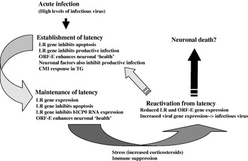
Viral particles, or perhaps subviral particles, enter the peripheral nervous system via cell–cell spread. If infection is initiated via the oral cavity, nasal cavity or ocular orifice, the primary site for latency is sensory neurons within TG. Relatively high levels of viral gene expression (Schang and Jones, Reference Schang and Jones1997) or infectious virus (Inman et al., Reference Inman, Lovato, Doster and Jones2002) can be detected in TG from 1–6 days after infection (see Fig. 3, for a summary of the latency–reactivation cycle of BHV-1). Viral gene expression and detection of infectious virus are subsequently extinguished, but viral genomes can be detected in TG (establishment of latency). In contrast to productive infection in cultured cells, a significant number of infected neurons survive, and these neurons harbor latent genomes. A hallmark of latency is the abundant level of transcription that occurs from the latency related (LR) gene (Jones, Reference Jones1998, Reference Jones2003; Jones et al., Reference Jones, Geiser, Henderson, Jiang, Meyer, Perez and Zhang2006) and ORF-E (Inman et al., Reference Inman, Zhou, Webb and Jones2004). It appears that the LR transcript is the first viral transcript expressed in infected neurons because we have detected a spliced LR transcript in TG at 24 h after calves were infected (Devireddy and Jones, Reference Devireddy and Jones1998). In contrast, we were not able to detect IE or E gene expression at the same time using sensitive RT-PCR assays, suggesting that LR gene products play a pivotal role in programming the outcome of virus infection in sensory neurons. Support for this prediction comes from the finding that LR gene products promote establishment of latency by inhibiting apoptosis (Ciacci-Zanella et al., Reference Ciacci-Zanella, Stone, Henderson and Jones1999; Henderson et al., Reference Henderson, Perng, Nesburn, Wechsler and Jones2004) and viral gene expression (Bratanich et al., Reference Bratanich, Hanson and Jones1992; Geiser et al., Reference Geiser, Inman, Zhang and Jones2002). ORF-E may promote efficient establishment of latency because it can induce neurite-like outgrowth in mouse neuroblastoma cells (Perez et al., Reference Perez, Meyer, Henderson, Jiang, Sherman, Doster, Inman and Jones2007). Consequently, ORF-E may enhance restoration of mature neuronal functions following infection. During maintenance of latency, infectious virus is not detected using standard virological methods. Since the LR gene and ORF-E are abundantly expressed during the maintenance of latency, they are predicted to play an active role in maintaining latency.
Summary of BHV-1 latency–reactivation cycle. For details, see the subsection ‘Summary of the latency–reactivation cycle in cattle’.

Elevated corticosteroid levels (stress) and/or immune suppression can initiate reactivation from latency. The stress associated with moving cattle from one location to another is one obvious stimulus that can trigger reactivation from latency and BRDC. During reactivation from latency, three significant events occur: (i) productive viral gene expression is readily detected in sensory neurons, (ii) ORF-E and LR gene expression decrease dramatically and (iii) infectious virus is secreted from nasal or ocular swabs (Jones, Reference Jones1998, Reference Jones2003; Jones et al., Reference Jones, Geiser, Henderson, Jiang, Meyer, Perez and Zhang2006). Administration of dexamethasone to calves or rabbits latently infected with BHV-1 reproducibly leads to activation of viral gene expression and reactivation from latency (Rock et al., Reference Rock, Lokensgard, Lewis and Kutish1992; Jones, Reference Jones1998; Reference Jones2003; Jones et al., Reference Jones, Newby, Holt, Doster, Stone, Ciacci-Zanella, Webster and Jackwood2000, Reference Jones, Geiser, Henderson, Jiang, Meyer, Perez and Zhang2006; Inman et al., Reference Inman, Lovato, Doster and Jones2002). Although many latently infected neurons do not apparently produce infectious virus, a much higher number of neurons are detected in which viral gene expression occurs (Rock et al., Reference Rock, Lokensgard, Lewis and Kutish1992), suggesting that virus-producing neurons are not common.
Non-neural sites of latency–persistence in cattle
Although establishment of latency in ganglionic neurons is the main site of latency for BHV-1 and other α-herpesvirinae subfamily members, latent or persistent infections also occur in non-neural sites, tonsils and lymph nodes for example. BHV-1 DNA is consistently detected in tonsils (Winkler et al., Reference Winkler, Doster and Jones2000), peripheral blood cells (Fuchs et al., Reference Fuchs, Hubert, Detterer and Rziha1999), lymph nodes and spleen of latently infected calves, even when infectious virus is not detected (Mweene et al., Reference Mweene, Okazaki and Kida1996). Pseudorabies virus (Sabo and Rajanci, Reference Sabo and Rajanci1976; Cheung, Reference Cheung1995), equine herpesvirus type 4 (Borchers et al., Reference Borchers, Wolfinger and Ludwig1999) and canine herpesvirus type 1 (Miyoshi et al., Reference Miyoshi, Ishii, Takiguchi, Takada, Yasuda, Hashimoto, Okasaki and Kida1999) DNA is also detected in lymphoid tissue during latency. It remains to be seen what non-neural cell types are latently infected with BHV-1, and whether viral genes are expressed in these latently infected cells. In contrast to latency in sensory neurons, LR-RNA is not abundantly expressed in latently infected lymphoid tissue (Winkler et al., Reference Winkler, Doster and Jones2000). Infectious virus can be detected when germinal centers from tonsil of latently infected calves are explanted (Perez et al., Reference Perez, Inman, Doster and Jones2005) adding support to the hypothesis that BHV-1 has established a latent or persistent infection in cells of lymphoid origin.
The LR gene is abundantly expressed during latency and is necessary for the latency–reactivation cycle
As discussed above, LR-RNA is abundantly transcribed in latently infected neurons (Rock et al., Reference Rock, Beam and Mayfield1987, Reference Rock, Lokensgard, Lewis and Kutish1992; Kutish et al., Reference Kutish, Mainprize and Rock1990), and is antisense relative to the bICP0 gene (Fig. 1A). The lytic start site for LR-RNA is downstream of the start site used in TG (Bratanich et al., Reference Bratanich, Hanson and Jones1992; Hossain et al., Reference Hossain, Schang and Jones1995) (Fig. 1B). The LR gene has two ORFs (ORF-1 and ORF-2), and two reading frames that lack an initiating ATG (RF-B and RF-C) (Fig. 1B). A peptide antibody directed against ORF-2 recognizes a protein encoded by the LR gene (Hossain et al., Reference Hossain, Schang and Jones1995; Jiang et al., Reference Jiang, Hossain, Winkler, Holt, Doster and Jones1998, Reference Jiang, Inman, Zhang, Posadas and Jones2004). A fraction of LR-RNA is polyadenylated and alternatively spliced in TG, suggesting that more than one LR protein is expressed (Hossain et al., Reference Hossain, Schang and Jones1995; Devireddy and Jones, Reference Devireddy and Jones1999). LR gene products inhibit cell proliferation, bICP0 RNA expression (Bratanich et al., Reference Bratanich, Hanson and Jones1992; Schang et al., Reference Schang, Hossain and Jones1996; Geiser et al., Reference Geiser, Inman, Zhang and Jones2002), and apoptosis (Ciacci-Zanella et al., Reference Ciacci-Zanella, Stone, Henderson and Jones1999). LR protein expression is necessary for inhibiting apoptosis, but not cell growth (Geiser and Jones, Reference Geiser and Jones2005) or bICP0 expression (Bratanich et al., Reference Bratanich, Hanson and Jones1992; Schang et al., Reference Schang, Hossain and Jones1996; Geiser et al., Reference Geiser, Inman, Zhang and Jones2002).
A mutant BHV-1 strain with three stop codons at the N-terminus of ORF-2 (Inman et al., Reference Inman, Lovato, Doster and Jones2001b) does not express ORF-2 or RF-C following infection of bovine cells (Jiang et al., Reference Jiang, Inman, Zhang, Posadas and Jones2004). As expected, wt or the LR rescued virus express ORF-2 and RF-C. Calves infected with the LR mutant virus exhibit diminished clinical symptoms, and reduced shedding of infectious virus from the eye, TG or tonsil (Inman et al., Reference Inman, Lovato, Doster and Jones2001b, Reference Inman, Lovato, Doster and Jones2002; Perez et al., Reference Perez, Inman, Doster and Jones2005). The LR mutant virus does not reactivate from latency following treatment with dexamethasone, whereas all calves latently infected with wt virus or the LR rescued virus shed infectious virus following dexamethasone treatment (Inman et al., Reference Inman, Lovato, Doster and Jones2002). Thus, the LR gene is necessary for the latency–reactivation cycle in calves. It is not clear whether LR proteins are necessary for stimulating reactivation from latency or whether these products establish and maintain latency in a pool of neurons that are capable of supporting reactivation from latency.
Previous studies determined that the LR transcript is alternatively spliced in TG during acute versus latent infections (Devireddy and Jones, Reference Devireddy and Jones1998; Devireddy et al., Reference Devireddy, Zhang and Jones2003). A novel alternatively spliced cDNA is expressed in TG at 7 days after infection (7d cDNA), which is the transition from acute infection to establishment from latency. The 7d cDNA has the potential to encode the N-terminal domain of ORF-2 fused to ORF-1, suggesting that this novel fusion protein promotes establishment of latency. The 7d cDNA was used in a two-hybrid screen to identify cellular proteins that interact with the novel LR fusion protein encoded by the 7d cDNA (Meyer et al., Reference Meyer, Perez, Geiser, Sintek, Inman and Jones2007). The LR fusion protein interacts with two proteins that can induce apoptosis (Bid and Cdc42), and with the CCAAT enhancer-binding protein α (C/EBP-α). Bid links the extrinsic pathway of apoptosis to the intrinsic pathway because following cleavage by caspase 8, Bid interacts with mitochondria (Li et al., Reference Li, Zhu, Xu and Yuan1998), which leads to cytochrome c and Smac/Diablo release (Wang, Reference Wang2001). Bid is also specifically cleaved by granzyme B (Barry et al., Reference Barry, Heibein, Pinkoski, Lee, Moyer, Green and Bleackley2000; Alimonti et al., Reference Alimonti, Shi, Baijal and Greenberg2001; Pinkoski et al., Reference Pinkoski, Waterhouse, Heibein, Wolf, Kuwana, Goldstein, Newmeyer, Bleackley and Green2001), suggesting that the ability of LR proteins to interact with Bid impairs cytotoxic T-lymphocyte (CTL)-induced death of infected neurons. Neuronal stress, including removal of nerve growth factor from neuronal cultures (Kanamoto et al., Reference Kanamoto, Mota, Takeda, Rubin, Miyazono, Ichijo and Bazenet2000; Xu et al., Reference Xu, Maroney, Dobrzanski, Kukekov and Greene2001), induces Cdc42 activity, a Rho GTPase family member (Harwood and Braga, Reference Harwood and Bragga2003). When PC12 cells (rat neuronal-like cell line) are treated with nerve growth factor, they differentiate and behave like mature sympathetic neurons (Greene and Tischler, Reference Greene and Tischler1976; Greene et al., Reference Greene, Aletta, Rukenstein and Green1987). If PC12 cells or primary sympathetic neurons are deprived of nerve growth factor, Cdc42 is activated resulting in apoptosis (Kanamoto et al., Reference Kanamoto, Mota, Takeda, Rubin, Miyazono, Ichijo and Bazenet2000; Xu et al., Reference Xu, Maroney, Dobrzanski, Kukekov and Greene2001). Since dexamethasone is a potent stressor and reproducibly induces BHV-1 reactivation from latency (Sheffy and Davies, Reference Sheffy and Davies1972; Ackermann et al., Reference Ackermann, Peterhans and Wyler1982; Homan and Easterday, Reference Homan and Easterday1983; Ackermann and Wyler, Reference Ackermann and Wyler1984; Brown and Field, Reference Brown and Field1990; Rock et al., Reference Rock, Lokensgard, Lewis and Kutish1992; Jones et al., Reference Jones, Newby, Holt, Doster, Stone, Ciacci-Zanella, Webster and Jackwood2000; Winkler et al., Reference Winkler, Doster, Sur and Jones2002), Cdc42 may be activated in TG.
The interaction between the LR fusion protein and C/EBP-α may also influence BHV-1 gene expression because C/EBP-α stimulates lytic gene transcription of other herpesviruses, EBV, HHV-8 and HVS for example (Wang et al., Reference Wang, Wu, Yu and Hayward2003b; Wu et al., Reference Wu, Wang, Chen, Wang, Hayward and Hayward2004; Wakenshaw et al., Reference Wakenshaw, Walters and Whitehouse2005; Huang et al., Reference Huang, Liao, Chen, Wu, Hutt-Fletcher, Hayward and Hayward2006). Three lines of evidence support the finding that C/EBP-α enhances BHV-1 productive infection: (i) C/EBP-α protein expression is stimulated during productive infection, (ii) there is a positive correlation between viral gene expression in TG and C/EBP-α protein expression and (iii) wt C/EBP-α, but not a DNA binding mutant, enhances BHV-1 plaque formation (Meyer et al., Reference Meyer, Perez, Geiser, Sintek, Inman and Jones2007). It will be of interest to determine whether C/EBP-α directly stimulates viral promoters or if it indirectly activates productive infection.
The BHV-1 LR gene and HSV-1 LAT (latency-associated transcript) share structural and functional properties. For example, LR-RNA and LAT are transcribed in an antisense direction of bICP0 and ICP0 respectively, and both are abundantly expressed during latency (Rock et al., Reference Rock, Beam and Mayfield1987; Kutish et al., Reference Kutish, Mainprize and Rock1990; Hossain et al., Reference Hossain, Schang and Jones1995; Schang and Jones, Reference Schang and Jones1997; Devireddy and Jones, Reference Devireddy and Jones1998; Winkler et al., Reference Winkler, Doster and Jones2000). Furthermore, the LR gene (Ciacci-Zanella et al., Reference Ciacci-Zanella, Stone, Henderson and Jones1999) and LAT (Perng et al., Reference Perng, Jones, Ciacci-Zanella, Stone, Henderson, Yukht, Slanina, Hoffman, Ghiasi, Nesburn and Wechsler2000; Inman et al., Reference Inman, Perng, Henderson, Ghiasi, Nesburn, Wechsler and Jones2001a; Ahmed et al., Reference Ahmed, Lock, Miller and Fraser2002) inhibit apoptosis. To test whether the LR gene restores spontaneous reactivation to a HSV-1 LAT deletion mutant (dLAT2903), a 2 kb fragment containing the LR promoter and LR coding sequences was inserted into the LAT locus of dLAT2903, and the recombinant virus designated CJLAT (Perng et al., Reference Perng, Maguen, Jin, Mott, Osorio, Slanina, Yukht, Ghiasi, Nesburn, Inman, Henderson, Jones and Wechsler2002). Insertion of the LR gene into the HSV-1 LAT locus restores high levels of spontaneous reactivation in the rabbit eye model and in explant-induced reactivation. Rabbits infected with CJLAT have higher levels of recurrent eye disease (stromal scarring and detached retinas). Further evidence for the expanded pathogenic properties of CJLAT came from the finding that CJLAT is more lethal in mice relative to LAT+ or LAT− strains of HSV-1. In contrast, insertion of the LR gene with stop codons into dLAT2903 generated a virus that behaves like the parental LAT-null mutant adding further support that LR protein expression regulates the latency–reactivation cycle (Mott et al., Reference Mott, Osorio, Jin, Brick, Naito, Cooper, Henderson, Inman, Jones, Wechsler and Perng2003).
ORF-E encodes a protein that is expressed in TG of latently infected calves
A small ORF that is present within the LR promoter has been designated ORF-E (Fig. 1A). ORF-E is antisense to the LR transcript and downstream of the bICP0 ORF. A transcript that encompasses ORF-E is expressed in productively infected bovine cells and TG of latently infected calves (Inman et al., Reference Inman, Zhou, Webb and Jones2004). When ORF-E protein coding sequences are fused in frame with green fluorescent protein (GFP) sequences, GFP protein expression is detected in the nucleus of mouse or human neuroblastoma cells. In contrast, the ORF-E–GFP fusion protein is expressed throughout rabbit skin cells.
Polyclonal antiserum directed against an ORF-E peptide or the entire ORF-E protein specifically recognizes the nucleus of sensory neurons in TG of latently infected calves and mouse neuroblastoma cells transfected with an ORF-E-expressing plasmid (Perez et al., Reference Perez, Meyer, Henderson, Jiang, Sherman, Doster, Inman and Jones2007). ORF-E also induces neurite-like projections in these same mouse neuroblastoma cells. In contrast, neurite-like projections are not observed following transfection of mouse neuroblastoma cells with an empty vector, suggesting that ORF-E induces changes in the physiology of neuronal cells. It will be necessary to construct a BHV-1 mutant that does not express the ORF-E protein to test whether ORF-E plays a role in the latency–reactivation cycle.
Immune response to BHV-1 following acute infection
Although BHV-1 has the potential to immune-suppress cattle, a potent immune response ultimately occurs, which prevents systemic infection. With respect to BRDC, this implies that immunosuppression initiated by BHV-1 is short-lived.
The host immune response to BHV-1 infection includes innate and adaptive immune responses (Rouse and Babiuk, Reference Rouse and Babiuk1978). Innate immune responses include the antiviral action of IFN, alternative complement pathway and local infiltration of lymphoid cells, macrophages, neutrophils or natural killer (NK) cells for example. Following BHV-1 infection, IFN-α and IFN-β are detectable in nasal secretions as early as 5 h post-infection, reach a maximum level at 72–96 h post-infection and can persist for up to 8 days after infection (Straub and Ahl, Reference Straub and Ahl1976). During early times after infection, IFN-α and IFN-β promote leukocyte migration, activate macrophages and increase NK cell activity (Babiuk et al., Reference Babiuk, Bielefeldt, Gifford, Czarniecki, Scialli and Hamilton1985; Lawman et al., Reference Lawman, Gifford, Gyongyossy-Issa, Dragan, Heise and Babiuk1987; Jensen and Schultz, Reference Jensen and Schulz1990). Activation of macrophages and increasing NK cell activity stimulate cytolytic activities against virus-infected cells (Rouse and Babiuk, Reference Rouse and Babiuk1978). NK cells are a diverse population of non-adherent effecter cells that lack T- and B-cell markers (Cook and Splitter, Reference Cook and Splitter1989). These cells require a long incubation period with the target cell for optimum lysis (Campos and Rossi, Reference Campos and Rossi1986). In addition, NK-like cytotoxicity is also associated with a population of CD3+ CD45+, Fc receptor-positive lymphocytes, which may represent a subset of γδ T-cells (Amadori et al., Reference Amadori, Archetti, Vernadi and Berneri1992).
Adaptive or humoral immune responses lead to production of neutralizing antibodies that bind virus particles and inhibit productive infection. Envelope glycoproteins gB, gC, gD and gH are the most potent inducers of virus neutralizing antibodies (Van Drunen Littel-van den Hurk, Reference Van Drunen and Babiuk1986; Marshall et al., Reference Marshall, Israel and Letchworth1988). In addition, non-neutralizing antibody may mediate the destruction of enveloped virus or cells expressing viral proteins on the cell membranes, and this process is referred to as antibody-mediated cell cytotoxicity (Van Drunen Littel-van den Hurk et al., Reference Van Drunen, Tikoo, Liang and Babiuk1993). Neutralizing and non-neutralizing antibodies produced against envelope proteins can also inhibit virus infection by several distinct mechanisms: (i) membrane attack complex (MAC) lysis of virus envelope and virus-infected cells mediated by antibody and complement and (ii) antibody-mediated cell cytotoxicity in which either IgG interacts with Fc receptor-positive cells (macrophages) or (iii) binding of C3b to IgM mediates binding to C3b receptor-positive cells (lymphocytes, macrophages). In all cases, virus-infected cells are lysed (Rouse et al., Reference Rouse, Wardley and Babiuk1976, Reference Rouse, Grewal and Babiuk1977a, Reference Rouse, Wardley and Babiukb; Rouse and Babiuk, Reference Rouse and Babiuk1977, Reference Rouse and Babiuk1978). Production of virus neutralizing and non-neutralizing antibodies (IgG) can be detected 8–12 days post-infection (Rouse and Babiuk, Reference Rouse and Babiuk1978). Local/mucosal immunity depends on secreted neutralizing antibodies (IgA molecules) and systemic humoral immunity depends on IgG.
Cell-mediated immune (CMI) responses play an important role in killing virus-infected cells that express viral antigens on the cell surface (Van Drunen Littel-van den Hurk, Reference Van Drunen2007). For example, a CD8+ CTL response is an important defense against BHV-1 because cell-to-cell spread in upper respiratory epithelium occurs before hematogenous spread (Van Drunen Littel-van den Hurk, Reference Van Drunen2007). Cytotoxic and proliferatve T lymphocyte responses occur in the blood approximately 8 days after infection. CTL responses have been induced by gB and gD DNA vaccines in mice (Deshpande et al., Reference Deshpande, Ambagala, Hegde, Hariharan, Navaratnam and Srikumaran2002; Huang et al., Reference Huang, Babiuk and van Drunen2005). Further, gC and gD have been identified as targets for CTL responses in cattle (Denis et al., Reference Denis, Slaoui, Keil, Babiuk, Ernst, Pastorer and Thiry1993) and gB DNA vaccines elicit a CTL response in cattle (Huang et al., Reference Huang, Babiuk and van Drunen2005). Other structural and non-structural viral proteins may also play a role in CMI response because only a limited number of BHV-1 proteins have been evaluated for CTL activity (Van Drunen Littel-van den Hurk, Reference Van Drunen2007). In addition to destruction of infected cells, T-lymphocytes release a number of lymphokines that modulate specific and non-specific immune response. For example, IFN-γ and other factors that further activate macrophages (Campos et al., Reference Campos, Bielefeldt, Hutchings, Rapin and Babiuk1989) are produced. BHV-1 proteins (gB-, gC-, gD- and VP8) are recognized by CD4+ T helper cells from immune cattle (Hutchings et al., Reference Hutchings, van Drunen and Babiuk1990). Cells expressing gB, gC or gD on their membranes have also been identified as targets for CD4+ T-cells (Leary and Splitter, Reference Leary and Splitter1990; Tikoo et al., 1995a, b). Finally, CD4+ T-cells are important for the development of antibody response and for developing effective CD8+ T-cell memory (Janssen et al., Reference Janssen, Lemmens, Wolfe, Christen, von Herrath and Schoenberger2003; Shedlock and Shen, Reference Shedlock and Shen2003). Therefore, both CD4+ and CD8+ T cells as well as antibodies are required for long-term protection.
A prolonged CMI response occurs in the peripheral nervous system during latency
Several independent studies have demonstrated that T-cells, CD8+ T-lymphocytes in particular, control HSV-1 infection in sensory ganglia during latency (Nash et al., Reference Nash, Jayasuriya, Phelan, Cobbold, Waldmann and Prospero1987; Simmons and Tscharke, Reference Simmons and Tscharke1992; Simmons et al., Reference Simmons, Tscharke and Speck1992; Cantin et al., Reference Cantin, Hinton, Chen and Openshaw1995; Shimeld et al., Reference Shimeld, Whiteland, Nicholls, Grinfeld, Easty, Gao and Hill1995; Halford et al., Reference Halford, Gebhardt and Carr1996; Liu et al., Reference Liu, Tang and Hendricks1996; Shimeld et al., Reference Shimeld, Whiteland, Williams, Easty and Hill1996, Reference Shimeld, Whiteland, Williams, Easty and Hill1997). Viral antigen-positive neurons surrounded by non-neural cells expressing TNF-α, IL-2, IL-6, IL-10 or IFN-γ can also be detected in mice latently infected with HSV-1 (Halford et al., Reference Halford, Gebhardt and Carr1996; Liu et al., Reference Liu, Tang and Hendricks1996; Shimeld et al., Reference Shimeld, Whiteland, Williams, Easty and Hill1997). No cells with lymphoid cell morphology are detected in TG of uninfected mice. A persistent infiltration of lymphoid cells in sensory ganglia and spinal cord is also detected in mice or guinea pigs latently infected with HSV-2 (Milligan et al., Reference Milligan, Meador, Chu, Young, Martin and Bourne2005). Following infection of calves with BHV-1, foci of infiltrating lymphocytes can be readily detected during acute infection (Winkler et al., Reference Winkler, Doster, Sur and Jones2002). Finally, in human TG latently infected with HSV-1, a chronic immune response has also been observed (Theil et al., Reference Theil, Derfuss, Paripovic, Herberger, Meinl, Schueler, Strupp, Arbusow and Brandt2003; Verjans et al., Reference Verjans, Hintzen, van Dun, Poot, Milikan, Laman, Langerak, Kinchington and Osteerhaus2007). Collectively, these studies indicate that long-term persistence of T-cells in sensory ganglia during latency has the potential to regulate the latency–reactivation cycle.
Persistence of immune cells in TG may be due to viral protein expression in a rare neuron or satellite cell. Careful examination of TG neurons for viral gene expression in HSV-1 latently infected mice (37–47 days after infection) demonstrated that abundant viral transcripts, viral protein and viral DNA replication occur in approximately 1 neuron per 10 TG (Feldman et al., Reference Feldman, Ellison, Voytek, Yang, Krause and Margolis2002). Infectious virus is not detected in these mice, confirming that they are latently infected. Neurons expressing high levels of HSV-1 transcripts are invariably surrounded by foci of infiltrating white blood cells. The term ‘spontaneous molecular reactivation’ has been coined to describe these rare neurons (Feldman et al., Reference Feldman, Ellison, Voytek, Yang, Krause and Margolis2002). Rare cells in TG of calves latently infected with BHV-1 can be detected that express viral transcripts and proteins (Winkler et al., Reference Winkler, Doster, Sur and Jones2002), suggesting that molecular spontaneous reactivation occurs during BHV-1 latency.
CD8+ T-cells that produce IFN-γ are believed to play an important role in preventing reactivation from latency in sensory neurons in mice latently infected with HSV-1 (Liu et al., Reference Liu, Khanna, Chen, Fink and Hendricks2000, Reference Liu, Khanna, Carriere and Hendricks2001). Two independent studies have also concluded that IFN-α and IFN-γ control recurrent herpetic lesions (Cunningham and Mikloska, Reference Cunningham and Mikloska2001; Mikloska and Cunningham, Reference Mikloska and Cunningham2001). In addition to IFN, lymphocyte-mediated cytotoxicity could inhibit virus spread in TG. Lymphocyte-mediated cytotoxicity induces two potent apoptotic pathways: the granule exocytosis and the Fas–Fas ligand pathway (Kagi and Hengartner, Reference Kagi and Hengartner1996; Shresta et al., Reference Shresta, Pham, Thomas, Graubert and Ley1998). The granule exocytosis pathway is employed predominantly by CD8+, NK and lymphokine-activated killer cells. It appears that the BHV-1 LR gene plays a role in regulating lymphocyte infiltration because enhanced infiltration of lymphocytes occurs in TG of calves acutely infected with the LR mutant virus (Perez et al., Reference Perez, Lovato, Zhou, Doster and Jones2006). The ability of dexamethasone to induce apoptosis of inflammatory cells in TG of calves latently infected with BHV-1 correlates with reactivation from latency (Winkler et al., Reference Winkler, Doster, Sur and Jones2002).
BHV-1 vaccines
Many commercially available vaccines directed against BHV-1 can cause disease in small calves, in part because these vaccines are immunosuppressive. Consequently, certain vaccines have the potential to induce BRDC in small calves. A discussion of the current status of BHV-1 vaccines and how they can be improved is included in the following paragraphs.
Commercially available vaccines
Commercially available vaccines directed against BHV-1 consist of modified live attenuated virus (MLV) or killed whole virus (KV) (Van Drunen Littel-van den Hurk, 2005). In many cases, MLVs are derived by serial passage in tissue culture. KV vaccines are usually produced by chemical treatments, such as formaldehyde, β-propiolactone or binary ethyleneimine. MLVs generally induce both humoral and cellular immune response because virus replication in infected cells leads to presentation of viral antigen on MHC class I+ and II+ molecules. Safety is a concern for MLVs because these strains can establish latency and upon reactivation can be transmitted to pregnant cows, which can lead to abortion. MLVs can also be pathogenic in small calves because their immune system is not fully developed, and most MLVs can be immunosuppressive. KV vaccines are usually safe but they are not as efficacious because they usually produce only humoral immunity but no cellular immune responses. Additionally, KV vaccines always require more than one injection to achieve acceptable neutralizing antibody levels. In the case of formaldehyde-inactivated KV, antigens may also be denatured, which may affect the immunogenicity of vaccine preparations. KV also requires suitable adjuvant formulations and some adjuvants may induce injection site reactions.
Commercially available BHV-1 vaccines are primarily evaluated on induction of neutralizing antibodies and the duration as well as level of virus shedding following challenge of vaccinated animals. As discussed earlier, the CMI response against the virus is important to prevent cell-to-cell spread of the virus. Therefore, a direct measure of CMI directed against a vaccine virus is important. Traditionally, IFN-γ levels in vaccinated calves have been an indicator of cellular immune responses. However, a recent study demonstrated that there is an increase in CD25 by CD4+, CD8+ and γδT lymphocytes from a BHV-1 MLV-vaccinated group (Endsley et al., Reference Endsley, Quade, Terhaar and Roth2002). Therefore, increased expression of CD25, in addition to IFN-γ production by T-cells after vaccination, appears to be a useful cellular immunity marker (Van Drunen Littel-van den Hurk, Reference Van Drunen2007).
Recently, there has been an apparent increase in IBR outbreaks in vaccinated feedlot cattle (commonly referred to as vaccine outbreaks) (Van Drunen Littel-van den Hurk et al., Reference Van Drunen, Myers, Doig, Karvonen, Habermehl, Babiuk, Jelinski, Van Donkersgoed, Schlesinger and Rinehart2001; Ellis et al., Reference Ellis, Waldner, Rhodes and Ricketts2005). Many of these vaccine outbreaks occur in feedlots that have used several different BHV-1 MLVs without serological markers, suggesting that they are not vaccine-specific. Since the vaccines used in the feedlots did not have any serological marker, it is difficult to determine by serology whether an animal is infected with an MLV or field strain. Thus, determining the source of origin from a particular vaccine break is not possible nor is it possible to test whether these outbreaks were due to changes in the vaccine strain. It is also possible that there is the emergence of a new IBR strain that is not covered by existing MLVs. A study investigating several isolates from such outbreaks determined that at least one such isolate had mutations within sequences comprising the gD-specific neutralizing epitope (Van Drunen Littel-van den Hurk et al., Reference Van Drunen, Myers, Doig, Karvonen, Habermehl, Babiuk, Jelinski, Van Donkersgoed, Schlesinger and Rinehart2001).
Development of genetically engineered gene-deleted BHV-1 vaccines
During the last 10–15 years, the usefulness of genetically engineered gene-deleted vaccines has become increasingly apparent because they can be attenuated and serologically distinguished from wt field strains (Van Drunen Littel-van den Hurk, Reference Van Drunen2005). Numerous viral mutants (gC-, gE-, gG-, Us9-deleted (Kaashoek et al., Reference Kaashoek, Fijsewijk, Ruuls, Keil, Thiry, Pastoret and Van Oirschot1998; Chowdhury et al., Reference Chowdhury, Ross, Lee, Hall and Chu1999; Butchi et al., Reference Butchi, Jones, Perez, Doster and Chowdhury2007), thymidine kinase (TK)-deleted (Chowdhury, Reference Chowdhury1996; Kaashoek et al., 1996a, b) and LR gene mutant virus (Inman et al., Reference Inman, Lovato, Doster and Jones2002)) have been constructed and their in vivo pathogenic properties, reactivation properties and immunogenicity analyzed. Based on recent studies, both gE- and Us9-deleted viruses were safe in calves because they do not reactivate from latency and they are highly attenuated (Kaashoek et al., 1996a, b, Reference Kaashoek, Fijsewijk, Ruuls, Keil, Thiry, Pastoret and Van Oirschot1998; Chowdhury et al., Reference Chowdhury, Ross, Lee, Hall and Chu1999; Butchi et al., Reference Butchi, Jones, Perez, Doster and Chowdhury2007; Liu et al., Reference Liu, Coats, Jones and Chowdhury2007). Studies with gC-, gG- or TK-deleted viruses showed that they either reactivate from latency and/or they retain some degree of virulence (Kaashoek et al., Reference Kaashoek, van Engelenburg, Moerman, Gielkens, Fijsewijk and van Oirschot1996a, Reference Kaashoek, Rijsewijk and Oirschotb, Reference Kaashoek, Fijsewijk, Ruuls, Keil, Thiry, Pastoret and Van Oirschot1998). Studies with a LR mutant virus showed that the virus does not reactivate from latency and has reduced pathogenicity in calves (Inman et al., Reference Inman, Lovato, Doster and Jones2002). Therefore, considering the virulence and reactivation properties of gene-deleted vaccine strains, gE-, Us9-deleted or the LR mutant virus have the potential to be safer vaccine candidates. Comparative vaccine efficacy studies showed that relative to gC- and gG-deleted viruses, gE-deleted virus is less efficacious (Kaashoek et al., Reference Kaashoek, Fijsewijk, Ruuls, Keil, Thiry, Pastoret and Van Oirschot1998). However, the gE-deleted vaccine has been used successfully to eradicate IBR from a number of European countries. Although the gE-deleted marker vaccine is not as efficacious as others noted above, it will be used until a better genetically engineered vaccine is developed.
Recent efforts to improve the IBR marker vaccine are directed to delete viral gene sequences that are immunosuppressive. As noted above, gN inhibits TAP and down-regulates MHC-I antigen presentation. Therefore, a vaccine virus lacking the gN TAP binding domain may stimulate better cellular immune responses. Our recent studies show that a gE cytoplasmic tail truncated virus is similarly attenuated in calves infected with the virus as the entire gE ORF-deleted virus. Notably, like the gE-deleted virus, the gE cytoplasmic tail truncated virus does not reactivate from latency (Liu et al., Reference Liu, Coats, Jones and Chowdhury2007). Since the cytoplasmic tail-specific amino acid sequences generate antibodies that immunoprecipitate gE, antibodies specific to the cytoplasmic tail sequence may serve as a serological marker to distinguish vaccinated versus infected calves (Whitbeck et al., Reference Whitbeck, Knapp, Enquist, Lawrence and Bello1996; Chowdhury et al., Reference Chowdhury, Lee, Ozkul and Weiss2000). Furthermore, the Us9 gene is located immediately downstream of the gE cytoplasmic tail gene; therefore, a recombinant BHV-1 can be constructed in which the gE cytoplasmic tail and the Us9 coding regions are deleted. A vaccine virus that lacks US9, the cytoplasmic tail of gE, and gN TAP binding domain may be a superior vaccine candidate because it: (i) should stimulate better cellular immune responses against BHV-1, (ii) will incorporate a serological marker and (iii) should not be pathogenic. Concerns have been raised about recombination occurring between two vaccine strains, which could lead to the presence of virulent viral strains in vaccinated herds (Thriy et al., Reference Thriy, Muylkens, Meurens, Gogev, Thiry, Vandelplasschen and Schynts2005). Although it is clear that recombination can occur, the process requires that both viruses must replicate at the same time in the same cell of an infected calf.
Vectored vaccine strains
Intranasal vaccination of young calves with a replication competent (E3-deleted) bovine adenovirus (BAV)-3 that expresses full-length BHV-1 gD induced protective gD-specific neutralizing antibodies and cellular immune responses (Zakhartchouk et al., Reference Zakhartchouk, Pyne, Mutwiri, Papp, Baca-Estrada, Griebel, Babiuk and Tikoo1999). Several studies also concluded that gD-specific bacterial plasmid-based DNA vaccines can serve as an effective subunit vaccine. Specifically, an expression plasmid that encodes the secreted form of gD (tgD) induces significant protection against BHV-1 challenge following intradermal injection (Van Drunen Littel-van den Hurk et al., Reference Van Drunen, Braun, Lewis, Karvonen, Baca-Estrada, Snider, Cartney, Watts and Babiuk1998; Braun et al., Reference Braun, Babiuk, Loehr and van Drunen1999). Relative to killed BHV-1 vaccine, the tgD DNA vaccine induced significant IFN-γ production in PBMCs. Regardless of maternal antibody levels, the plasmid encoding tgD also triggered neutralizing antibodies, lymphoproliferative responses and IFN-γ-secreting cells in newborn lambs, suggesting that this approach may be efficacious for neonate immunization (Van Drunen Littel-van den Hurk et al., Reference Van Drunen, Braun, Lewis, Karvonen, Babiuk and Griebel1999).
Mucosal delivery of gB-based plasmid DNA vaccines has also shown promising results. Delivery of plasmid- based BHV-1 gB DNA vaccine in the female vulvomucosal epithelium induced gB-specific antibodies and cellular immune responses. Furthermore, vaccinated animals have significantly reduced genital lesions upon genital challenge (Huang et al., Reference Huang, Babiuk and van Drunen2005). Recent studies using a combined gB–gD DNA vaccine in mice indicated that mice immunized with the combined vaccine containing the secreted forms of the respective glycoproteins developed higher titers of anti-BHV-1 neutralizing antibodies and increased cellular immune response compared to single plasmid injected groups (Caselli et al., Reference Caselli, Boni, Di Luca, Salvatori, Vita and Cassai2005).
Use of BHV-1 as a vaccine vector
BHV-1-based vectors have significant advantages relative to other potential virus vectors for immunization of cattle against economically important viral and bacterial diseases, including those that initiate BRDC. Cattle are routinely immunized against BHV-1 and, therefore, the use of BHV-1 as a vector would not pose a new risk to cattle or humans. Gene-deleted BHV-1 strains could be used to express other bacterial and viral gene coding sequences. BHV-1 expressing the BVD (bovine diarrhea virus) gP53 (E2) protein has been constructed (Kweon et al., Reference Kweon, Kang, Choi and Kang1999; Wang et al., Reference Wang, Whitbeck, Lawrence, Volgin and Bello2003a). While the protective efficacy of the BHV-1 vectored vaccine against BVD challenge has not yet been reported, BHV-1 expressing the E2 protein induced BVD gP53-specifc neutralizing antibody in vaccinated calves (Kweon et al., Reference Kweon, Kang, Choi and Kang1999). BVD is well known to be immunosuppressive and, like many RNA viruses, can readily mutate (Peterhans et al., Reference Peterhans, Jungi and Schweizer2006). Such complications from modified live BVD vaccines could be avoided by using BHV-1 as live attenuated vector to deliver BVD-specific protein(s). Similarly, BHV-1-expressing bacterial proteins could be used to immunize against bacterial diseases that affect dairy and beef cattle. These may include Mannheimia hemolytica protective antigens, which are a significant cause of pneumonia that occurs as a result of shipping fever. Similarly, Mycobacterium avium subspecies paratuberculosis (MAP) is a slow growing bacterium with zoonotic potential (Grant, Reference Grant2005). Development of an effective vaccine is hindered due to its extremely slow growth and difficulties in protective bacterial antigen purification. Several mycobacterial proteins trigger cellular immune responses (Rossels et al., Reference Rossels, Marche, Roupie, Govaerts, Godfroid, Walravens and Huygen2006), which are important for protective immunity against MAP. BHV-1-expressing MAP proteins would certainly have a potential as a vaccine against MAP and BHV-1. Although BHV-1 can be used as a potential vector for delivering foreign antigens to calves, a thorough understanding of the immune-suppressive nature of BHV-1 and the latency–reactivation cycle is essential.
Acknowledgments
The laboratory of CJ is supported by two USDA grants (2005-01554 and 2006-01627), a grant from NIAID (R21AI069176) and, in part, a Public Health Service grant 1P20RR15635 to the Nebraska Center for Virology. The laboratory of SC is supported by two USDA grants (00-02103 and 2004-35204-14657).